|
|
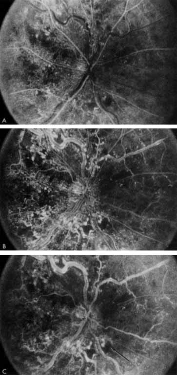
| Fig. 4 Fluorescein angiogram after moderately ischemic central retinal vein occlusion. A. Early venous phase. The capillary bed is dilated and engorged. Punctate areas of fluorescence represent microaneurysms or small areas of capillary leakage. B. Midvenous phase. There is considerable delay in venous return and an increase in and coalescence of punctate areas of extravascular fluorescence. C. Late venous phase. Fluorescence staining along the vein margins and scattered areas of capillary nonperfusion (arrow) are present. |