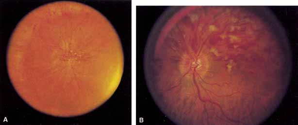
Fig. 7
Equator-plus
(A)
and 30° photograph
(B)
of a hemispheric branch retinal vein occlusion.