Chapter 29
Neuro-ophthalmic Aspects of Orbital Disease
MANOJ M. THAKKER and JAMES C. ORCUTT
Main Menu   Table Of Contents

Search

THE AFFERENT SYSTEM
THE EFFERENT SYSTEM
OTHER CRANIAL NERVES
CASE EXAMPLES
REFERENCES

The orbit is a unique structure, a bony cave of approximately 1-fluid ounce volume, opened widely at the entrance and tapering to an opening smaller than a pencil at the apex. Through this space runs the most complex nerve of the body, the optic nerve, which contains more than a million individual nerve fibers. More than half our sensory knowledge about our environment travels to the brain through the 2 optic nerves. In addition, there are 10 other nerves, 4 major blood vessels, and the origins of 5 muscles surrounding the optic nerve at the orbital apex—all in a space no larger than a dime. It is not surprising that examination of these nerves, vessels, and muscles can help the ophthalmologist exquisitely localize an orbital disease process.

The techniques of neuro-ophthalmic examination and orbital examination are both discussed elsewhere in Volume 2. The readers of this chapter should acquire an appreciation of the usefulness of neuro-ophthalmic examination techniques applied to orbital diagnosis. This chapter is divided into three sections: the afferent system—vision and sensation; the efferent system—eye movement, autonomic nerves and facial innervation; and examples of clinical cases demonstrating the applications of the neuro-ophthalmic examination to orbital diagnosis. Anatomy remains the bridge between examination and diagnosis and so is reviewed with each section. Additional anatomic detail is available elsewhere in these volumes as well as in other texts.1–3

Back to Top
THE AFFERENT SYSTEM

ANATOMY

Orbit

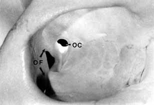
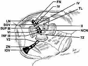
The orbital apex has two major openings, the orbital fissure and the optic canal (Fig. 1). The orbital fissure is divided into a superior and inferior portion by the tendon of Zinn, the inferior insertion for the tendinous anulus of Zinn (Fig. 2).1,4 (The anulus of Zinn is completed by the origins of the rectus muscles and the superior tendon of Lockwood.) Structures emanating through the optic canal (the optic nerve, ophthalmic artery, and orbital sympathetic nerves) as well as some structures coursing through the superior orbital fissure (the superior and inferior branches of the oculomotor nerve (cranial nerve III), the abducens nerve (cranial nerve VI) and the nasociliary branch of the ophthalmic division (cranial nerve V1) of the trigeminal nerve pass through the anulus of Zinn.

Fig. 1 Two openings are present at the orbital apex: the orbital fissure (OF) and the optic canal (OC).

Fig. 2 Structures at the orbital apex. The anulus of Zinn is composed of the tendon of Zinn (TZ), tendon of Lockwood (TL), and the insertions of the rectus muscles. Structures exiting the superior orbital fissure outside the anulus of Zinn include the lacrimal nerve (LN), superior ophthalmic vein (SOV), the frontal nerve (FN), the trochlear nerve (IV). and the anastomotic branch between the middle meningeal artery and lacrimal artery (not shown). Structures passing through the anulus of Zinn include the superior (SUP III) and the inferior (INF III) branch of the oculomotor nerve, the nasociliary nerve (NCN), the abducens (VI), and the optic nerve (II) with its associated ophthalmic artery and sympathetic nerves. Structures exiting through the inferior orbital fissure include the infraorbital nerve (V2), the inferior ophthalmic vein (IOV), and the zygomatic branch of the infraorbital nerve (ZN). (Courtesy of Deborah D. Sherman, MD, Nashville, TN)

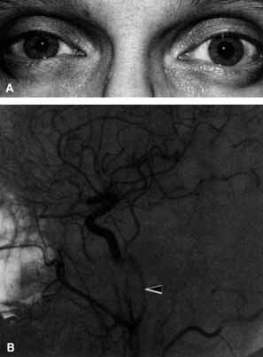
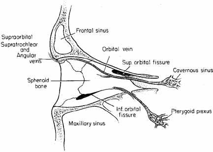
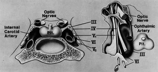
The orbital vascular supply is provided primarily by the ophthalmic artery (Fig. 3), and its branches, the central retinal artery, muscular branches, posterior ciliary arteries, posterior and anterior ethmoidal arteries, and the lacrimal artery. Venous drainage of the orbit is primarily by the superior ophthalmic vein into either the cavernous sinus or the pterygoid plexus (Fig. 4). A variable but generally lesser amount of venous outflow leaves through the inferior orbital vein into the pterygoid plexus. Numerous anastomoses are generally present between the superior and inferior ophthalmic veins.

Fig. 3 Orbital arterial supply derives from the internal carotid artery (InCA) by the first major intracranial branch, the ophthalmic artery (OA). The OA branches into the central retinal artery, muscular branches, posterior ciliary artery (PCA), posterior ethmoidal artery (PEA), anterior ethmoidal artery (AEA), and the lacrimal artery (LA) eventually terminating as the supratrochlear artery (STA) and supraorbital artery (SOA). (Courtesy of Deborah D. Sherman. MD, Nashville, TN)

Fig. 4 Orbital venous drainage occurs primarily by the superior ophthalmic vein (SOV) through the superior orbital fissure into the cavernous sinus, A lesser venous drainage occurs by the inferior orbital vein (IOV) through the inferior orbital fissure into the pterygoid plexus. Numerous anastomoses are common between these veins.

Cranial Nerve II (Optic Nerve)

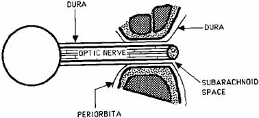
The optic nerve is contained within a dural sheath, which is continuous at the orbital apex with the periosteum of the optic canal. Intracranially the periosteum is continuous with the dura, thus providing access of cerebrospinal fluid to the sub-arachnoid space surrounding the optic nerve (Fig. 5). Elevated cerebrospinal fluid pressure, meningeal inflammation, or air from pneumocephalus can extend into the orbit within the optic sheath (Fig. 6).5

Fig. 5 Longitudinal section of the optic nerve through the optic canal. The intracranial subarachnoid space is continuous with the subarachnoid space surrounding the optic nerve. The subarachnoid space extends anteriorly to the level of the lamina cribrosa.

Fig. 6 T2-weighted magnetic resonance imaging (MRI) shows cerebrospinal fluid (CSF) surrounding the optic nerve within the subarachnoid space (arrow). The CSF extends to the posterior pole of the globe. An optic nerve meningioma is present on the contralateral side.

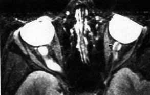
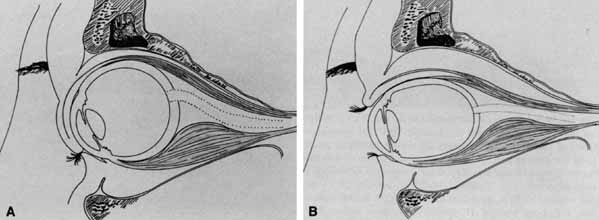
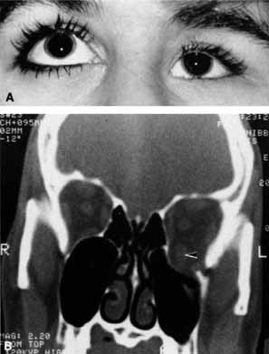
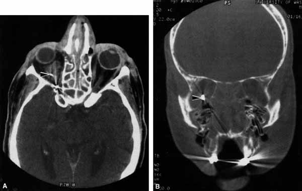
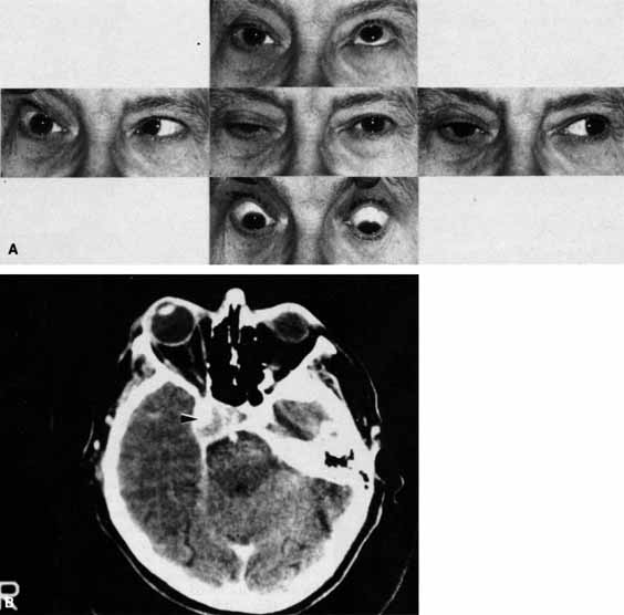
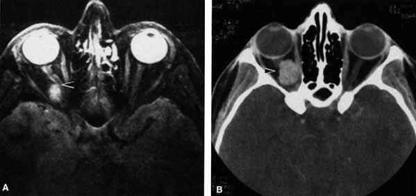
The optic nerve remains vulnerable to compression within the bony confines of the orbit. Compressive optic neuropathy is most likely to occur where the optic nerve is tethered to the bone, that is, at the orbital apex (Fig. 7A). The optic nerve in other areas of the orbit can be displaced markedly without suffering vision loss (Fig. 7B).

Fig. 7 A. A cavernous hemangioma measuring 1.5 cm in longest dimension is located at the orbital apex displacing the optic nerve (arrow). Visual acuity is 20/40. there is a relative afferent pupil defect, and visual field shows a central scotoma. B. A cavernous hemangioma measuring 2 cm in longest dimension is located in the midorbit displacing the optic nerve (arrow). Visual acuity is 20/20. There is no relative afferent pupil defect and the visual field is normal.

Cranial Nerve V (Trigeminal Nerve)

The ophthalmic branch of the trigeminal sensory nerve (cranial nerve V1) arises in the trigeminal ganglion, travels forward within the infralateral wall of the cavernoussinus and divides into its three major branches, the nasociliary, lacrimal, and frontal branches (Fig. 8).

Fig. 8 Anatomy of motor and sensory nerves within the cavernous sinus.

The lacrimal and frontal nerves pass above the anulus of Zinn along with the trochlear nerve (cranial nerve IV), the superior ophthalmic vein, and the anastomotic branch of the middle meningeal artery to the lacrimal artery. The frontal branch of cranial nerve V1, provides sensory input from the upper eyelid and forehead, while the lacrimal branch receives its input from the lacrimal gland and the superior temporal eyelid. The lacrimal branch is unique because its terminal branches carry the efferent parasympathetic fibers for lacrimal gland secretion, received from the zygomaticotemporal nerve near the lateral orbital wall.

The nasociliary branch of cranial nerve V1 gives off branches to the globe (long and short posterior ciliary nerves) before terminating as the infratrochlear nerve that provides sensory innervation to the superior medial eyelid. A small, external nasal branch is also given off with the anterior ethmoidal artery that extends under the nasal mucosa to innervate the tip of the nose.

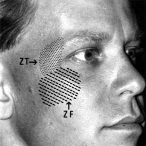
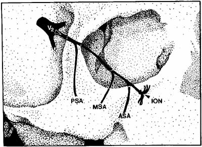
The infraorbital fissure transmits the infraorbital nerve, a branch of the maxillary division (cranial nerve V2) of the trigeminal nerve (see Fig. 2). Branches of the infraorbital nerve rise in the lateral orbit, providing sensation to the temple (zygomaticotemporal nerve) and the lateral malar area (zygomaticofacial nerve) (Fig. 9). The infraorbital nerve continues anteriorly within a bony tunnel until it appears through the maxillary bone to innervate the ipsilateral face to the midline from the lower eyelid to the upper lip. Prior to innervating the face, the infraorbital nerve sends three major descending branches to the upper teeth. The most proximal branch is given off prior to entering the infraorbital fissure (superior posterior alveolar nerve) and innervates the upper molars; the next two branches (middle superior, and anterior superior alveolar nerves) are given off within the infraorbital fissure and canal. These nerves innervate the remainder of the ipsilateral teeth to the midline (Fig. 10). The infraorbital fissure also transmits the inferior orbital vein that drains the inferior orbit into the pterygoid plexus (see Fig. 4).

Fig. 9 Peripheral distribution of the zygomaticotemporal (ZT) and zygomaticofacial (ZF) nerves.

Fig. 10 The infraorbital nerve (ION) is the major branch of the maxillary division (V2) of the trigeminal nerve. Three branches to the upper teeth originate prior to facial innervation. The posterior superior alveolar nerve (PSA) decussates from the infraorbital nerve prior to entering the infraorbital fissure or canal, while the middle superior alveolar nerve (MSA) and the anterior superior alveolar nerve (ASA) decussate within the orbital portion of the infraorbital nerve.

OPTIC NERVE

The tempo and character of vision loss can be important diagnostically. The tempo of visual acuity deterioration varies from optic nerve tumors with characteristically chronic and progressive loss to that of cellulitis or idiopathic orbital inflammation which may be apoplectic. Characteristics of visual change are also important; for instance, gaze-evoked vision loss is most commonly associated with intraconal tumors such as [MT1]optic nerve meningiomas or cavernous hemangiomas.6 This symptom may also occur in patients with orbital fractures, intraconal foreign bodies, or extraconal tumors that compress the central retinal artery.7,8 Transient obscurations of vision, a fleeting blurring of vision lasting seconds, may be associated with papilledema.9 This symptom can be differentiated from amaurosis fugax (complete loss of vision of moderate duration), which is more likely to be secondary to embolic occlusion of the ophthalmic or central retinal artery.10

Every patient with a suspected orbital abnormality deserves a complete eye examination, whether he or she needs it or not.11 Visual acuity with current correction, a manifest refraction, and a cycloplegic refraction are important to detect induced hyperopia from an intraconal tumor. A rough approximation is 3 D of induced hyperopia for each millimeter of globe shortening. Evaluation of afferent visual function should include tests of color vision, relative afferent pupillary defect, and visual field.

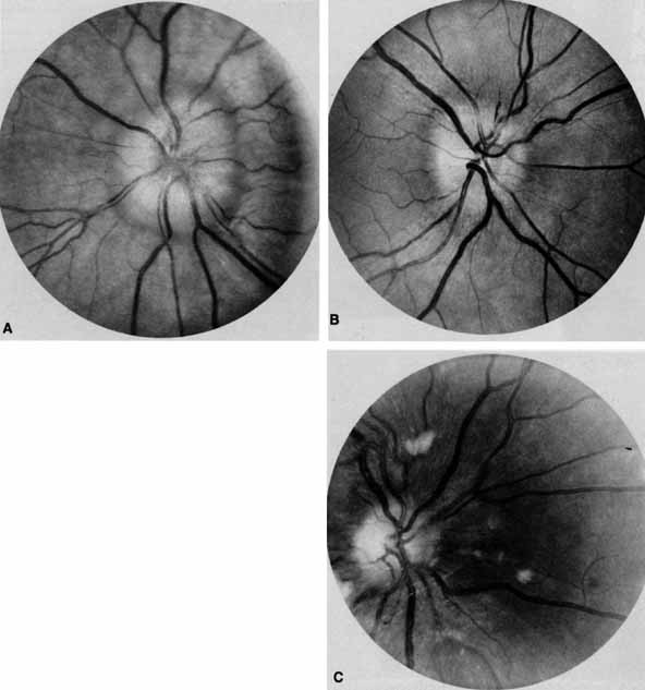
Ophthalmoscopy can be very helpful in orbital diagnosis. Optic disc edema may be caused either by papilledema (increased intracranial pressure) (Fig. 11A), papillitis (anterior optic neuritis) (Fig. 11B), or vasculitis (e.g., lupus erythematosus, sarcoidosis, Wegener's granulomatosis, and giant cell arteritis) (Fig. 11C).12 Papilledema (Fig. 11A) is usually present with near-normal visual function, although some patients may lose vision.9 In contrast, papillitis (Fig. 11B) characteristically leads to reduced visual function, commonly is associated with orbital pain, and is associated with inflammatory cells in the vitreous overlying the disc. Optic disc vasculitis (Fig. 11C) may be present with other findings of vasculitis, and commonly is associated with poor acuity and evidence of vascular occlusion (e.g., cotton-wool spots, hemorrhages). The differential diagnosis of the swollen, erythematous disc is discussed elsewhere in these volumes.

Fig. 11 A. Papilledema caused by pseudotumor cerebri. Visual acuity is 20/15 and the visual field is normal. B. Papillitis of anterior optic neuritis. Visual acuity is 20/400 with a relative afferent pupillary defect and a central scotoma. C. Vasculitis of optic disc caused by lupus erythematosus. Visual acuity is 20/200, there is a relative afferent pupillary defect, and a cecocentral scotoma. Note cotton-wool spots.

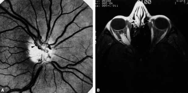
Optociliary shunts (Fig. 12A) develop as a result of long-term obstruction of the central retinal vein. This may occur secondary to optic nerve meningiomas,13 optic nerve gliomas,14,15 optic nerve meningoceles,16 central retinal vein occlusions,17 optic nerve sarcoidosis,18 craniosynostosis,19 optic nerve arachnoid cysts,20 optic disc drusen,21 and papilledema.15 Congenital optociliary shunts, which are exceedingly rare, have not been associated with optic neuropathy or orbital disease.22,23 The presence of optic nerve pallor, poor acuity and optociliary shunts generally implies an optic nerve meningioma in the absence of other funduscopic findings (Fig. 12B).13

Fig. 12 A. Optociliary shunt vessels (arrow) are present on the surface of an atrophic optic disc. The patient's visual acuity was hand motions. The patient demonstrates 2 mm of proptosis. B. Gadolinium-enhanced, fat-suppressed magnetic resonance imaging (MRI) demonstrates an intraorbital optic nerve sheath meningioma (arrow).

Visual field testing can also be helpful in diagnosis of orbital processes. Optic disc–associated visual field changes are usually altitudinal because of the structure of the horizontal raphé (Fig. 13). This visual field defect is typically a result of anterior ischemic optic neuropathy (AION).24 Compressive lesions of the orbit or retrobulbar inflammation are more likely to produce a central or cecocentral scotoma. However, one must be aware that there is a significant degree of overlap between various types of optic nerve lesions and visual field abnormalities.25 In these cases, neuroimaging may be a useful adjunct in differentiating between different neuro-ophthalmic and orbital processes.26

Fig. 13 Pathway of ganglion cell axons that originate temporal to the macula (A) do not cross the horizontal meridian. Any damage to the superior ganglion cell axons at the optic disc will generate atrophy within the superior nerve fiber layer (B). The visual field defect will be inferior but not crossing the horizontal plane. (Hoyt WF, Rios-Montenegro EN, Behrens MM, et al: Homonymous hemioptic hypoplasia: funduscopic features in standard and red-free illumination in three patients with congenital hemiplegia. Br J Ophthamol 56:537, 1972)

The relationship of visual function to orbital findings often helps in the differential diagnosis. For instance, patients with cavernous hemangiomas may have significant proptosis but retain normal, best-corrected visual function (see Fig. 7B), but patients with intrinsic optic nerve meningiomas may demonstrate minimal proptosis with significant vision loss (see Fig. 12B).

Trigeminal Sensation

Historically, the progression of numbness may be helpful in diagnosis of orbital disease. For instance, adenoid cystic carcinoma of the lacrimal gland27,28 and some melanomas29 have a predilection for perineural invasion. The presence of a lacrimal gland mass, which is followed by numbness in the distribution of the lacrimal (see Fig. 2), zy-gomaticotemporal, or zygomaticofacial nerves (Fig. 9), can indicate perineural extension of an adenoid cystic carcinoma.

Corneal sensation should always be tested prior to anesthetizing the cornea for applanation tonometry. Corneal sensation can be roughly quantified using an anesthesiometer; however, asymmetry of corneal sensation may be the most important finding in unilateral orbital disease. A cotton wisp, pulled from a cotton-tip applicator, is lightly drawn from the conjunctiva across the limbus onto the cornea. The blink should occur with corneal but not conjunctival touch. Sensory testing of cranial nerves V1 and V2 with the cotton wisp can be performed at the same time. The cutaneous distribution of the zygomaticofacial and zygomatico-temporal branches should not be ignored (see Fig. 9). The upper teeth should also be tested by tapping with the cotton-tip applicator stick, comparing right to left sides and the front teeth to the molars. Blowout fractures that involve the infraorbital nerve commonly result in numbness of the upper teeth because the anterior and middle superior alveolar nerves arise from the infraorbital nerve within the infraorbital canal (see Fig. 10). However, because the posterior superior alveolar nerve arises from the infraorbital nerve before it enters the infraorbital canal, if the molars are also numb, one must consider the possibility of an associated basilar skull fracture.

Pain is a helpful diagnostic symptom. There are five types of pain associated with the eye and orbit11,19,30:

  1. Pain with eye movement
  2. Pain with palpation (tenderness)
  3. Ocular pain
  4. Orbital pain
  5. Referred pain

Structures that are pain-sensitive within the orbit are the optic nerve sheath, the extraocular muscle sheaths, and the intermuscular septae. Stretching of any of these structures, if they are inflamed, will produce pain on ocular movement. Optic neuritis, myositis, sinusitis with secondary myositis, or cellulitis may result in pain with eye movement. Most of these entities may also lead to constant orbital pain that increases with eye movement.

Tenderness is characteristic of anterior orbital disease that either distends or inflames periostium or Tenon's capsule. Orbital masses that are commonly tender are leaking dermoid cysts, mucoceles, dacryoadenitis, and orbital pseudotumor involving Tenon's capsule (scleritis).

Examples of ocular pain secondary to ocular disease include photophobia caused by keratitis, the aching of uveitis, and the pain of angle-closure glaucoma. These diagnoses are generally evident on complete eye examination and are discussed in their respective chapters.

Orbital pain caused by orbital disease is constant, deep-seated behind the eye, moderate to severe in intensity, and usually unilateral, but not migratory. There is usually little doubt in either the patient's or the examiner's mind about the presence of true orbital pain, as compared with referred pain (see further on in text). Orbital diseases that commonly produce pain include adenoid cystic carcinoma of the lacrimal gland, cellulitis, idiopathic orbital inflammation, and acute orbital hemorrhage.

Referred pain (Table 1) is the most common cause of orbital pain (see case 6). Tension headache is probably the most common cause of referred orbital pain. Tension headaches are generally localized to the periorbital region, often to the brows, and are more diffusely localized than true orbital pain.11 The pain is commonly bilateral or may migrate from side to side. The pain is usually mild, rarely awaking the patient from sleep, and is controlled by mild analgesics.


TABLE 1. Referred Orbital Pain


Tension headache
Occipital neuralgia
Nasopharyngeal carcinoma
Intracranial or intracavernous aneurysms
Cavernous arteriovenous malformation
Cavernous sinus inflammation (e.g., Tolosa-Hunt syndrome)


Fleeting, stabbing, sharp pains so commonly mentioned by patients are usually referred pains. In the face of a normal ophthalmic and neurologic examination these fleeting pains are unassociated with a pathologic process.30

Greater occipital neuralgia may also lead to referred pain. The trigeminal nerve nucleus, which has its afferent input in the pons, has a spinal extension to the level of C1–2. An afferent input from the greater occipital nerve at C1 or C2 can stimulate the spinal nucleus of the trigeminal nerve by ephaptic transmission resulting in referred orbital pain.31

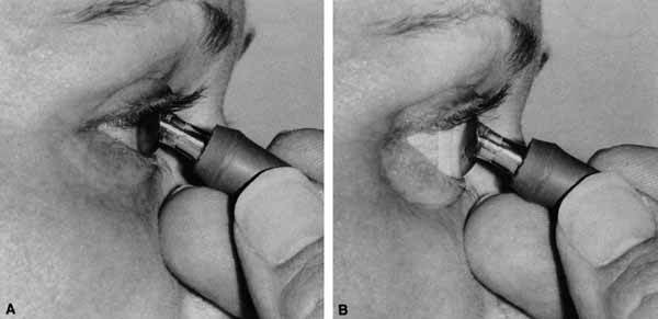
Branches of the ophthalmic division of the trigeminal nerve also contribute to the afferent arc of the oculocardiac reflex. Sudden pressure on the eyeball, acute elevation of intraocular pressure or stretching the extraocular muscles can cause symptoms ranging from nausea and syncope to potentially life-threatening bradycardia. Afferent fibers from cranial nerve V1 pass through polysynaptic pathways in the reticular formation before synapsing with visceral motor nuclei of the vagus nerve, which activate efferent parasympathetic fibers that slow the heart rate.32 Clinical scenarios where an ophthalmologist may encounter this include manipulation of extraocular muscles during strabismus surgery, acute angle-closure glaucoma, herniation of rectus muscles after blowout fractures,33 and acute retrobulbar hemorrhages. In addition to administering intravenous atropine to pharmacologically increase the heart rate, the acute management of these patients may require emergent decompression of the orbit by releasing the lateral canthal tendon, immediate reduction of orbital fractures and reposition of herniated orbital contents,33 and peripheral iridotomy for acute angle-closure glaucoma.

Back to Top
THE EFFERENT SYSTEM

ANATOMY

Cranial Nerve III (Oculomotor Nerve)

The oculomotor nerve, after leaving the brain stem in the interpeduncular fossa crosses between the posterior cerebral and the superior cerebellar artery, passing forward parallel to the posterior communicating artery. The nerve then penetrates the cavernous sinus where it lies in the lateral wall (see Fig. 8). The oculomotor nerve divides into a superior and inferior branch a variable distance behind the globe, usually within the cavernous sinus. Both branches enter the orbit through the superior orbital fissure and anulus of Zinn (see Fig. 2). The superior branch innervates the levator and superior rectus muscles. The inferior branch innervates the inferior rectus muscle, medial rectus muscle, the ciliary ganglion parasympathetics, and the inferior oblique muscle.

Cranial Nerve IV (Trochlear Nerve)

The trochlear nerve has a long intracranial course, exiting the brain stem dorsally after decussating and runs forward under the free edge of the tentorium until it enters the cavernous sinus. Fixation to the rigid tentorium makes the nerve vulnerable to trauma. The trochlear nerve, like the oculomotor, is also contained within the lateral wall of the cavernous sinus (see Fig. 8), but is unique in that it is the only motor nerve to the extraocular muscles that enters the orbit through the superior orbital fissure outside the anulus of Zinn (see Fig. 2). The trochlear nerve terminates as it innervates the trochlear muscle.

Cranial Nerve VI (Abducens Nerve)

The abducens nerve leaves the brain stem at the pontomedullary junction and ascends along the clivus until reaching the petrous bone. Here the nerve turns forward at nearly a right angle, and passes under the petroclinoid ligament (Dorello's canal) before perforating the cavernous sinus. The abducens nerve is the only nerve to run freely within the substance of the cavernous sinus (see Fig. 8) before entering the orbit through the superior orbital fissure and the anulus of Zinn to innervate the lateral rectus muscle from its medial aspect (see Fig. 2).

Sympathetic Nerves

Sympathetic input to the orbit arises within the hypothalamus and descends in the spinal cord until leaving the spinal cord in the lower cervical and upper thoracic nerve roots to join the sympathetic chain (Fig. 14). Sympathetic fibers to the orbit synapse in the superior cervical ganglion. At this point, some fibers are given off to ipsilateral sudoriferous glands of the face. The remaining sympathetic fibers to the eye rise with the internal carotid artery sheath. A contribution of sympathetic fibers from the carotid artery join the ophthalmic division of the trigeminal nerve within the temporal bone while other fibers remain with the carotid artery. Sympathetic fibers enter the orbit with the ophthalmic artery or the nasociliary branch of cranial nerve V1. At this point, the branches are given off to the sympathetic muscles of the upper and lower eyelid. The remainder of the sympathetic fibers that entered the orbit with the nasociliary nerve run forward within the long posterior ciliary nerves to the iris dilator muscle. Some sympathetic fibers also enter the globe with the short posterior ciliary nerves reaching the blood vessels of the ciliary body.

Fig. 14 Sympathetic innervation to the orbit. Sympathetic nerves innervating the pupil enter the orbit with the nasociliary nerve, while those innervating the eyelid retractors enter with the ophthalmic artery as well as the nasociliary nerve. Sudomotor fibers branch at the superior cervical ganglion and innervate the ipsilateral facial sweat glands.

Parasympathetic Nerves

The parasympathetic fibers to the pupil and ciliary muscle arise in the parasympathetic subnucleus of cranial nerve III (Edinger-Westphal nucleus). The parasympathetic fibers travel within the oculomotor nerve until entering the orbit, where they remain with the inferior branch of the oculomotor nerve (Fig. 15). The parasympathetic nerves leave the oculomotor nerve in midorbit to synapse in the ciliary ganglion, which lies lateral to the optic nerve. Parasympathetic fibers exit the ciliary ganglion as the short, posterior ciliary nerves, the majority of the fibers innervating the ciliary body and a few in number (under 3%) reach the pupillary sphincter muscle.34 Parasympathetic innervation to the lacrimal gland traverses the orbit only in its final aspect as it jumps from the zygomaticotemporal branch of cranial nerve V2 to the lacrimal branch of cranial nerve V1 just prior to innervation of the lacrimal gland.

Fig. 15 Parasympathetic innervation to the pupillary constrictor fibers originates within the Edinger-Westphal subnucleus (EW) of the oculomotor nucleus (3). Parasympathetic axons enter the orbit as part of the inferior branch of the oculomotor nerve. Synapse occurs in the ciliary ganglion (CG) before finally innervating the pupillary constrictor muscle. (SC, superior colliculus; PTN, pretectal nucleus; LGN, lateral geniculate; OT, optic tract; CHI, chiasm; ON, optic nerve; RET, retina).

OPHTHALMOPLEGIA

Abnormalities of cranial nerves III, IV, and VI (neurogenic) cause a misalignment of the visual axes resulting in diplopia. Diplopia may also occur if there is an orbital disease resulting in a restriction of eye movement (restrictive) or associated with an abnormally contracting muscle (myogenic). The differential diagnosis between neurogenic, myogenic, and restrictive causes of diplopia requires a thorough eye movement examination (Table 2).


TABLE 2. Eye Movement Examination


  Forced duction Saccadic velocity Intraocular pressure with gaze Fatigue
Restrictive+Normal
Neurogenic
Myogenic+


Restrictive ophthalmoplegia can be evaluated by several means: forced duction testing, elevation of intraocular pressure on attempted gaze,35 or saccadic velocity testing.11 Forced ductions are performed by topically anesthetizing the insertion of the rectus muscle opposite the field of reduced movement. The patient is instructed to look in the direction of limited movement while the examiner grasps the muscle insertion with forceps and attempts to forcefully move the eye. Forced duction testing is useful under anesthesia; however, it is often difficult to perform because of patient apprehension. The use of a forced duction device makes the testing much easier.11 The forward traction test is performed to determine if an enophthalmic eye is reducible surgically. The test is performed either by grasping opposite extraocular muscle insertions with forceps or by using the suction device and pulling forward (Fig. 16).11

Fig. 16 A. Forward traction test is performed by placing the suction device over the anesthetized cornea. B. The globe is easily pulled forward into normal anatomic position.

Restrictive ophthalmoplegia results in increased intraocular pressure when an attempt is made to move the globe in the direction of limited movement.35 With an attempted movement, the restricted noncompliant muscle acts as a tether, squeezing the globe between itself and the contracting agonist muscle, elevating intraocular pressure (Fig. 17). In contrast, if movement is limited from either a myogenic or neurogenic cause, the intraocular pressure does not increase but, in fact, the pressure may decrease, due to relaxation of the antagonist muscle during attempted duction.

Fig. 17 A. Sagittal section of the orbit shows an enlarged, noncompliant inferior rectus muscle. B. Contraction of the superior rectus muscle squeezes the globe against the noncompliant inferior rectus muscle, elevating intraocular pressure.

Saccadic velocity can be assessed clinically by asking the patient to rapidly refixate from one finger to another, testing only within the range the eye can move. The speed of movement can be compared to that of the normal contralateral eye. This test is best performed by looking at the bridge of the patient's nose and comparing the velocity of each eye simultaneously, using the examiner's parafoveal visual field. Saccadic velocities will generally be normal within the range the eye can move if the etiology is restrictive. Neurogenic or myogenic ophthalmoplegia demonstrates a reduced saccadic velocity.

Restrictive ophthalmoplegia may caused by any orbital process that mechanically limits an extraocular muscle from contraction. Congenital orbital fibrosis, muscle entrapment in a fracture, tumor bulk, muscle trauma, muscle inflammation (e.g., pseudotumor, thyroid myopathy), muscle fibrosis, tension pneumo-orbitus or engorgement of extraocular muscles with blood36 can all lead to a restrictive ophthalmoplegia. Consult the index for chapters that cover these diagnoses in detail.

Blowout fractures of the orbit may result in ophthalmoplegia on either a restrictive or neurogenic basis.37 Restriction most often occurs due to entrapment of muscles, entrapment of orbital septae,38 or intrasheath hemorrhage with reduction of muscle compliance (Fig. 18). Less commonly, orbital emphysema secondary to blowout fractures, air hose injuries to the eye,39 or even subcutaneous emphysema entering the orbit may cause a restrictive ophthalmoplegia.40 It is also not uncommon for the inferior nerve branch of cranial nerve III to the inferior rectus and inferior oblique muscles to be damaged at the time of fracture. Entrapment is easily diagnosed with forced ductions, and the reduced velocity of infraducting saccades can help determine damage to the inferior nerve branch of cranial nerve III. Combined restrictive and neurogenic ophthalmoplegia may occur, clinically presenting as a blowout fracture with tissue herniation through a fractured orbital floor limiting upgaze. The muscle is extricated surgically from the fracture site and often, postoperatively, a large hypertropia develops. The inferior nerve branch of cranial nerve III is damaged, however, and the infraducting saccades may not have been checked prior to surgery. The ductions may well improve over time or resolve with strabismus surgery, but the patient may claim the damage to the third cranial nerve occurred intraoperatively. The moral is to always do a complete eye movements examination preoperatively.

Fig. 18 A. A 27-year-old woman sustained a blow to the left eye from a motor vehicle accident 3 days prior to this examination. Upgaze is limited on the left; the globe is ptotic and enophthalmic. B. Computed tomography scan demonstrates a large orbital floor fracture and a large inferior rectus muscle (arrow) that is not entrapped within the fracture. The extraocular movements improved to normal over the following 10 days but the enophthalmos increased.

Neurogenic ophthalmoplegia is covered extensively elsewhere in these volumes. The orbital diagnostician should not ignore examination of cranial nerves III, IV, and VI. It is not uncommon, especially in orbital trauma, that a restrictive and neurogenic ophthalmoplegia coexists.37

Myogenic ophthalmoplegia associated with orbital disease is unusual. Myasthenia gravis, chronic progressive external ophthalmoplegia, myotonic dystrophy, and orbital fibrosis syndrome are described elsewhere in these volumes. Examination characteristics that should alert the examiner to myasthenia gravis include fatigability, variability, ptosis, the Cogan eyelid twitch (upward overshoot of the eyelid on upward saccades), and improvement with edrophonium. Graves' disease occurs in 5% of patients with myasthenia gravis.41,42 The presence of a nonrestrictive component to ophthalmoplegia, variability of ophthalmoplegia, and fatigability should alert the examiner to further evaluate the possibility of myasthenia gravis in patients with Graves' disease.

Parasympathetic Nerves

Abnormalities in parasympathetic innervation to the orbit result in decreased reflex tearing, a dilated pupil, or reduced accommodation. Reflex tearing is reduced when parasympathetic fibers to the lacrimal gland are damaged. The stimulated Schirmer's test can be used to test reflex tearing. Anesthetic drops are placed in the eye, which is then dried. The nose is stimulated with a cotton-tipped applicator and Schirmer test strips are placed for 2 minutes. Asymmetry of tear production may indicate a reduced reflex tear secretion.

Lateral orbitotomy may disrupt the anastomotic parasympathetic branch between cranial nerves V2 and V1, with subsequent reduction in reflex tearing. Tumors invading the orbit from the maxillary sinus or pterygopalatine fossa may disturb the parasympathetic nerve for reflex tearing. Damage to the cranial nerve VII intracranially, such as an acoustic neuroma, may inhibit reflex tearing in combination with an ipsilateral peripheral seventh cranial nerve palsy.

Pupillary dilation and reduction of accommodation occur when parasympathetic innervation to the globe is damaged. When the damage occurs at or distal to the ciliary ganglion, Adie's pupil results. The pupil contracts poorly to light but better to accommodation. The pupil movements are slow and poorly coordinated (vermiform). Generally there is a reduction in accommodation range. The pupil in such cases develops a supersensitivity to parasympathomimetics such as 0.1% pilocarpine. Orbital trauma, inflammation, and intraocular laser treatment may result in Adie's pupil. Deep orbital dissection lateral to the optic nerve in the region of the ciliary ganglion or around the anterior optic nerve where the short ciliary nerves run may also produce Adie's pupil. The denervation may only be sectoral, resulting in an abnormally contoured pupil which demonstrates slow tonic contracture only in the involved sector. Because the parasympathetics have diverged from cranial nerve III prior to entering the ciliary ganglion, pupillary dilation which contracts with 0.1% pilocarpine is not usually associated with neurogenic ophthalmoplegia (Fig. 19).

Fig. 19 A. A patient underwent endoscopic exploration of the right ethmoid and maxillary sinus for persistent epistaxis. Postoperatively the patient demonstrates right proptosis, restricted extraocular movements of the right eye, and a dilated right pupil. Instillation of 0.1% pilocarpine resulted in miosis on the right and no change on the left. Computed tomography (CT) shows a vessel clip lateral to the optic nerve (arrow), near the position of the ciliary ganglion. B. Coronal CT scan showing the clip lateral to the optic nerve (arrow).

Damage to parasympathetic fibers proximal to the ciliary ganglion also results in a dilated pupil but without the susceptibility to 0.1% pilocarpine. Trauma, surgery, inflammation, and intracranial aneurysms may produce a dilated pupil. Damage to the parasympathetics proximal to the ciliary ganglion is usually associated with ptosis and third cranial nerve ophthalmoplegia.

Sympathetic Nerves

Damage to sympathetic fibers produces an oculosympathetic paresis (Horner's syndrome) comprising ipsilateral ptosis, miosis, and anhidrosis. The sympathetic fibers innervating the sudoriferous glands of the face diverge at the superior cervical ganglion; therefore, damage to the sympathetic fibers due to orbital disease generally does not demonstrate anhidrosis (see case 6). Supersensitivity occurs when the sympathetic damage occurs distal to the superior cervical ganglion, such that weak solutions of neosynephrine drops (0.5%) results in eyelid elevation and pupillary dilation. In fact, the prescribing of dilute sympathomimetics is useful therapeutically in some patients with postganglionic Horner's syndrome.

The pupillary sympathetic fibers enter the orbit with the nasociliary nerve,43 while the parasympathetics enter with the oculomotor nerve, both passing through the superior orbital fissure within the anulus of Zinn (see Fig. 2). Therefore a pure, superior orbital fissure syndrome or orbital apex syndrome may present with a miotic, dilated, or midposition pupil, depending on the balance of damage to the opposing innervation.

Back to Top
OTHER CRANIAL NERVES

CRANIAL NERVE VII (FACIAL NERVE)

Cranial nerve VII has no intraorbital component and so is rarely affected by pure orbital disease. However, damage to cranial nerve VII can result in orbital findings which aid in localization. Examples include the decreased reflex tearing of intra-cranial or proximal seventh cranial nerve damage, lagophthalmos associated with myotonic and muscular dystrophy, and essential blepharospasm. Seventh cranial nerve function may also be damaged by orbital surgery. Neurosurgical coronal flaps can produce weakness of the frontalis branch of cranial nerve VII if the incision is carried too far over the zygomatic arch, elevation is too superficial, or extensive traction is applied to the flap. Branch nerve palsy of cranial nerve VII can also occur if a lateral orbitotomy incision is carried too posterior over the zygomatic arch.

CRANIAL NERVE I (OLFACTORY NERVE)

Anosmia caused by damage of cranial nerve I may be a helpful symptom of ethmoid or sphenoid sinus tumors invading the orbit (Fig. 20).44 Olfactory groove meningiomas and esthesioneuroblastomas may also present with anosmia prior to developing vision loss.45 Trauma that involves the orbit and is associated with anosmia should be considered a basilar skull fracture with potential for cerebrospinal fluid leak until proven otherwise (Fig. 21).

Fig. 20 A 13-year-old child presented with a 2-week history of sinusitis and anosmia. Her visual acuity decreased to to 20/400 1 day prior to admission. Computed tomography shows a large ethmoid tumor extending through the cribriform plate and optic canal. A rhabdomyosarcoma was diagnosed by biopsy.

Fig. 21 A 21-year-old man sustained a skull fracture from a motor vehicle accident 2 months prior to this examination. The patient describes anosmia since the accident. Computed tomography discloses a large medial wall orbital fracture extending through the cribriform plate (arrow). Orbital reconstruction was performed through a frontal craniotomy where dura was found incarcerated within the fracture. The orbital fracture was repaired by cranial bone grafting.

Back to Top
CASE EXAMPLES

CASE 1

A 70-year-old woman presented with a 6-month history of progressive diplopia and ptosis. She demonstrated complete right third, fourth, and sixth cranial nerve palsies, and hypesthesia of cranial nerve V1 (Fig. 22A). The right pupil was dilated. Computed tomography (CT) disclosed an intracavernous aneurysm (see Fig. 22B).

Fig. 22 A. A 70-year-old woman with complete right third, fourth, and sixth cranial nerve palsies. Hypesthesia of cranial nerve V1 was also present. B. CT scan demonstrates an intracavernous aneurysm (arrow).

The so-called superior orbital fissure syndrome occurs when an infiltrative, inflammatory, or ischemic event occurs within the superior orbital fissure, but not in the orbital apex.4 A complete superior orbital fissure syndrome occurs when all the neurovascular components passing through the superior orbital fissure are damaged, producing a total ophthalmoplegia, ptosis, and anesthesia of cranial nerve V1 (see Fig. 22A). The pupil may be dilated, miotic, or midposition and fixed, depending on the balance of parasympathetic and sympathetic damage. The superior ophthalmic vein, best seen on CT, may be dilated if venous outflow from the orbit is obstructed. Clues to venous outflow obstruction are increased intraocular pressure, fullness of the upper eyelid, and hyperemia of the deep Tenon's vessels. Ophthalmoscopically, the retinal veins may be dilated. The effect of a lesion in the superior orbital fissure or the anterior cavernous sinus cannot be differentiated clinically (see Fig. 22B). When the posterior cavernous sinus becomes involved, hypesthesia of cranial nerve V2 may also be present.

The only difference between a superior orbital fissure syndrome and an orbital apex syndrome is the presence of visual loss caused by optic nerve involvement. Visual acuity, color vision, or the visual field are abnormal. An ipsilateral relative afferent pupil defect is present.

CASE 2

A 30-year-old man noted diplopia and right ptosis for the past month. The patient demonstrated normal vision and symmetric pupils but had a neurogenic ptosis (Fig. 23A) and limited right supraduction (Fig. 23B). Otherwise his extraocular movements were full. CT demonstrated an intracranial aneurysm of the posterior communicating artery aneurysm (Fig. 23C).

Fig. 23 A. A 30-year-old man demonstrates a neurogenic right ptosis. The pupils are symmetric. B. Supraduction of the right eye is limited. Paresis of the superior branch of cranial nerve III was diagnosed. C. Computed tomography (CT) demonstrates a posterior communicating artery aneurysm (arrow).

Anatomically, the third cranial nerve branches into its superior and inferior divisions as it enters the orbit through the superior orbital fissure. Superior branch damage results in ptosis (levator muscle) and decreased supraduction (superior rectus muscle). Inferior branch damage results in decreased adduction (medial rectus), decreased infraduction (inferior rectus), decreased excycloduction (inferior oblique), and a dilated pupil (parasympathetic). Anatomically, a cranial nerve III branch nerve lesion seems to imply an anterior cavernous sinus or orbital apex localization. However, functionally, the third cranial nerve may bifurcate in the intracranial portion of the nerve, so cranial nerve III branch nerve palsies have been demonstrated with intracranial lesions. The localizing finding of a cranial nerve III branch nerve lesion is therefore not absolute (see Fig. 23).46

CASE 3

A 12-year-old child was referred by his school nurse because he had been complaining intermittently of double vision and the nurse had noted intermittent right ptosis. The child presented with a right ptosis and limited infraduction of the left eye (Fig. 24A). The ptosis worsened with prolonged up-gaze. A Cogan lid twitch was noted on the right with upward saccades. Injection of 1 mg of edrophonium resulted in complete resolution of diplopia and ptosis (Fig. 24B). The diagnosis was myasthenia gravis.

Fig. 24 A. A 12-year-old child presented with right ptosis and limited infraduction of the left eye. B. The ptosis resolved and eye movements were normal after injection of 1 mg of edrophonium. The diagnosis was myasthenia gravis.

Most orbital and cavernous sinus lesions produce afferent and efferent palsies in an anatomic pattern, for example, the superior orbital fissure has cranial nerves III, IV, VI and V1. When the pareses are not localized anatomically, or when there is variability in findings over time, one should include carcinomatosis or myasthenia gravis (see Fig. 24) in the differential diagnosis. Fatigability and a Cogan lid twitch strongly suggest myasthenia gravis.

CASE 4

A 52-year-old woman presented with diplopia when looking to her right. She had noted a red right eye for the past 3 months (Fig. 25A). Visual acuity was 20/20 in both eyes and the pupils were normal. The right eye was limited in abduction. Forced duction testing showed abduction of the right eye to be restricted. Intraocular pressures were 23 mm Hg OD, and 15 mm HG OS. CT showed a dilated right superior ophthalmic vein (Fig. 25B) which was shown to be caused by a dural cavernous fistula. The right medial rectus muscle was also enlarged (Fig. 25C) because of blood engorgement, reducing muscle compliance, which accounted for the restricted abduction.

Fig. 25 A. A 52-year-old woman with a right dural cavernous fistula demonstrates a red right eye. B. Computed tomography (CT) scan demonstrates dilated right superior ophthalmic vein (arrow) secondary to the dural cavernous fistula. C. Enlarged right medial rectus muscle (arrow).

Carotid cavernous fistulas can develop either as a result of trauma or spontaneously.47 A direct carotid cavernous fistula results from a tear in the intracavernous carotid artery with arteriolization of the cavernous sinus and superior orbital vein. Concomitant cranial nerve III, IV, VI, V1 and V2 paresis, elevated intraocular pressure, proptosis, and distention of the deep Tenon's vessels anteriorly (see Fig. 25A) with neuroradiologic evidence of an enlarged superior ophthalmic vein are common (see Fig. 25B). Ophthalmoplegia may also occur on a restrictive basis because of engorgement of the extraocular muscles with blood (see Fig. 25C).36 Dural carotid cavernous fistulas occur where small branches of the intracavernous carotid rupture, allowing access of arterial blood to the cavernous sinus. Typically, the findings are similar to a direct carotid cavernous fistula but of less magnitude. There is no history of trauma, and typically occurs in older persons. However, there is crossover between these two types of carotid cavernous fistula. An excessive ocular pulse tonographically may be helpful in the diagnosis of a carotid cavernous fistula in such cases.48

CASE 5

A 51-year-old woman was referred by her internist for left proptosis. The patient had noted diplopia for the past month but denied blurred vision or pain. Past history was significant for breast carcinoma requiring mastectomy. Vision was 20/20 in both eyes and the pupils were normal. Extraocular movements of the right eye were limited. Forced ductions were restricted. Exophthalmometry readings were 14 mm OD and 18 mm OS (Fig. 26A). CT disclosed a mass encircling the right globe (Fig. 26B). Biopsy disclosed metastatic scirrhous breast carcinoma retracting the right globe.

Fig. 26 A. Right enophthalmos caused by metastatic scirrhous breast carcinoma. B. Computed tomography (CT) scan shows a mass encircling the right globe.

Pseudoproptosis may be caused by ophthalmoparesis, in which the lax muscles allow the globe to prolapse forward. Proptosis should be of minimal degree and the globe is easily reducible. High myopia, contralateral enophthalmos (see Fig. 26), orbital asymmetry, blepharoptosis, or eyelid retraction can also produce pseudoproptosis.11

CASE 6

A 40-year-old man developed severe, unrelenting, right-sided headache 1 month after falling from a roof. He noted ptosis and a small pupil coincident with onset of the headache (Fig. 27A). The arteriogram disclosed a narrowed internal carotid artery compatible with the diagnosis of a carotid artery dissection (Fig. 27B).

Fig. 27 A. Right-sided Horner's syndrome (miosis and ptosis) caused by an internal carotid artery dissection. B. Arteriogram demonstrates a narrowed right internal carotid artery (arrow) compatible with a carotid artery dissection.

An example of referred pain and Horner's syndrome is the carotid artery dissection syndrome.49 When the dissection occurs the patient generally develops unilateral referred orbital pain, ptosis, and a miotic pupil but without anhidrosis (see Fig. 27A). Typically, dissection occurs in the older, vasculopathic patient or days to weeks after head trauma. The symptoms are similar to those of cluster headache, that is, recurrent unilateral severe headache and oculosympathetic paresis, the difference in carotid artery dissection being unrelenting pain and unresponsiveness to normal migraine therapy. Arteriogram or magnetic resonance imaging are useful for documentation of the carotid artery dissection (see Fig. 27B).

Back to Top
REFERENCES

1. Wobig JL: The orbital adnexa. In Reeh MJ, Wobig JL, Wirtschafter JD, eds: Ophthalmic Anatomy: A Manual with Some Clinical Applications. San Francisco: American Academy of Ophthalmology, 1981:11–852

2. Doxanas MT, Anderson RL: Clinical Orbital Anatomy. Baltimore: Williams & Wilkins, 1984

3. Zide BM, Jelks GW: Surgical Anatomy of the Orbit. New York: Raven Press, 1985

4. Kline LB: The Tolosa-Hunt syndrome. Surv Ophthalmol 27:79, 1982

5. Agarwal SA, Maloof AJ, Gomes LJ: Pneumatization of the intraorbital optic nerve after severe head trauma. Ophthal Plast Reconstr Surg 18:469, 2002

6. Orcutt JC, Tucker WM, Mills RP, et al: Gaze-evoked amaurosis. Ophthalmology 94:213, 1987

7. Otto CS, Coppit GL, Mazzoli RA, et al: Gaze-evoked amaurosis: a report of five cases. Ophthalmology 110:322, 2003

8. Knapp ME, Flaharty PM, Sergott RC, et al: Gaze-induced amaurosis from central retinal artery compression. Ophthalmology 99:238, 1992

9. Orcutt JC, Page NGR, Sanders MD: Factors affecting visual loss in benign intracranial hypertension. Ophthalmology 91:1303, 1984

10. Fisher CM: Observations of the fundus oculi in transient monocular blindness. Neurology 9:333, 1959

11. Orcutt JC: Orbital Tumors. In Kohn R, ed: Ophthalmic Plastic and Reconstructive Surgery. Philadelphia: Lea & Febiger, 1988:287–320

12. Friedman DI: Papilledema and pseudotumor cerebri. Ophthalmol Clin North Am 14:129, 2001

13. Frisen L, Hoyt WF, Tengroth BM: Optociliary veins, disc pallor and visual loss. Acta Ophthalmol (Copenh) 451:241, 1973

14. Henkind P, Benjamin JV: Vascular anomalies and neoplasms of the optic nerve head. Trans Ophthalmol Soc UK 96:418, 1976

15. Eggers HM, Sanders MD: Acquired optociliary shunt veins in papilledema. Br J Ophthalmol 64:267, 1980

16. Miller NR and Solomon S: Retinochoroidal (optociliary) shunt veins, blindness and optic atrophy: a non-specific sign of chronic optic nerve compression. Aust N Z J Ophthalmol 19: 105, 1991

17. Henkind P, Wise GN: Retinal neovascularization, collaterals and vascular shunts. Br J Ophthalmol 58:413, 1974

18. Mansour AM: Sarcoid optic disc edema and optociliary shunts. J Clin Neuro Ophthalmol 6:47, 1986

19. Krohel GB, Stewart WB, Chavis RM, eds: The 6 P's of orbital clinical evaluation: pain. In Orbital Disease: A Practical Approach. New York: Grune & Stratton, 1981:9–20

20. Miller NR, Green WR: Arachnoid cysts involving a portion of the intraorbital optic nerve. Arch Ophthalmol 93:1117, 1975

21. Karel J, Otradovels J, Peleska M: Fluorescence angiography in circulatory disturbances in drusen of the optic disc. Ophthalmologica 164:949, 1972

22. Kraupa E: Die Anastomosen an Papillen-und Netzhautvenen. Arch Augenheilk 78:182, 1915

23. Anderson SF, Townsend JC, Selvin GJ, et al: Congenital optociliary shunt vessels. J Am Optom Assoc 62:109, 1991

24. Rizzo JF, Lessell S: Optic neuritis and ischemic optic neuropathy. Overlapping 25. clinical profiles. Arch Ophthalmol 109:1668, 1991

26. Gerling J, Meyer JH, Kommerell G: Visual field defects in optic neuritis and anterior ischemic optic neuropathy: distinctive features. Graefes Arch Clin Exp Ophthalmol 236:188, 1998

27. Rizzo JF, Andreoli CM, Rabinov JD: Use of magnetic resonance imaging to differentiate optic neuritis and nonarteritic anterior ischemic optic neuropathy. Ophthalmology 10:1679, 2002

28. Lee DA, Campbell RJ, Waller RR, et al: A clinicopathologic study of primary adenoid cystic carcinoma. Ophthalmology 92:128, 1985

29. Jakobiec FA, Font RL: Lacrimal gland tumors. In Spencer WB, ed: Ophthalmic Pathology: An Atlas and Textbook. Philadelphia: WB Saunders, 1985:2496–2525

30. Reed RJ, Leonard DD: Neurotropic melanoma: a variant of desmoplastic melanoma. Am J Surg Pathol 3:301, 1979

31. Kalina RE, Orcutt JC: Ocular and periocular pain. In Bonica J, ed: The Management of Pain in Clinical Practice, 2nd ed. Philadelphia: Lea & Febiger, 1990:759–768

32. Bode DD: Ocular pain secondary to occipital neuritis. Ann Ophthalmol 11:589, 1979

33. Miller NR, ed: Sensory innervation of the eye and orbit: Trigeminal nerve, facial pain, and headache. In Clinical Neuroophthalmology. Baltimore: Williams & Wilkins, 1985:379–380

34. Sires BS, Stanley RB, Levine LM: Oculocardiac reflex caused by orbital trapdoor fracture: an indication for urgent repair. Arch Ophthalmol 116:955, 1998

35. Warwick R: The ocular parasympathetic nerve supply and its mesencephalic sources. J Anat 88:71, 1954

36. Gamblin GT, Harper DO, Galentine P, et al: Prevalence of increased intraocular pressure in Graves' disease: evidence of frequent subclinical ophthalmopathy. N Engl J Med 308:420, 1983

37. Leonard TJK, Moseley IF, Sanders MD: Ophthalmoplegia in carotid cavernous sinus fistula. Br J Ophthalmol 68:128, 1984

38. Lyon DB, Newman SA: Evidence of direct damage to extraocular muscles as a cause of diplopia following orbital trauma. Ophthalmic Plast Reconstr Surg 5:81, 1989

39. Koornneef L: Eyelid and orbital fascial attachments and their clinical significance. Eye 2:130, 1988

40. Kaiserman I: Large subconjunctival emphysema causing diplopia and lagophthalmos. Eur J Ophthalmol 13:86, 2003

41. Fox RJ, Teener JW, Liu GT: Orbital emphysema and diplopia following thoracotomy. Eur J Ophthal 9:309, 1999

42. Milliken CH, Haines SF: The thyroid gland in relation to neuromuscular disease. Arch Intern Med 92:5, 1953

43. Barton JJ, Fouladvand M: Ocular aspects of myasthenia gravis. Semin Neurol 20: 7, 2000

44. Miller NR, ed: Pupil: Embryology, Anatomy, Innervation, and Reflex movements. In Clinical Neuro-Ophthalmology. Baltimore: Williams & Wilkins, 1985:400–441

45. Lindquist TD, Orcutt JC, Gown AM: Monoclonal antibodies to intermediate filament proteins: diagnostic specificity in orbital pathology. Surv Ophthalmol 32:421, 1988

46. Argiris A, Dutra J, Tseke P, et al: Esthesioneuroblastoma: the Northwestern University experience. Laryngoscope 113:155, 2003

47. Guy J, Savino PJ, Schatz NJ, et al: Superior division paresis of the oculomotor nerve. Ophthalmology 92:777, 1985

48. Keltner JL, Satterfield D, Dublin AB, et al: Dural and carotid cavernous sinus fistulas-diagnosis, management and complications. Ophthalmology 94:1585, 1987

49. Miller NR: The value of pneumotonography in the diagnosis and management of carotid cavernous fistula. Presented at the 8th International Neuro-ophthalmology Symposium, Winchester, England, June, 1990

Back to Top