|
|
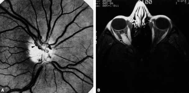
| Fig. 12 A. Optociliary shunt vessels (arrow) are present on the surface of an atrophic optic disc. The patient's visual acuity was hand motions. The patient demonstrates 2 mm of proptosis. B. Gadolinium-enhanced, fat-suppressed magnetic resonance imaging (MRI) demonstrates an intraorbital optic nerve sheath meningioma (arrow). |