|
|
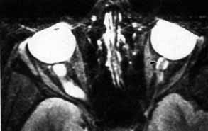
| Fig. 6 T2-weighted magnetic resonance imaging (MRI) shows cerebrospinal fluid (CSF) surrounding the optic nerve within the subarachnoid space (arrow). The CSF extends to the posterior pole of the globe. An optic nerve meningioma is present on the contralateral side. |