Chapter 13
Electrical Signals of the Retina and Visual Cortex
PAUL A. SIEVING
Main Menu   Table Of Contents

Search

RETINAL NEURONS AND THEIR GENERAL ORGANIZATION
ELECTRICAL SIGNALS FROM THE RETINA
RETINAL INFORMATION PROCESSING
REFERENCES

The retina is a neurosensory tissue whose task is to convert light into vision. Whereas light is a physical stimulus with properties of intensity and wavelength, vision is a neurosensory event with properties of brightness, color, contrast, motion, and spatial extent. The photoreceptor cells are specialized for detecting light and responding to wavelength. The remaining sensory vision properties are encoded by neural processing of the various cellular layers of the retina. Much of what is known about retinal sensory processing comes from studying the electrical signals of the several types of specialized retinal cells, particularly by impaling individual cells for intracellular recordings. Of course, one cannot insert an intracellular electrode into the retina of a clinical patient. There are techniques of extracellular recording, however, that provide a powerful—albeit indirect—adjunct to the study of retinal neurotransmission: the electroretinogram (ERG) and electro-oculogram (EOG) record electrical activity at the surface of the eye; visual evoked cortical potentials (VECPs) provide a means to study vision signals that reach the cortex.

This chapter describes the generation of electrical signals beginning with the photoreceptors and then follows the signals through the retinal microcircuit. This chapter attempts to provide a synthesis of information from many sources. Research by many investigators has contributed to the wealth of information regarding visual signal processing. For additional information, the interested reader may wish to refer to the list of suggested readings at the end of the chapter.

Back to Top
RETINAL NEURONS AND THEIR GENERAL ORGANIZATION

OVERVIEW

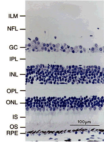
The retina is composed of millions of neurons organized into several primarily cellular layers (Fig. 1). With light microscopy, one can readily recognize three cellular layers, which contain the cell bodies of neurons: the photoreceptor-outer nuclear layer, the inner nuclear layer, and the ganglion cell layer. Connections between these neural layers are made by processes that lie in the outer and inner plexiform layers. The retinal pigment epithelium (RPE) is a single layer of contiguous cells lying beneath the photoreceptors.

Fig. 1. Radial section of the primate retina near the fovea. Note predominance of cones, with only a few rods. RPE = retinal pigment epithelium; OS = outer segments (of photoreceptor); IS = inner segments; ONL = outer nuclear layer; OPL = outer plexiform layer; INL = inner nuclear layer; IPL = inner plexiform layer; GC = ganglion cell layer; NFL = nerve fiber layer; ILM = inner limiting membrane.

Vision is initiated when the photoreceptors are activated by quanta of light. The resulting visual signals pass through the plexiform layer of neural processes and reach the bipolar cells in the inner nuclear layer. The signal is then passed through the inner plexiform layer into the ganglion cell layer and transmitted through the axons that form the optic nerve, and hence to the visual cortex.

In the primate retina, the photoreceptors of the outer nuclear layer number about six million cones and 120 million rods. In addition to containing several types of bipolar cells, the inner nuclear layer contains horizontal cells and amacrine cells, both of which mediate lateral signal interactions that are important for spatial processing of vision. The horizontal cells lie in a row closest to the outer plexiform layer, and the amacrine cells lie in a row closest to the inner plexiform layer.

The primate retina is not homogeneous from center to periphery. The central retina is specialized for cone vision, whereas the peripheral retina is organized better for rod vision. Cones are concentrated within the central-most retina and form the fovea, which is a rod-free area. The fovea is further specialized in that it contains only the photoreceptors; the second- and third-order retinal neurons are pushed to the side. This organization accomplishes two criteria that are important for fine vision:

  1. The image that is focused on the foveal cones is the sharpest and least degraded because light passes through the fewest layers of the inner retina.
  2. Tight packing provides minimal space between the cones in the fovea and thus allows the sharpest reconstruction of fine image detail, corresponding to the finest grain of a digitized photograph.

Meanwhile, the bipolar, horizontal, and amacrine cells that subserve vision processing from the densely packed foveal cones are displaced sideways into a parafoveal mound of neural tissue.

Whereas cones are packed most densely in the fovea, the rod photoreceptors reach the highest concentration 20° to 30° from the fovea. Although the retinal neurocircuitry associated with rods does not allow for acuity of better than about 20/200, the high density of rods in the midperiphery still provides the maximal sensitivity for dim-light vision at this region. The retina is thinnest in the fovea, thickest in the parafovea and macula, and of intermediate thickness in the peripheral retina. One misconception is that cones are limited to the fovea and macula. This is obviously incorrect, since we can perceive color across our entire field of vision, from edge to edge. Since color is mediated exclusively by cones, this reinforces the concept that cones exist in the peripheral retina, although at a considerably reduced number compared to the macula.
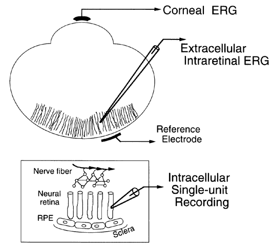
PHOTORECEPTORS

Rod and cone photoreceptors are specialized for converting light into neural signals (Fig. 2). The earliest information about electrical activity of the photoreceptors came with Granit's identification of the PIII response of the ERG as deriving from this layer of cells. In the corneal ERG, PIII is the negative-going response that forms the a-wave when elicited with stimuli of medium to bright intensity. The a-wave is the earliest and negative wave of the ERG elicited with relatively bright flashes. During the 1960s, Brown and associates inserted a microelectrode into the monkey fovea and identified the ERG a-wave signal produced by cone photoreceptors. This provided the first information about the shape of the cone response when these cells hyperpolarize to a light flash. The next advance came from impaling individual photoreceptors, and subsequently by suction electrode recordings performed by Baylor and colleagues.

Fig. 2. Schematic of a rod photoreceptor. Free-floating discs in the outer segment contain rhodopsin. The sodium channel is open in the dark and allows a dark current to flow between the inner and outer segments. Closure of the channel by light interrupts the dark current, which changes the electrical voltage outside the cell. This generates the a-wave, which is the fast, corneal-negative ERG component.

Note that when a cell hyperpolarizes, the inside becomes relatively more negative compared to the outside. All this is relative, since cells normally are negative inside compared to the extracellular space. Thus the terms hyperpolarization and depolarization are with reference to the normal resting potential of the cell membrane. A depolarizing cell becomes less negative inside upon stimulation, whereas a hyperpolarizing response causes the cell membrane to become more negative.

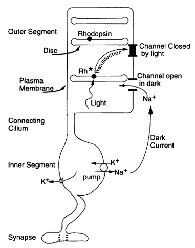
Photoreceptor “Dark Current”

The process of translating light into electrical signals is termed transduction. The biochemical steps that are involved occur within the photoreceptor outer segments, beginning with isomerization of the vitamin A retinal from the cis to the trans form within the visual pigment. The protein portion of rhodopsin is called opsin and is arranged in the shape of seven helixes that span the membrane of the free-floating discs in the rod outer segment. Isomerization of retinal from cis to trans form opens up the space of the opsin helixes and allows rhodopsin to interact with transducin. The subsequent cascade of events leads to a decrease of cyclic guanosine monophosphate (cGMP) which closes the ionic channels on the plasma membrane of the rod outer segment. This channel normally is open, which allows entry of sodium into the rod outer segment in the dark, with a compensatory extrusion of potassium from the rod inner segment to balance the net charge within the cell. This circulating “dark current” between the inner and outer segments maintains the rod in a relatively depolarized state. Closure of the cGMP-sensitive channel by light interrupts the dark current. Interrupting the influx of sodium, which carries positive charge into the cell, causes the photoreceptor to hyperpolarize. This intracellular voltage change is conveyed to the synaptic terminal, where the visual signal is transmitted to the bipolar and horizontal cells.

To a first-time student of retinal neurophysiology, these events initiated by light may appear the reverse of what would be expected. Rather than initiating a positive response in the rod, light actually suppresses a current that exists in the dark. Furthermore, in the dark, glutamate is released from the photoreceptor synapse (its neurotransmitter), and glutamate release is stopped upon signaling by light. The outcome of signaling light, however, is the same in that light causes a signaling difference to reach the bipolar cells as the message that vision was initiated.

Rod photoreceptors are highly efficient and can signal the isomerization event from a single photon of light. Each rod contains approximately 109 rhodopsin molecules, and the probability is remarkably high that every photon will be absorbed as it passes through the length of the rod outer segment. Fortunately, rhodopsin molecules are very stable in the dark and have a 400-year half-life before they spontaneously isomerize. Occasionally, however, a rhodopsin molecule will undergo spontaneous isomerization even in the absence of light, and this thermal noise results in a spurious signal being transmitted as “vision.” Fluctuations in the photocurrent of single rods can be observed in the dark, with a frequency of approximately one every few minutes in single unit recordings of rods. These spurious events are indistinguishable from rod responses initiated by true light and are called dark-light (alternatively called dark-noise). One can experience dark-light as the infrequent, spontaneous speckles seen after being in a fully darkened room for at least 1 hour. Dark-light is a temperature-dependent event and is more frequent with higher body temperature.

When they are in constant light, rod photoreceptors become less sensitive and do not respond as readily to individual photons. The rod response saturates in bright sustained light and renders the rod incapable of further signaling. Half-saturation occurs when each rod absorbs approximately 30 photons/second. Bright room lighting, equivalent to standard office lighting with fluorescent bulbs, is sufficient to saturate human rods.

Visual Transduction

The general scheme of visual transduction in rods is as follows. Light activates rhodopsin which couples to transducin. Transducin increases the activity of phosphodiesterase which, in turn, decreases cGMP by converting it to GMP. The high concentration of cGMP in the dark normally maintains the membrane channel in an open configuration. With light activation, cGMP decreases, and the channel closes. In darkness, the open channel allows influx of sodium. When the channel closes, the sodium influx decreases and interrupts the dark current flow that is maintained between the rod inner segment and outer segment. This, in turn, causes electrical hyperpolarization of the rod and produces the a-wave.

Closure of membrane channels by light also interrupts the normal influx of calcium into the outer segment. Since calcium is important for maintaining phosphodiesterase levels, the calcium change interrupts the degradation of cGMP to GMP. This, in turn, partially nullifies the light-activated decrease of cGMP and allows channels to reopen, terminating the light response and causing light adaptation.

The transduction process requires a number of constituents. It has recently been appreciated that genetic mutations that alter the molecular structure of the proteins and enzymes involved in transduction frequently lead to retinal degeneration. Rhodopsin mutations are found in 20% to 40% of persons with autosomal dominant retinitis pigmentosa. Retinal dystrophies are also associated with mutations in phosphodiesterase, in the outer segment channel protein, and in transducin. A detailed understanding of how these genetic mutations result in death of rods and widespread retinal degeneration remains an unsolved problem. One particular mutation in the rhodopsin molecule at codon 90 (glycine-90-aspartate) was found to alter the stability of rhodopsin and to increase the level of dark-light in human subjects with a special form of congenital night blindness.

PARALLEL PATHWAYS: VISION IN STARLIGHT AND DAYLIGHT

Separate Rod and Cone Signals

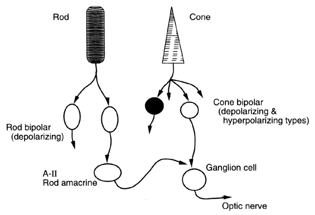
For the most part, the tasks of the rods and cones are quite different, and their signals remain separate within the retinal circuitry (Fig. 3). Rods respond to extraordinarily dim light equivalent to starlight, and a single photon is sufficient to activate a rod photoreceptor. The sensitivity of the rod pathway is further increased by pooling of signals, with signals from as many as 1000 rods all converging onto a single ganglion cell. This effectively enhances the sensitivity of rod vision in very dim light.

Fig. 3. Cartoon of retinal wiring for rods and cones. Cones make contacts onto second-order bipolar cells of both the depolarizing and hyperpolarizing type. In turn, these synapse onto ganglion cells, which transmit the visual signal down the optic nerve. Cones in the primate fovea each contact approximately 30 bipolar and horizontal cells. By comparison, each rod synapses onto only two bipolar cells of the depolarizing type. The rod circuit adds an extra stage, in that rod signals are sent to amacrine cells. The A-II cell, also known as the “rod amacrine” cell, is in the rod pathway and makes presynaptic contact onto ganglion cells through the cone bipolar pathway, and the signal is transmitted through the optic nerve.

By comparison, each cone acts relatively independently of other cones: they do not pool signals substantially. This autonomy is necessary to ensure the precise spatial discrimination required for fine visual acuity. Pooling of cone signals would blur vision.

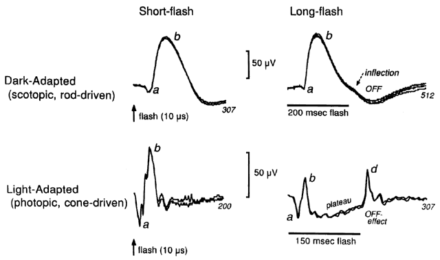
Each cone photoreceptor is activated only when struck by several photons simultaneously. This leads to an intrinsic difference in sensitivity from rods, which are activated by single photons. The dimmest light range of sensitivity is called scotopic, and implies that vision is mediated exclusively by rods. For bright stimuli, rods saturate and no longer respond significantly to further light, which leaves the cone circuit to mediate vision in the photopic range. The mesopic range overlaps the range of dim and bright light, and is therefore a transition zone between scotopic rod vision and photopic cone vision. Both rods and cones can respond under mesopic adaptation conditions, which correspond to the dim light at dusk and dawn. Patients with retinal disease frequently experience their worst vision impairment under mesopic conditions.

Rod Circuit in Starlight

The rod microcircuitry employs two clever tricks to increase the sensitivity of vision in starlight. A single rod connects to two depolarizing bipolar cells (DBCs). This divergence effectively amplifies the rod signal by a factor of two. Further amplification occurs because the rod synapse onto these DBCs has an intrinsic gain of about fivefold. These two rod DBCs make synaptic contact onto five amacrine cells, which further spreads the signals from each individual rod cell.

Rod signal sensitivity also benefits from convergence, in that signals from as many as 1000 rod photoreceptors are pooled onto a single ganglion cell. The convergent pooling of rod signals causes a loss of spatial discrimination, since a single photon captured by any of the 1000 rod photoreceptors in the pool will signal the same set of ganglion cells for transmission to the brain. Although this design may appear to be sloppy, it provides a tremendous increase in visual sensitivity in very dim light, with the tradeoff of reduced spatial discrimination.

Cone Circuit in Daylight

The cone circuit operates inherently differently from rods. Whereas individual rod photoreceptors are activated by absorption of a single photon, activation of an individual cone requires four to six simultaneous quantal hits. An immediate advantage to this scheme is that cones will be insensitive to vision in dimmest light and will not interfere with rod vision, since they will not send overlapping signals to the brain. The cone microcircuitry is also more complicated even at the first stage of vision, since individual cones synapse onto as many as 30 postsynaptic cells, including horizontal cells and several classes of bipolar cells. Some cone bipolar cells function by depolarizing when stimulated (depolarizing bipolar cells [DBCs]) whereas others hyperpolarize when stimulated (hyperpolarizing bipolar cells [HBCs]). This design is useful for daylight vision, in which both increments and decrements of light are important. Cone circuitry exhibits convergence to a lesser extent than rod circuitry.

Output Pathway from Retina to Brain

Despite the seemingly independent rod and cone circuitry of the retina, only a single set of ganglion cells actually transmits retinal signals toward the brain. Thus, signals from both rod and cone photoreceptors ultimately must feed into the same ganglion cells. In the cone circuit, transmission is from cones to cone bipolar cells to ganglion cells. The rod pathway is more circuitous. The rod bipolar cells do not synapse onto ganglion cells directly but only indirectly through the amacrine cells. Rods synapse onto bipolar cells and then onto amacrine cells that pass the signal to ganglion cells. The retina is rich in amacrine cells; the cat and rabbit are known to have 30 to 40 types. The connection from the rod bipolar cell to the ganglion cell is primarily through the AII amacrine cell, which has multiple feedback and feedforward control mechanisms through the other types of amacrine cells. This amacrine network ultimately controls signal transmission from the rod pathway to the ganglion cell by controlling the AII amacrine cell. From an engineering perspective, the AII amacrine cell serves as a switch that can disconnect the rod pathway from the cone pathway in bright light photopic vision, where the cone pathway needs to have exclusive access to the vision outflow to the brain, without interference from rod signals.

INTERACTION BETWEEN ROD AND CONE SIGNALS

Rod and cone signals are maintained relatively separate under purely scotopic and purely photopic conditions. For the intermediate realm of the mesopic vision in twilight conditions, however, there is evidence that the rod and cone signals can interact. One example involves detection of flickering-light stimuli by the cones, which becomes less sensitive when the rods begin to adapt to the dark and regain their sensitivity in dimmer light. Evidently, rods can have a considerable impact on the cone circuit under this particular condition. Another example involves color and spatial discrimination. Both are mediated by cone systems but are partially suppressed under mesopic conditions in which rods are active.

To explain rod and cone interactions, it must be demonstrated that there is some anatomic connection between these two pathways. One site of interaction would be through the AII amacrine cell, where rod signals can be fed onto the cone bipolar cells and thereby impinge on the cone signals. Another site is through the horizontal cells that lie immediately adjacent to both rods and cones, in the microcircuitry of the outer retina. Furthermore, it has been demonstrated that rod signals in the primate retina are found in cone photoreceptors, which indicates the presence of electrical coupling between rods and cones.

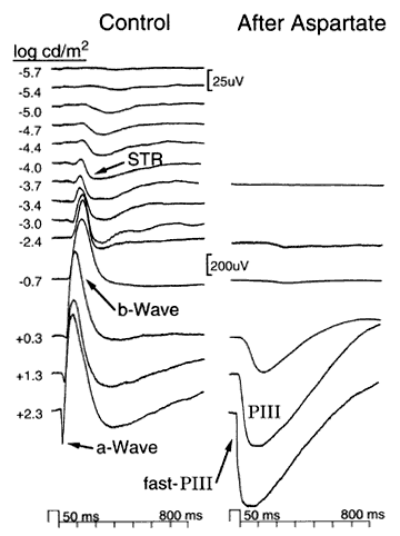
Signals that are conducted through the rod AII amacrine cell can be studied by recording the scotopic threshold response (STR) of the ERG. This is a tiny, negative-going ERG wave, best recorded under extremely dim light conditions and after complete dark adaptation of 1 hour or more. The STR is present in the ERG of a variety of species, including mouse, rat, cat, dog, sheep, monkey, and human. Although the full diagnostic value of the STR remains to be determined, the STR is absent in some types of congenital stationary night blindness (CSNB) patients and is diminished in glaucoma.

RETINAL NEUROTRANSMITTERS

As befits the neural complexity of the retina, all of the major neurotransmitters have been identified in this tissue. Glutamate is the principal transmitter from photoreceptors onto bipolar and horizontal cells. Glutamate is also used by bipolar cells to communicate with amacrine cells. At both rod and cone photoreceptors, glutamate is continuously released in the synaptic cleft in darkness, and its release is interrupted by the light response of the photoreceptor. The postsynaptic machinery is of two principal types:

  OFF-bipolar cells (HBCs) receive direct chemical synaptic input from glutamate. The presence of glutamate in the dark depolarizes the OFF-bipolar cells, whereas the interruption of glutamate in the light hyperpolarizes these cells.
  ON-bipolar cells (DBCs) work in reverse. These cells are hyperpolarized in their unexcited state in the dark during glutamate release, whereas cessation of glutamate release in the light causes these bipolar cells to depolarize. This involves inverting the signaling process and indicates that a second messenger system is involved at the synapse, which can reverse the normal chemical signaling and cause bipolar cells to depolarize only upon withdrawal of synaptic glutamate during the light-evoked photoreceptor response.

The sign-inverting synapse between the rods and the depolarizing rod bipolar cell uses a metabotropic glutamate receptor that can be blocked by 2-amino-4-phosphonobutyric acid (APB). This particular APB-sensitive mGluR6 glutamate receptor was molecularly cloned in 1993. Although multiple subtypes of the glutamate receptor have been identified, the APB-sensitive form appears to be unique to the retina. Experiments show that applying APB to the retina of many species, including primates, will block synaptic transmission and cause a loss of the dark-adapted b-wave (i.e., it blocks bipolar depolarization) without altering the a-wave (from rod photoreceptors). This particular ERG change mimics quite closely the ERG found in the type of CSNB patients who have rod photoreceptors and rod visual pigment, but who still cannot see at night, presumably because of faulty synaptic transmission. It may be that genetic mutations that alter the function of this metabotropic receptor could result in CSNB abnormality.

Inner retinal neurons utilize a variety of neurotransmitters, including gamma-aminobutyric acid (GABA), glycine, and dopamine. Specialized NMDA receptors have been identified in the proximal retina of many species. Dopamine is involved in setting the dark- or light-adaptation state of the retina. Dopamine can modulate the coupling or uncoupling of photoreceptors as well as produce retinomotor movement of the pigment epithelial sheaths that surround the rod and cone photoreceptors. Dopamine is released from dopaminergic amacrine cells in the proximal retina and diffuses to the outer retina, where it works in exocrine fashion on the photoreceptors and RPE. Work with experimental myopia in the chick model has implicated dopamine in development of myopia.

Back to Top
ELECTRICAL SIGNALS FROM THE RETINA

RECORDING CONFIGURATIONS

Intracellular Recordings

To perform intracellular recordings, individual retinal neurons are impaled with a microelectrode that penetrates the retina (Fig. 4). One can record directly from these neurons, and then afterward identify the specific cell for histologic study by injecting dye through the microelectrode that impales the cell. Although intracellular recordings give detailed information about individual neurons, this technique does not elucidate the bigger picture of how individual neurons interact and how they contribute in aggregate to the complex spatial properties of vision.

Fig. 4. Recording configurations used to observe electrical signals of the retina. Corneal electroretinographic (ERG) recordings employ an electrode touching the surface of the eye with the reference electrode placed at the canthus or on the forehead. Extracellular responses are recorded by sliding a fine-tipped electrode into the retina alongside the neurons. As depicted here, the electrode frequently consists of glass pulled to a fine tip a few micrometers in diameter and filled with conductive fluid. The retinal depth of the tip position gives information about which cell type contributes most to the response. The reference electrode is placed behind the sclera for intraretinal recordings. Intracellular recordings are made by impaling a cell with an electrode. After recording single-unit activity, one can inject marker dye into the cell to identify the exact cell studied and to examine its projections onto neighboring cells. RPE = retinal pigment epithelium.

If two or more neurons are impaled simultaneously, each with separate microelectrodes, the input-output relationship of a pair of cells can be studied. By this approach, the light response of a bipolar cell can be studied while simultaneously recording from the rod or cone photoreceptor cell that drives the response. Double electrode recordings are important for deducing the transmission properties of the synapses that connect different cells, such as between photoreceptor and bipolar cells. Recently, multielectrode arrays have been etched onto microchips that are floated on the surface of the retina to record simultaneously from many adjacent ganglion cells. This has provided a wealth of information about spatial vision properties.

Extracellular Intraretinal Recordings

Extracellular recordings are used to study electrical activity within the retina, but outside the cell bodies. This technique is a bridge between intracellular recordings and corneal recordings. For intact eyes in vivo, an electrode can be advanced through the sclera and into the retina. When the microelectrode is placed adjacent to a ganglion cell, the rapid firing of an individual spike discharge is observed. If the electrode is advanced into the subretinal space adjacent to photoreceptor outer segments, the extracellular voltage resulting when rods and cones hyperpolarize upon stimulation by light can be observed. This approach provides a way to localize ERG components that arise from the different classes of neurons in the separate cellular layers of the retina. As an alternative, the eye can be removed and hemisected, and the neurosensory retina then can be gently lifted out, placed in a superfusion chamber, and maintained for extended periods of study via an extracellular electrode.

Corneal Recordings

Corneal recordings are used for the ERG. The ERG is the cumulative, total extracellular output of neural retinal signaling. The ERG provides a noninvasive way to record from human subjects. Historically, the ERG was the earliest method used to sample neural retinal signals. Although this technique has considerable power, it cannot provide detailed information about individual cells within the retina. Rather, it provides a picture of the aggregate of activity from the various retinal cellular layers and from classes of retinal neurons, such as all of the rods, or cones, or bipolar cells, or amacrine cells together. Understanding some of the ERG potentials provided the first insight into overall organization of the neural retina and resulted in a Nobel prize for Granit in 1967. His book is an ERG classic and well worth reading.

These three recording configurations—intracellular, extracellular, and corneal ERG—provide complementary information. Although intracellular recordings can provide a detailed picture of the activity of individual neurons, it is difficult to build up a general overview of retinal activity only by observing one cell at a time. Extracellular recordings can sample the massed responses of many neurons simultaneously, such as the rod photoreceptors when recording in subretinal space; however, this technique is still invasive and consequently is not applicable for clinical studies. With noninvasive corneal ERG recordings, one can deduce which signals are from the photoreceptors, bipolar cells, or amacrine cells. Consequently, the ERG is invaluable for the clinical diagnosis of retinal disease. The ERG is limited, however, because signals from different cells frequently overlap in time and interfere with the observation of single cell types in isolation.

ELECTRORETINOGRAM: FIELD POTENTIALS FROM THE RETINA

The ERG represents the massed extracellular response of the retina (Fig. 5). The ERG can be recorded either (1) by inserting a microelectrode into the retina; or (2) by placing an electrode on the cornea, remote from the retina. When recorded with a corneal electrode, the ERG provides an important noninvasive tool of considerable power for diagnostic assessment of retinal disease.

Fig. 5. Electroretinogram of a rat over an 8 log-unit intensity range. The brightest flash elicits a sharp, initial, negative a-wave followed by a larger, positive b-wave. As the stimulus is made dimmer, the a-wave is diminished first, and then the b-wave is reduced, until only a slow, negative scotopic threshold response (STR) is seen for the dimmest flashes. The a-wave reflects activity of rod photoreceptors, the b-wave of bipolar cells, and the STR of amacrine cells. Aspartate was injected into the vitreous of the eye. Aspartate blocks transmission at the photoreceptor synapse, so that bipolar and amacrine cells can no longer respond to light. Because the b-wave is no longer present after aspartate, the a-wave is larger. After aspartate, the a-wave is the initial and leading edge of PIII.

Signals from many different classes of retinal cells can be observed by the ERG, including responses of the RPE, rod photoreceptors, cone photoreceptors, DBCs and HBCs, and some types of amacrine cells. The main limitation of ERG analysis involves the difficulty of isolating the contribution from a single class of neurons, since the individual contributions overlap each other and sum together into a single complex waveform.

Despite this long and venerable history of ERG studies extending back for many decades, major new advances continue to occur in understanding the origins of ERG waves, and new components of the ERG continue to be identified. One strategy for separating the many different ERG components lies in manipulating the stimulus conditions, including stimulus of color, flicker rate, intensity, duration, and background adaptation. With dim flashes in complete darkness, the ERG originates from the rod system. Rod responses are suppressed by light adaptation, and recording in the presence of a constant bright background will elicit ERG responses exclusively from the cone system. Blue stimuli favor rod responses, whereas longer wavelength red stimuli favor the cone system. Rapid flicker elicits primarily cone system activity, since rod photoreceptors cannot respond to flicker much above 10 to 20 Hz. For higher flicker rates, the individual rod wavelets fuse together into a sustained response.

Methods of Localizing Electroretinographic Components

Initial ERG research was limited to recording with an electrode touching the cornea. ERG waves were then separated by manipulating stimulus parameters, including intensity, adaptation state, color, duration, and repetition rate. Subsequently, drugs or anoxia were found to alter one wave preferentially over another. This led to Granit's classic description of ERG processes: PI, PII, and PIII. PII was most susceptible to anoxia and was thought to originate from the middle layers of the retina that are perfused by the retinal circulation. The b-wave is formed by the leading edge of PII, in classic terminology. By comparison, PIII was highly resistant to anoxia and was believed to reflect outer retinal activity of photoreceptors that are supplied metabolically through the choroidal circulation. The leading edge of PIII (termed fast-PIII) forms the negative-going photoreceptor a-wave. PIII persists beyond the a-wave, and is termed slow-PIII, which results from Müller cell depolarization by a potassium decrease in the subretinal space.

With the advent of microelectrodes, these were inserted into the retina and placed adjacent to individual cells or classes of cells. When adjacent to photoreceptors in the distal retina, the microelectrode could record photoreceptor hyperpolarization by light. This confirmed that the a-wave originates from rod photoreceptors, at least for modestly bright flashes in a dark-adapted state. Similarly, the b-wave is maximal when the microelectrode is at middle retinal depths, and the STR is maximal for an electrode in the proximal retina.

Early Receptor Potential

As the name implies, this response is earlier than the membrane potential (a-wave) of the rod and cone photoreceptors. The ERP is elicited with extremely bright flashes that activate (“bleach”) the majority of rhodopsin molecules simultaneously. The response has a small, corneal-positive R1 component that is followed by a larger negative R2 component. The entire ERP waveform is complete within 1 millisecond. Maximal amplitude of the human ERP is normally about 200 μV.

The ERP signal results from the cis-trans isomerization of the pigment molecules, during which the conformational change causes electrical charges to move to a new position across the lipid membrane of the outer segment discs. The ERP amplitude is proportional to the amount of visual pigment bleached by each flash. Consequently, the ERP can provide a direct measure of visual pigment quantity of the eye in vivo. The ERP has been used clinically to demonstrate reduced visual pigment quantity in photoreceptor degeneration seen in retinitis pigmentosa, which indicates that there is a loss of the photoreceptor outer segments that normally contain rhodopsin and the cone pigments. By tracking the rate of recovery of ERP amplitudes after an extensive bleach, the rate of recovery of visual pigment can be determined in clinical patients. With this approach, visual pigment regeneration has been found to be retarded in some retinal dystrophies, such as fundus albipunctatus.

a-Wave

The a-wave is defined classically as the initial negative wave of the ERG under either dark- or light-adapted conditions (Fig. 6). The a-wave generally is attributed to photoreceptors directly when they hyperpolarize in response to a flash of light. Recent studies have shown that the scotopic a-wave can be used to deduce detailed function of the rod photoreceptors in clinical patients. Even properties of signal transduction within the rods can be teased out of the a-wave signal by proper analysis. For moderately intense light flashes, under dark-adapted conditions, it appears that the a-wave does originate primarily from rod photoreceptors. This simple explanation for the a-wave origin, however, is now known to be correct only under these mesopic conditions of moderately bright flashes. Under scotopic conditions, the initial negative response is an STR. The STR is a small negative wave of the ERG. It originates from AII amacrine cell activity, not from rod photoreceptors. Furthermore, under light-adapted conditions, in addition to the cone photoreceptors, HBCs also contribute to the photopic a-wave.

Fig. 6. Human electroretinographic recordings under dark-adapted scotopic conditions and light-adapted photopic conditions from a 43-cd/m2 background light. The scotopic flash is dim and elicits only a small a-wave, with a larger b-wave following it. The waveforms are similar for the short, 10-microsecond and for the long, 200-millisecond stimuli in the scotopic condition; only a small inflection occurs at the termination of the long stimulus. For the 10-microsecond flash, photopic condition, the a-wave and b-wave are found. With the 150-millisecond stimulus, however, both the b-wave at stimulus appearance and the d-wave at stimulus termination are present. The number at the ends of each trace gives trace duration in milliseconds.

b-Wave

The b-wave is a positive-going ERG response to moderately bright stimuli under either dark- or light-adapted conditions. The b-wave occurs immediately after the photoreceptor a-wave. For dimmer light responses, however, the b-wave is recorded even in the absence of an a-wave, indicating that the b-wave has greater sensitivity to a light flash than does the a-wave. Obviously, photoreceptors must be stimulated for a b-wave to occur. Thus, an a-wave must always be present to some minute degree, but its size is negligibly small compared to the larger and more sensitive b-wave. The b-wave can be recorded from both the rod and cone circuit. Dim light and dark adaptation favors rod b-waves, whereas light adaptation and bright stimuli favor cone b-waves.

The b-wave results from activity of depolarizing bipolar cells, either directly or indirectly through Müller cells. The DBCs release potassium when stimulated, and there is evidence that this is taken up by Müller cells, which also depolarize and produce a transretinal ERG potential. This leads to confusion in the terminology: bipolar cell activity is requisite for the b-wave, but the b-wave voltage is actually mediated through Müller cell depolarization from potassium. As glial cells, Müller cells are highly responsive to potassium, which causes them to depolarize and give a transretinal voltage of the correct magnitude and polarity to account for the b-wave. Experiments have monitored potassium in the extracellular space and have found good correlation between the b-wave time course and the accumulation of potassium. Furthermore, direct microelectrode recordings of Müller cells during ERG flash stimuli showed a response consistent with the b-wave voltage.

Nevertheless, the issue of direct Müller cell involvement in b-wave production is not fully resolved. Recent new evidence implicates the DBCs directly in b-wave production. In this case, the b-wave voltage may directly reflect the depolarization of bipolar cells in response to a light flash, and b-wave production may not require Müller cells to complete the circuit. This issue remains to be settled in the future.

The b-wave is a very useful ERG response for clinical diagnosis. Because of its great sensitivity, it is reduced even in early stages of retinal degeneration. Selective suppression of the b-wave response, but retention of the photoreceptor a-wave response, is found in particular types of CSNB, which is a nonprogressive form of retinal dystrophy that causes far less ultimate vision loss than retinitis pigmentosa. Consequently, it is clinically important to distinguish CSNB from retinitis pigmentosa. The b-wave is also selectively reduced, with the a-wave preserved, in juvenile X-linked retinoschisis which is thought to involve Müller cells.

Although the b-wave is classically described as originating from activity involving DBCs, recent evidence indicates that, in the light-adapted response, HBCs also contribute. Both DBCs and HBCs are activated by light under photopic conditions. Because these cells undergo opposite voltage changes when stimulated, their extracellular activity will tend to cancel their individual contributions to the cone-driven photopic b-wave. Consequently, the photopic b-wave has a smaller amplitude and shorter duration than the rod-driven b-wave, for which activity of rod DBCs are the primary ERG event. Additional research currently is ongoing that undoubtedly will clarify our understanding of the ERG in the future.

c-Wave

The c-wave is a slow, positive-going ERG response that occurs after the b-wave and originates, in part, from the RPE. Unfortunately, Müller cells can also contribute an ERG response with a similar time course but opposite polarity, and this subtracts from the RPE c-wave component. Cells of the RPE do not respond directly to light. Rather they hyperpolarize when potassium in the subretinal space decreases around the photoreceptor upon stimulation with light. This same potassium decrease also affects the Müller cell processes, and this glial cell also contributes to the slow corneal-negative PIII.

The c-wave has not been used widely for clinical recordings for two reasons. First, the c-wave develops later than the b-wave and is susceptible to contamination from artifacts caused by a patient's eye movements during its slower time course. The c-wave can be recorded in a research setting with trained observers, but patients rarely maintain steady fixation sufficiently long to observe a high-quality c-wave. Second, the relative c-wave contributions from the RPE and Müller cells appear to vary among subjects. Some normal subjects have very small c-waves, presumably because of a close balance between contributions from the RPE and Müller cells.

d-Wave

The human light adapted ERG exhibits a positive-going d-wave at the termination of a prolonged light stimulus lasting 100 milliseconds or more. The phasic, positive d-wave is recorded only under photopic conditions because its mechanism is cone driven. The d-wave originates from at least two different cell types. With cessation of a light stimulus, cone photoreceptors return to their resting potential and contribute a positive-going return to baseline as part of the d-wave. Second, the HBCs also return to their resting potential at cessation of a photopic stimulus, and they contribute to the d-wave also. Dimmer, but still photopic, stimuli favor the bipolar cell components by virtue of their greater activation from the synaptic gain that exists between the cones and the bipolar cells. Since the b-wave (at light onset) and the d-wave (at light offset) are both phasic positive waves, they may combine to form a single larger response when very short flashes are used, as is frequently done for the clinical ERG.

Scotopic Threshold Response

The STR is a small, negative-going ERG response recorded with very dim stimuli after prolonged total dark adaptation. The STR originates from activity of amacrine cells, which extrude potassium upon stimulation and cause depolarization of Müller cells. The AII amacrine cell is probably one of the cells involved. The STR is recorded with stimuli too dim to elicit cone pathway signals or even to see the rod b-wave. Consequently, the STR provides a unique index of rod pathway activity under the darkest conditions and with the dimmest stimuli. As mentioned above, because the negative STR can be the initial response seen in the ERG, it can be confused with a photoreceptor a-wave for dim flashes. If one follows the classic terminology of calling the initial and negative ERG wave the “a-wave,” then the STR qualifies for this designation. It has been demonstrated by several methods, however, that the STR does not originate from photoreceptors but rather from amacrine activity in the proximal retina. One can use the STR to track rod vision experimentally while manipulating neurotransmitters in the retina. In juvenile X-linked retinoschisis, the STR is absent, whereas the photoreceptor a-wave is present, further indicating Müller cell involvement in the STR origin.

Pattern Electroretinogram

The pattern ERG (PERG) utilizes a checkerboard or bar pattern stimulus to evoke an ERG response. The pattern periodically alternates the brighter and darker areas, but the overall light output from this stimulus remains constant. This seem to tap into spatial mechanisms of the retina. Because spatial mechanisms are developed most in the proximal retina, the PERG was conceived as a way to study vision processing of the proximal retina. PERG responses are diminished or abolished by loss of ganglion cells or damage to the proximal retina. PERG abnormalities are found in cases of glaucoma and from optic nerve pathology that causes retrograde damage to ganglion cells. Unfortunately, the pattern response can be tiny and difficult to record. The pattern stimulus is complex because one can manipulate the size of the checks, the overall brightness of the stimulus, and the contrast difference between bright and dark checks. The PERG response is complex because the stimulus taps into retinal mechanisms of luminance, contrast, spatial size, and temporal processing rates. Nevertheless, PERG appears to have clinical utility for glaucoma and optic nerve disease, both of which are otherwise difficult to assess with objective electrophysiologic methods.

RETINAL PIGMENT EPITHELIUM POTENTIALS: THE ELECTRO-OCULOGRAM

Retinal Pigment Epithelium

The retinal pigment epithelium (RPE) cells form a monolayer underneath the photoreceptors. These cells have highly specialized functions that serve the metabolic needs of the rods and cones. These cells are polarized, with the basal membrane lying adjacent to the choriocapillaris. The other side, adjacent to the photoreceptors, forms apical processes that extend into the subretinal space and insheath the individual rod and cone photoreceptors. Consequently, RPE cells respond to ionic trafficking of photoreceptors during light and darkness, as well as serum osmolyte concentrations in the choriocapillaris. Potassium decreases in the subretinal space when photoreceptors are stimulated, and the RPE responds to this change.

The polarity of the RPE monolayer can produce and maintain voltage changes that appear at the cornea in the ERG. These responses originate from the trans-epithelial potential that is initiated by ionic fluxes from the photoreceptors. The ERG c-wave results from a potassium change around the photoreceptors that causes hyperpolarization of the RPE apical membrane. In addition, a delayed hyperpolarization of the basal membrane, on the far side of the cell, modifies the c-wave voltage slightly.

Electro-Oculogram

The RPE is primarily responsible for the EOG potential. Because the RPE cells are connected by tight junctions that form a contiguous sheet, ionic imbalances between the basal and apical sides contribute to a constant potential (standing potential) across the tissue. Any change in retinal illumination, from light to dark and vice versa, causes a slow oscillation in the standing potential during a 1/2-hour period. This voltage change across the RPE is the primary response that contributes to the EOG voltage.

Because the c-wave and EOG are both generated in part by the RPE, these can be compared for clinical patients. A complete loss of the c-wave is seen in incomplete CSNB, whereas the EOG is unchanged. A reduced c-wave but normal EOG can be observed in Stargardt's disease, juvenile X-linked retinoschisis, and in some varieties of cone dystrophies.

Recording the Electro-Oculogram

The RPE standing potential can be recorded directly by placing electrodes on either side of it. In this way, the difference between the apical and basal potentials can be monitored. Although this can be done experimentally in animals, it is not possible in clinical patients. Much of the changes in standing potential in human subjects, however, can be monitored via the EOG, as follows:

  1. Electrodes are placed at the nasal and lateral canthi, and the patient is asked to change his or her gaze position periodically from left to right and back again.
  2. As the front of the eye rotates periodically toward each of the two electrodes in turn, the electrodes sample the net voltage across the eye. This is monitored in the dark for 15 minutes and then for 15 minutes in bright light.
  3. Because only a fraction of the total RPE standing voltage is measured, the absolute size of the EOG voltage that is measured has no direct meaning. As the RPE standing potential changes with illumination, however, the relative ratio of amplitudes in dark and light gives an index of the RPE response to ionic changes. This ratio of the light-peak/dark-trough normally is 2 or greater. A ratio of 1.5 or less indicates a clearly abnormal RPE response. Generally a ratio of about 1.8 is considered the demarcation between normal and abnormal.

VISUAL EVOKED CORTICAL POTENTIAL

The visual evoked cortical potential (VECP) is recorded by placing electrodes on the scalp and stimulating vision with a flash of light or a pattern reversal stimulus. The VECP represents mass activity of all cells in the visual cortex. A complex wave is generated with discernible positive and negative peaks that occur at predictable latency times after the visual stimulus.

Electrodes are placed on the scalp at anatomically fixed positions. Several schema have been devised for electrode placement, but the inion is the most essential location. The inion is the long ridge just above the base of the skull, and this position lies slightly below the visual cortex. The electrode positions are designated, in accordance with the underlying brain areas, as occipital, temporal, parietal, or frontal. Electrodes are placed over the right and left hemispheres to monitor information from the major decussation in the optic chiasm. In albinism, the decussation of fibers through the optic chiasm is abnormal. VECP stimulation of the right versus the left hemispheric visual field can document this abnormality, and this VECP profile appears pathognomonic of albinism.

Stimuli are either flashes of light or patterns of checks or bars. Patient cooperation is particularly important for pattern stimuli because visual inattention can alter the reliability of the VECP. VECP signals are remarkably small, and averaging of multiple traces is required to reduce noise and increase reliability.

The VECP measures the massed response of cortical neurons at a distance, through the scalp and skull. Consequently, the signals are greatly reduced, and the amplitudes are not a reliable index of normal cortical function. The peaks and troughs of a response to light stimulus do occur, however, at a predictable latency. Pathology of the retina or visual tracts will delay the signal transmission and alter the waveform latencies. In cases of poor vision without evident retinal disease, VECP can be used to probe the integrity of the optic nerve and cortical tracts. The fovea and macula are heavily represented in cortical vision, with relatively less representation of peripheral vision from the peripheral retina. Consequently, the VECP is more useful in evaluating cases of reduced visual acuity versus constricted visual fields.

Back to Top
RETINAL INFORMATION PROCESSING

COLOR PROCESSING

The neural circuitry for cone vision must multiplex signals for both acuity and color vision. Acuity is an achromatic function for which the task is to preserve the finest detail, irrespective of the color of the object. Neural circuitry for color vision must take sums and differences of signals from adjacent cones of different color. The foveal cone signals are channeled into midget bipolar cells, whereas parafoveal and peripheral cones are served by the midget-like bipolar cells. Signals from the foveal cones travel through a “dedicated line” of the midget bipolar cells, with very little pooling of signals from adjacent cones.

The 19th-century Young-Helmholtz hypothesis suggested that three different color receptors subserve to trichromatic color vision. This hypothesis was proven correct by microspectrophotometry of individual cone photoreceptors in the 1960s. Three classes were identified: red (L) cones responded best to long-wavelength light in the red color spectrum, green (M) cones to middle-wavelength light, and blue (S) cones to short wavelength bluelight. These three cone types were distinguished by the spectral absorbance maximum of the visual pigment that they contained. Subsequent cloning of the genes for these pigments showed that the L and M cone pigments were most similar, sharing a 96% homology among their 364 amino acid sequence. The genes for the L and M pigments are located at Xq28, near the tip of the long arm of the X-chromosome. The blue cone pigment, located on chromosome 7, has 296 amino acids and is more similar to rhodopsin than to the red and green opsins.

Light of essentially any color will activate all of these three cone types. Short-wavelength “blue” light is a stronger stimulus for the blue cone than is long-wavelength “red” light. The converse is true for the L cones. Each individual cone type does not perceive the color of the stimulus but merely responds to the intensity of stimulation at its particular wavelength. Consequently, at least two different color types of cone receptors must be present for color discrimination. In the case of a clinical patient with blue cone monochromasy (i.e., lacks the red and green pigments), this subject does not perceive “blue” color but merely is most sensitive to blue light. Blue cone monochromacy results from a gene defect that prevents expression of red and green color pigments but preserves expression of the gene for blue cone pigment on chromosome 7. The red and green color pigments are located on the long arm of the X-chromosome (Xq). Because absence of red and green color perception leads to blue cone monochromasy, this condition is an X-linked recessive trait that affects only males.

Color perception must involve some form of color triangulation conducted by the retinal neural circuitry, with input from the three color classes of cone photoreceptors. A neural circuit that takes the sum or difference of signals from L versus M cones can discriminate between different wavelengths of light. “Red” light stimulates L cones strongly and M cones weakly. Furthermore, L cone signals in the network will antagonize the M cone signal by partially suppressing neural response to M cone stimulation.

Color comparison circuitry can occur at several levels:

  1. Individual bipolar cells can receive input from two different cone color types.
  2. Horizontal cells mediate antagonistic surrounds with lateral information flowing between inputs to bipolar cells.
  3. Amacrine cells at the output of the bipolar cells can further mediate antagonistic lateral information pooling.
  4. Ganglion cells receive input from different cone types:
    1. The small parasol ganglion cells pool information from small groups of cones and project this to the parvocellular layers of the lateral geniculate nucleus.
    2. The large parasol ganglion cells pool information from larger populations of cones and project this to the magnocellular layer of the lateral geniculate nucleus.

{/NL}

To preserve the fine visual acuity of closely spaced cones in the fovea, each foveal cone transmits to a single midget bipolar cell and then to a single midget ganglion cell, which projects to the parvocellular layer of the lateral geniculate nucleus. This is the midget system. Parafoveal and peripheral cones are wired somewhat differently, with pooling of signals from the same or different cone types, into the midget-like system. This pooling causes some degradation of visual acuity, but it reduces the number of ganglion cells required to carry visual information to the brain. There are six million cones in the primate retina but only one million nerve fibers, so some degree of pooling and reduction of information is needed to conduct cone vision to the brain.

MOTION DETECTION

Motion detection in the visual system is thought to occur at or before the input to ganglion cells, and it probably involves lateral information spread through the amacrine cell network. Recordings from some ganglion cells show preferential detection of motion in one direction, with little response from the same cells to light moving in the opposite direction. A neural circuit has been hypothesized, in which antagonistic negative inputs are summed with the bipolar response into ganglion cells.

SPATIAL PROCESSING

Recordings from individual bipolar cells and ganglion cells have demonstrated a center-surround organization of visual information. A spot of light on the cells in the center of the field causes excitation of the bipolar or ganglion cell, whereas an annulus of light on the surround causes the opposite effect and inhibits bipolar or ganglion cell signaling. Two types of center-surround organization have been found: one with center ON and surround OFF, the other with center OFF and surround ON.

Each cell has a primary receptive field, in which a light stimulus will elicit a response. For photoreceptors, each cell forms an individual receptor field. For bipolars, however, center-surround organization occurs, with a central field to which the cell responds, and a surrounding field that inhibits the cell firing. This center-ON, surround-OFF organization results from pooling of signals from many photoreceptors, through the inhibitory circuit of horizontal cells. The direct coupling of a cone into an ON-bipolar cell will cause the central receptor field to turn on with a spot of light. Signals from surrounding cones are transferred through the inhibitory mechanism of horizontal cells and will turn this bipolar cell off when the surrounding cones are stimulated. This central-ON, surround-OFF organization of information is also found at the ganglion cell level. This organizational scheme allows edge detection to occur in the outer retina and motion detection in the proximal retina. The center-surround receptive fields are progressively more in the proximal retina. Pattern-ERG stimuli attempt to tap into this receptive field organization scheme. A checkerboard pattern stimulus will have a central square ON, with the surround squares turned OFF. With pattern reversal, the overall stimulus intensity does not change across the entire field of the pattern, but each square reverses from light to dark or dark to light. Consequently, pattern ERG recordings seem to elicit information about the integrity of the proximal retina, and recordings are useful in ganglion cell diseases such as glaucoma.

Back to Top
SUGGESTED READINGS

Armington JC: The Electroretinogram. New York, Academic Press, 1974This is an excellent starting point for reading on the foundations of ERG. A valuable book on the nature and origins of ERG signals, with information up to 1974.

Dowling JE: The Retina. Cambridge, MA, Belknap Press, 1987This book provides a wealth of information about the anatomy and physiology of the retina, both in terms of single cells and for ERG. Connectivity of neurons within the retinal pathways is discussed.

Gouras P: Retinal circuitry and its relevance to diagnostic psychophysics and electrophysiology. Curr Opin Ophthalmol 3:803, 1992A brief chapter by a well-known clinical retinal physiologist has particular insight into color vision processing in the retina and brain.

Granit R: Sensory Mechanisms of the Retina. New York, Hafner Publishing, 1963Originally published in 1947, this is Granit's synopsis of his studies of retinal physiology and contains his classical dissection of the ERG into processes PI, PII, and PIII.

Heckenlively JR, Arden GB (eds): Principles and Practice of Clinical Electrophysiology of Vision. St. Louis, Mosby-Year Book, 1991A comprehensive encyclopedic effort contains 114 chapters by 72 authors covering basic cellular aspects, techniques of ERG and psychophysical testing, and application for clinical vision studies. This is an excellent starting place for detailed knowledge about the tests and their applications.

Levick WR, Dvorak DR (eds): Special issue: Information processing in the retina. Trends Neurosci 9:188, 1986Trends in Neurosciences is published monthly. This particular issue provides an excellent introduction to topics on retinal vision processing, with brief and pertinent overviews in readable form.

Marmor MF, Arden GB, Nilsson SE, Zrenner E: Standards for clinical electroretinography. Arch Ophthalmol 107:816, 1989This presents a current standard for clinical ERG recordings. Particularly useful for setting up ERG clinical laboratories.

Marmor MF, Zrenner EL: Standards for clinical electro-oculography. Arch Ophthalmol 111:601, 1993This presents current standards for recording clinical EOG, and is valuable for setting up a new laboratory.

Osborne N, Chader G (eds): Progress in Retinal Research. New York, Pergamon Press, 1982-currentPublished serially since 1982, contains research review articles by experts on retinal anatomy, physiology, and cell biology. Each topic is short enough to be readable but long enough for explicit details. This is an excellent place to acquire knowledge about retinal structure and function.

Steinberg RH, Frishman LJ, Sieving PA: Negative components of the electroretinogram from proximal retina and photoreceptor. In Osborne N, Chader G (eds): Progress in Retinal Research, Vol 10, pp 121–160. New York, Pergamon Press, 1991A summary of ERG components that contribute negative waves to the response. The treatment helps to distinguish the a-wave from other negative contributions to the ERG.

Steinberg RH, Linsenmeier RA, Griff ER: Retinal pigment epithelial cell contributions to the electroretinogram and electro-oculogram. In Osborne N, Chader G (eds): Progress in Retinal Research, Vol 4, pp 33–66. New York, Pergamon Press, 1985A summary of how the RPE contributes to transretinal ERG signals and the EOG. Basic aspects are emphasized in considering the origins of responses.

Supported in part by NIH-EY06094 and by The Foundation Fighting Blindness, Hunt Valley, MD.

Back to Top