|
|
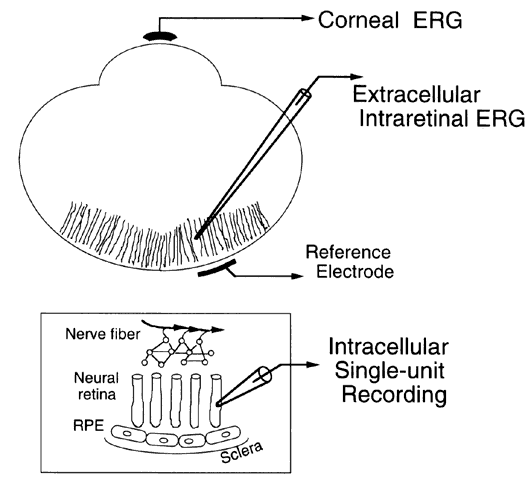
| Fig. 4. Recording configurations used to observe electrical signals of the retina. Corneal electroretinographic (ERG) recordings employ an electrode touching the surface of the eye with the reference electrode placed at the canthus or on the forehead. Extracellular responses are recorded by sliding a fine-tipped electrode into the retina alongside the neurons. As depicted here, the electrode frequently consists of glass pulled to a fine tip a few micrometers in diameter and filled with conductive fluid. The retinal depth of the tip position gives information about which cell type contributes most to the response. The reference electrode is placed behind the sclera for intraretinal recordings. Intracellular recordings are made by impaling a cell with an electrode. After recording single-unit activity, one can inject marker dye into the cell to identify the exact cell studied and to examine its projections onto neighboring cells. RPE = retinal pigment epithelium. |