|
|
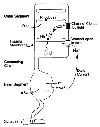
| Fig. 2. Schematic of a rod photoreceptor. Free-floating discs in the outer segment contain rhodopsin. The sodium channel is open in the dark and allows a dark current to flow between the inner and outer segments. Closure of the channel by light interrupts the dark current, which changes the electrical voltage outside the cell. This generates the a-wave, which is the fast, corneal-negative ERG component. |