|
|
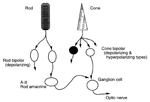
| Fig. 3. Cartoon of retinal wiring for rods and cones. Cones make contacts onto second-order bipolar cells of both the depolarizing and hyperpolarizing type. In turn, these synapse onto ganglion cells, which transmit the visual signal down the optic nerve. Cones in the primate fovea each contact approximately 30 bipolar and horizontal cells. By comparison, each rod synapses onto only two bipolar cells of the depolarizing type. The rod circuit adds an extra stage, in that rod signals are sent to amacrine cells. The A-II cell, also known as the “rod amacrine” cell, is in the rod pathway and makes presynaptic contact onto ganglion cells through the cone bipolar pathway, and the signal is transmitted through the optic nerve. |