Chapter 34
Anatomy of the Visual Sensory System
ALFREDO A. SADUN , JOEL S. GLASER and SWARAJ BOSE
Main Menu   Table Of Contents

Search

FUNCTIONAL ANATOMY
VISUAL ASSOCIATION AREAS AND INTERHEMISPHERIC CONNECTIONS
RETINOPTIC ORGANIZATION
NEW DIRECTIONS
VASCULAR SUPPLY
REFERENCES

The human primary visual sensory system comprises the retina, optic nerves, optic chiasm, optic tracts, lateral geniculate nuclei, geniculostriate radiations, striate cortex, visual association areas, and related interhemispheral connections. This specialized afferent system lies principally in a horizontal plane that crosses at right angles the major ascending sensory and descending motor systems of the cerebral hemispheres (Fig. 1). The anterior portion of the visual system is intimately related to the vascular and bony structures at the skull base and undersurface of the brain. The posterior portions are closely applied to the lateral aspects of the ventricular system that extend throughout the cerebral hemispheres. Thus, defects of the visual pathways, as revealed by visual field assessment or otherwise, have great localizing value in neurologic diagnosis. The dominant role of vision in humans may be expressed numerically by considering, for example, the number of axons in the human optic nerve (1.2 million) as compared with axons in the acoustic nerve (approximately 31,000).1–3 Thus, the ratio of afferent neurons in the peripheral visual apparatus to the number in the aural system is roughly 40 to 1.

Fig. 1. The visual sensory system. The left cerebral hemisphere has been removed with the exception of the occipital lobe and ventricular system. The left lateral geniculate body is hidden (arrow). Note the following relationships: optic nerve with internal carotid and anterior communicating arteries; chiasm in the floor of the third ventricle; forward sweep of temporal radiations around lateral ventricle; course of occipital radiations toward interhemispheral surface of occipital lobe. The cerebral falx and cerebellar tentorium are not illustrated.

The first distinct evidence of the human eye is found in the eight-somite stage of embryonic development, which occurs at about the third to fourth week of gestation.2,3 The two primordial optic bulbs extend to either side of the anterior end of the neural tube, the prosencephalon. A slight thickening between the two represents the torus opticus (that is, the primitive chiasmal anlage). The optic primordia evaginate to form the laterally placed cup-like vesicles, which contact overlying surface ectoderm and induce lens growth. The optic stalk and cups are notched ventrally (the “fetal” fissure) to permit entry of blood vessels, and the primitive retina is developing by about 5 weeks of gestation. Retinal ganglion cells differentiate and optic nerve fibers begin to fill the optic stalk, which is now surrounded by a cellular sheath. These afferent visual neurons reach the area of the chiasm at about the seventh week of gestation as the ventral fissure closes.

At approximately 50 days of gestation, the optic nerve contains a full complement of retinal ganglion cell axons, the optic disc and scleral opening are well defined, and endochondral ossification of the spheroid begins. Actually, the ultimate number of axons in the mature human optic nerve is the result of an initial overproduction of axons during the first half of gestation, followed by a 70% reduction (“die back”) between 16 and 30 weeks of gestation. Myelination of visual axons commences at the chiasm early in the seventh fetal month and progresses distally toward the lamina cribrosa until about the first postpartum month.

Retinal ganglion cells direct their axons to the proper regions of specific subcortical visual centers. Despite this remarkable specificity, surgical manipulations in the perinatal period can lead to retinofugal projections to nonvisual structures in hamsters.4 In the monkey, the retinal afferents from each eye overlap in the developing lateral geniculate nucleus.5 In the mouse, the contralateral projections to the lateral geniculate nucleus precede the ipsilateral retinofugal inputs, which may then displace them.6 The relative delay of ipsilateral retinal ganglion cell projections continues until after birth. Thus, a large number of the retinofugal axons that die back are probably of temporal retinal origin and displaced by nasal fibers from the contralateral retina.

Back to Top
FUNCTIONAL ANATOMY

RETINA

The functional organization of the visual sensory system begins at the retina. It is beyond the scope of this chapter to describe fully the complex vertical and horizontal organization of retinal elements, synaptic patterns, receptive field physiology, and other details of visual signal propagation. However, it is important to emphasize that there is a considerable degree of retinal processing that modifies neural signals before transmission to central structures such as the lateral geniculate nuclei and other subcortical visual areas. Ganglion cell subsets selectively encode specific aspects of visual information, such as acuity, color, image velocity and movement direction, and contrast; thus, visual signal processing takes place simultaneously but in separate “channels.”7 Visual information is already integrated at the retina and arrives at the optic nerve after considerable modulation.

In an excellent review of the primate retina, Rodieck8 describes the neuronal circuitry and synaptic organization within the 12 layers of the retina that contain at least 18 specific cell types. In the human retina, Rodieck describes four different percipient elements (rods and three types of cones), at least four types of bipolar cells, three types of amacrine cells, three types of horizontal cells, and at least three types of ganglion cells in addition to Muller cells. Humans, as all primates, have a nonlinear distribution of their ganglion cells across the retina.8 Axons from these ganglion cells form bundles in the nerve fiber layer en route to the optic disc. There is, at best, controversial evidence regarding the existence in primates of efferent fibers deriving from the optic nerve to terminate throughout the inner plexiform layer of the retina.9 If this pathway exists, the fibers probably are sympathetics that contact blood vessels.

Visual function across the retina is not uniform but takes a pattern of concentric zones that increase in sensitivity toward the central retina area, the fovea, which subserves the highest sensitivity. At progressively eccentric retinal locations there is a near-linear decrease in sensitivity (conversely, elevation of thresholds). Ultimately, retinal sensitivity is a manifestation of the underlying cytoarchitecture and the distribution of the percipient elements, the cones and rods. The fovea itself is essentially without rods; it is composed rather of about 100,000 compactly arranged slender cones. The entire posterior pole of the retina is dominated by the foveal and parafoveal cone system that occupies an area of approximately 1.5 mm in diameter. Small retinal ganglion cells subserving this central cone cell system send their small-caliber axons directly to the temporal aspect of the optic disc, forming the papillomacular nerve fiber bundle. This discrete bundle of nerve fibers is relatively isolated from other retinal nerve fibers that reach the optic disc by an arcuate course above and below the papillomacular bundle, forming dense superior and inferior bands (Fig. 2). Osterberg10 quantitatively examined the arrangement of photoreceptors in the human retina and found a skewed distribution: rod and cone populations are more dense in the superonasal retina and less dense in the inferotemporal retina. Van Buren11 performed retinal ganglion cell counts and showed the same eccentric pattern, especially in the one-cell-thick layer, which reached nearly twice as far on the nasal side of the fovea as on the temporal side. This asymmetric distribution of retinal elements about the fovea is reflected in the asymmetry of the nasal versus the temporal visual field and accounts for the relative foreshortening of the nasal periphery.12

Fig. 2. Retinal nerve fiber layer pattern. The dense temporal arcuate fiber bundles (solid arrows) are most easily seen. Nasal fibers (open arrows) take a more direct radial course. The papillomacular bundle (arrowheads) is most difficult to visualize.

In mammals, the inner aspects of cones form pedicles that are wide and flat and act as a platform for synaptic terminations. Each pedicle contains several invaginations that form synaptic contact with horizontal and bipolar neurons. Rods have a smaller synaptic termination at their inner end termed a spherule that contains only one invagination. Horizontal and bipolar cells synapse with pedicles and spherules in the outer plexiform layer. Amacrine and bipolar cells make synaptic contacts in the inner plexiform layer. Ultimately, visual information progresses centripetally to the retinal ganglion cells, whose dendritic processes extend into the inner plexiform layer and whose axons form the retinal nerve fiber layer. Most recently, a sixth class of retinal neuron, the interplexiform cell, has been described in monkeys and humans.13,14 These dopaminergic cells lie in the inner nuclear layer with processes that extend to both the inner and outer plexiform layers, providing an anatomical substrate for an inner to outer (centrifugal) retinal neural signal. In humans, retinal ganglion cells are stacked six to eight cells deep near the fovea but are found only sparsely in the peripheral retina. About 1.3 million retinal ganglion cells (down from two to three times that number before birth) contribute axons from 0.5 to 2.5 μm in diameter in the nerve fiber layer.15

Retinal ganglion cells have been classified since 1933 when Bishop suggested, based on electrophysiologic studies of the frog, that there were three groups of axons deriving from retinal ganglion cells that conduct at different velocities.16 Morphologically, Hopp had already demonstrated different shapes and sizes to retinal ganglion cells but had not put together a rigid classification scheme. However, Polyak, in 1941, did classify primate ganglion cells of four types, including parasol and midget cells.17,18 In 1966 the work of Enroth-Cugell and Robson19 clearly established the differences between X- and Y-cell types. In 1974, Boycott and Wassle20 were able to provide the corresponding morphologic classes of retinal ganglion cells in the cat.

Enroth-Cugell and Robson also first noted differences in the spatial frequencies with which high contrast sensitivity was obtained between X and Y cells. They noted that the receptive field centers of Y cells were larger than those of X cells and that X cells were more common than Y cells; they therefore concluded that X cells subserved high-resolution pattern vision and Y cells subserved movement vision. Boycott and Wassle described alpha, beta, and gamma cells based on distinctions in size, dendritic morphology, and axonal caliber. They suggested that the alpha cells corresponded to Y cells, the beta cells to X cells, and the gamma cells to W cells, which had by that time also been proposed by Rodieck.21

However, the retinal ganglion cell classification schemes for primates differs.22 Two types of morphologically distinct cells appear to project to different divisions of the lateral geniculate nucleus (LGN; the magnocellular and parvocellular layers). This segregation continues through the radiation projections to the cortex. The magnocellular LGN projects to 4C alpha. The parvocellular LGN projects to 4C beta and layers 2 and 3 of area V-1 and to the “pale stripe” region of area V-2. The segregation is continued beyond the synapses to areas MT, V-3, and V-4 of the cortex.23 However, as in the cat, there are physiologic differences between these systems as well. The parvosystem is characterized by color opponency, high spatial resolution, and low contrast sensitivity. The magnosystem, however, is characterized by color ignorance, low spatial resolution, fast temporal resolution, stereopsis, and high contrast sensitivity.24

In humans, Polyak17 believed that there were five or six different types of retinal ganglion cells. However, he used the overall classification scheme of large parasol ganglion cells that project to the magnocellular LGN (more recently referred to as the M-cell system) and smaller midget ganglion cells that project to the parvocellular LGN (P-cell system). Each of these two systems has a characteristic dendritic field diameter and a separate primary projection from LGN to striate cortex.25 However, to make things more complicated, midget, parasol, shrub, blue-cone, biplexiform, garland, small diffuse, giant, and displaced retinal ganglion cells have all been described.8 For many of these cells, differences in dendritic field size and orientation have been worked out in the monkey.26

In advanced primates, the fovea occupies the central 3 degrees of retina and is a roughly circular pit devoid of ganglion cells and surrounded by a multilayered annulus of densely packed small ganglion cells. Central projections, which have been traced by horseradish peroxidase, indicate that ipsilaterally projecting ganglion cells in the temporal foveal rim can generate 2 to 3 degrees of bilateral representation in the geniculocortical pathways, because of intermingling with contralaterally projecting cells on the nasal side of the foveal pit.27 These findings provide a potential retinal neural basis for foveal (fixational) “sparing” or “splitting” that is demonstrated by perimetry in the presence of lesions of the posterior visual pathways.

Ogden28 extensively studied horizontal and vertical retinotopy in the geographic bundles of ganglion cell axons as they traverse the retina to the optic disc. Despite previous reports that the nerve fiber layer is well organized, Ogden showed that horizontal retinotopic organization is present within nasal, but not temporal, nerve fiber bundles and that vertical retinotopic stratification is orderly within temporal but not nasal retinal bundles. In addition, temporal axons in the nerve fiber layer intermingle freely along the intraretinal course of the arcuate bundles. Therefore, most of the segregation of fibers to conform to a retinotopic distribution must occur at the optic disc or more posteriorly in the optic nerve. Species differences do occur, with the macaque retina apparently most resembling the human nerve fiber layer.

OPTIC DISC

The optic nerve head can be seen funduscopically end on and appears as a flat disc with a central depression of variable depth, called the optic cup. Papilla, a term that implies a nipple-like eminence, is used less frequently. The disc is the collective exit site of all retinal ganglion cell axons (i.e., the nerve fiber layer). The optic disc is located 3 to 4 mm nasal to the fovea and represents a 1.5 × 2.0-mm hiatus in the sclera, choroid, retinal pigment epithelium, and retina proper. The mean horizontal disc diameter is 1.76 ± 0.3 mm; the mean vertical diameter is 1.92 ± 0.3 mm; the mean horizontal cup to disc ratio is 0.39; and the mean vertical cup to disc ratio is 0.34. The number of nerve fibers appears to be positively correlated with the size of the optic nerve head: larger discs have relatively more fibers than smaller discs. However, the relationship is not linear. Smaller discs may show optic nerve head crowding.29,30 Further, the fiber population decreases with age, especially after 60.31a,31b There are no percipient elements in the area of the disc, which is represented in visual space as an absolute scotoma, the blind spot of Mariotte.

The retinal axons turn 90 degrees over the scleral disc margin and pass through the perforations of the lamina cribrosa. This fenestrated connective tissue is lined by astrocytes, is continuous with surrounding sclera, and partitions the nerve head into prelaminar, laminar, and retrolaminar compartments (Fig. 3). The extracellular matrix of the human lamina cribrosa contains collagen macromolecules resembling central nervous system basement membrane and does not resemble sclera in this regard.32 Axonal bundles are increasingly compartmentalized by glial-collagen pial septa as they traverse the lamina cribrosa into the distal portion of the optic nerve. The increase in interaxonal glial tissue density could play a possible role in the propagation of edema caused by ischemic optic neuropathies or when intraocular pressure is raised.33

Fig. 3. Schematic structure of optic disc and nerve. 1a, internal limiting membrane of retina; 1b, nerve fiber layer; 2, optic cup, lined by astrogial cells, and central retinal vessels; 3, ophthalmoscopically visible disc edge; 5, glial and connective tissue columns; 6, nerve fiber fascicles; 7, major portion of lamina cribrosa; 8, oligodendrocytes; Du, dura; Ar, arachnoid; Gl. M, glial mantle; Sep, pial septum. (Modified from Anderson DR, Hoyt WF: Ultrastructure of intraorbital portion of human and monkey optic nerve. Arch Ophthalmol 82;506, 1969)

OPTIC NERVE

The optic nerve may be regarded as consisting of four segments: intraocular (1 mm), intraorbital (25 to 30 mm), intracanalicular (9 to 10 mm), and intracranial (about 16 mm). Thus, the entire length of the optic nerve from the globe to the optic chiasm is 5 to 6 cm. The intraocular portion (optic disc) may be further divided into retinal, choroidal, and scleral levels as the retinal ganglion cell axons from the nerve fiber layer turn sharply posteriorly to exit the globe (see Fig. 3). Anderson34–37 and Minckler33 extensively described the microanatomical structure of the nerve as consisting of a neuroectodermal (nervous tissue proper) and a mesodermal component (providing support and nourishment). Mesodermal connective tissue includes the sclera, fibroblasts, meningothelial cells, and blood vessels. The collagenous component of the perforated lamina (also termed the lamina cribrosa scleralis) may be considered continuous, but not identical, with the sclera and also with the perioptic meninges. The more anterior aspects of the optic nerve head contain less connective tissue, with glial components predominating. The anterior optic nerve head consists of unmyelinated axons and astrocytes. It is unusual for oligodendrocytes or myelinated axons to be anterior to the laminar scleralis; funduscopically visible myelinated (medullated) nerve fibers are associated with anomalous rests of oligodendrocytes.

Behind the lamina cribrosa, the optic nerve abruptly increases in diameter from 3 to 4 mm. At this point, oligodendrocytes (responsible for the formation of myelin that ensheathes the axons) constitute approximately two thirds of the interstitial cells. In peripheral nerves, Schwann cells serve this same function. Therefore, the optic nerve must be considered analogous to white matter tracts of the brain rather than to peripheral nerves; this composition makes the optic nerve susceptible to diseases of central nervous system tracts, such as multiple sclerosis. The orbital and intracanalicular portions of the optic nerve contain a well-developed septal system derived from pia mater. The septa form porous cylindrical walls aligned along the long axis of the nerves and may contribute to flexible movement.38 The septa divide the nerve fibers into about one thousand parallel columns of variable shape and size. Astrocytes are intimately related to the pial septa and play a role in the support and nutrition of axons.

Approximately 1 cm posterior to the globe, a major branch of the ophthalmic artery pierces the inferior aspect of the meninges of the optic nerve, gains a central axial position, and emerges in the middle of the optic disc as the central retinal artery. The central retinal artery does not contribute significantly to the blood supply of the laminar and prelaminar portions of the optic nerve head. These areas of the optic nerve are supplied by an anastomotic arterial complex called the circle of Zinn-Haller. This structure receives contributions from the posterior ciliary arteries, the pial arterial network, and the peripapillary choroid (Fig. 4). Thus, the blood supply of the optic nerve head is derived primarily from choroidal and posterior ciliary vessels, as opposed to that of the retina, which is mediated by the central retinal artery.39,40

Fig. 4. Blood supply of the optic nervehead. 1, central retinal artery; 2, arterial circle of Zinn-Haller; 3, pial arterial network. Contribution to Zinn-Haller circle from posterior ciliary arteries, pial plexus, and peripapillary choroid. The latter also sends branches directly to prelaminar disc substance. (Modified from Kolker AE, Hetherington J Jr: Becker-Shaffer's Diagnosis and Therapy of the Glaucomas, 3rd ed. St. Louis, C.V. Mosby, 1970)

The intraorbital segment of the optic nerve (about 25 mm) exceeds the distance from the back of the globe to the orbital apex (less than 20 mm). Therefore, within the orbit, the optic nerve has redundancy in length and a sinuous course; this permits the nerve to move freely behind the globe during eye movements and also allows up to 6 or 8 mm of proptosis before the nerve begins to tether the back of the globe. At the orbital apex, the optic nerve enters the bony optic canal and is surrounded by the connective tissue origins of the superior, medial, and inferior recti muscles, which collectively constitute the so-called annulus of Zinn.

The optic canal runs posteromedially in the sphenoid bone, at an angle of approximately 35 degrees with the midsagittal plane (Fig. 5). The optic canal is 4 to 10 mm in length and contains not only the optic nerve but also the ophthalmic artery, branches of the carotid sympathetic plexus, and extensions of the intracranial meninges that form the sheaths of the optic nerve. The dural covering of the nerve and the periosteum of the canal are fused, but the arachnoid is continuous, permitting the subarachnoid space of the optic nerves to communicate freely with the intracranial subarachnoid space, both of which contain cerebrospinal fluid. However, variable amounts of trabecular connections between the arachnoid and the pia add resistance to the space. Further, the cul-de-sac of the anterior end of the subarachnoid space may occur more posteriorly than the back of the globe. As a consequence, some patients with raised intracranial pressure manifest assymmetric or even unilateral disc edema.

Fig. 5. The optic canal. A. Anterior view of left orbital apex. Orbital end of optic canal is vertically oval (solid arrows) and separated from the superior orbital fissure (open arrow) by the optic strut. Note transilluminated ethmoidal and sphenoidal air cells that form the medial orbital wall and the medial wall of the optic canal. B. Posterior view of the intracranial aspect of the left optic canal, demonstrating horizontally oval contour. The optic strut (OS) forms the ventrolateral margin of the canal and separates it from the carotid artery. In this preparation, the ethmoidal and sphenoidal air cells have been opened. AC, anterior clinoid; PL, planum; SPH, sphenoidal wing. C. Tomographic section of optic canals in upper diagram. Normal axial tomogram below. (C from Harwood-Nash DC: Optic gliomas and pediatric neuroradiology. Radiol Clin North Am 10:83, 1972)

The mesial surface of the optic canal protrudes into the superolateral aspect of the spheroid sinus, and according to Fujii and colleagues,41 the optic nerves are separated from the sinus cavity by only the nerve sheath and mucosa in some 4% of specimens; in 78%, less than a 0.5-mm thickness of bone separates the optic nerves from the sinus cavity. Manipulations at the lateral spheroid sinus wall during transsphenoidal surgical procedures may damage the optic nerves.

The optic nerves are fixed at the intracranial opening of the optic canals, the upper margins of which are formed by an unyielding falciform fold of dura. This constriction may notch the superior surface of the optic nerve when sellar-based adenomas, or internal carotid artery aneurysms, elevate the chiasm. From the internal (posterior) foramina of the canals, the optic nerves converge toward the chiasm in the anteroinferior floor of the third ventricle. The two nerves ascend toward the chiasm at an angle of approximately 45 degrees with the nasotuberculum line (Fig. 6); the intracranial nerve segment averages 17 ± 2.4 mm in length, so that the optic chiasm itself sits 10.7 ± 2.4 mm above the dorsum of the sella turcica.40 Occasionally, the intracranial optic nerves are shorter, and the chiasm may lie directly above the sella in a position that is called prefixed. More commonly, the optic chiasm is positioned 10 mm above the insertion of the diaphragma sella onto the dorsum.42 This being the case, it should be understood that pituitary tumors must extend well above the sella before the optic chiasm is encroached on. By the time chiasmal field defects can be found, pituitary tumors are already large and have major suprasellar extensions. Small tumors are detected clinically when signs of unilateral optic nerve compression evolve.

Fig. 6. Relationships of the optic nerves (ON) and chiasm (X) to the sellar structures and third ventricles (3). C, anterior clinoid; D, dorsum sellae; P, pituitary gland in sella.

The anterior perforated substance, the root of the olfactory tract, and the anterior cerebral artery lie superior to the optic nerve in its intracranial path. The internal carotid artery is below and then lateral to the nerve, and the ophthalmic artery enters the optic canal within the dural sheath of the nerve. The inferior surfaces of the frontal lobes (gyrus recti) of the cerebral hemispheres are above the optic nerves. The anterior cerebral and anterior communicating arteries lie between the frontal lobes and the optic nerves (see Fig. 1). Medial to the anterior clinoid process (see Fig. 5B), the optic nerve lies just above the siphon of the intracavernous portion of the internal carotid artery and is separated from the cavernous sinus by the optic strut. Thus, expanding lesions of the cavernous sinus, such as aneurysm or meningioma, may impinge on the optic nerve. At the origin of the ophthalmic artery, aneurysms may compress the nerve from a medial and ventral direction.

OPTIC CHIASM

The anatomy of the optic chiasm has been an intriguing subject since the time of Galen, who, in the 2nd century, likened the structure to the Greek letter chi, with subsequent additions and emendations by Leonardo da Vinci (1504), William Briggs (1676), Thomas Willis (1681), Rene Descartes (1686), Isaac Newton (1704), and Ramon y Cajal (1898), among others.43,44 However, only by the end of the 19th century was partial discussion of retinal axons in most mammalian chiasms ascertained histologically. Moreover, Ramon y Cajal saw the chiasm as a structure that corrected the inversion of sensory space imposed by the optical system of the eye. In other words, he understood that a discontinuity of the visual field would result in a physiologically scrambled cerebral pattern. These considerations of physiologic optics led Ramon y Cajal to elaborate a general theory of cerebral crossings that also provides the basis for contralateral motor and sensory projections.

The optic chiasm derives from the merging of the two optic nerves. The superior and posterior aspect of the optic chiasm is contiguous with the anteroin-ferior floor of the third ventricle. The optic chiasm measures approximately 8 mm from anterior to posterior notch, 15 mm in its horizontal diameter, and 4 mm in height. Like the optic nerve, it is also surrounded by meningeal sheaths and cerebrospinal fluid.45,46 The chiasm lies within the circle of Willis. The regional vascular relationships of the intracranial optic nerves and optic chiasm are critical, because aneurysms commonly involve the internal carotid arteries and the basal arterial circle. As the internal carotid arteries curve posteriorly and upward out of the cavernous sinuses, they lie immediately below the optic nerves. The carotid arteries then ascend vertically along the lateral aspects of the chiasm (see Fig. 1). The precommunicating portions of the anterior cerebral arteries are closely related to the superior surface of the chiasm and optic nerves.

According to Barber and coworkers,47 the human optic chiasm anlage is visible in the floor of the forebrain between the optic vesicles at the 3-mm stage of embryogenesis. Between 4 and 8 weeks of gestation, axons and retinal ganglion cells grow toward the brain into the floor of the third ventri-cle and partially decussate to form a true chiasm(30-mm stage). Kupfer48 calculated in the adult human that the ratio of crossed and uncrossed fibers in the optic chiasm was approximately 53 to 47, with the uncrossed portion being greater than that seen in any other primate. After the 8-week stage of development, retinofugal axons encircle the lateral aspects of the diencephalon to reach a collection of cells that differentiate from the dorsolateral portion of the thalamus to form the dorsal nucleus of the lateral geniculate body. During the second month, the eyes assume a frontal position, such that the optic nerves must pass upward and medially to gain the optic canals. At about the 10th week, uncrossed retinal fibers begin to appear. The primitive optic recess forms at the anterior aspect of the third ventricle; the recess extends variably a short distance into the proximal ends of the optic nerves but is eventually obliterated as more retinofugal axons reach the optic chiasm. Ultimately, the small optic recess represents the remnant of the proximal ends of the optic vesicles.

As the thalamic nuclear masses increase in size, the third ventricle becomes a vertical slit (at about 12 weeks or the 60-mm stage). At the beginning of the fourth month, the optic tracts continue to be formed by decussating fibers that pass around the thalamic nuclei. During the last few months of gestation, the development of the optic chiasm consists mainly of an increase in size owing to ingrowth of retinal axons, and its ultimate topographic configuration is dictated by the growth of contiguous structures and modification in the shape of the third ventricle. Myelination of these axons begins only after the visual pathways are otherwise complete. Sometime during the fifth month, myelination begins at the geniculate nucleus, reaches the chiasm during the sixth month, and finally progresses from a proximal to distal direction along the optic nerves during the eighth and ninth months of gestation.

There is evidence that each neuroretina has contributions from both sides of the prospective forebrain. Using cell-labeling techniques (induced chromosomal mosaicism or horseradish peroxidase), it has been shown in the frog embryo49 that there is reciprocal movement (translocation) of cells from each side of the primitive forebrain into the contralateral retinal anlage. This migration begins before neural tube closure and results in the formation of a primitive optic chiasm into which grow axons from each retina, destined for synaptic sites on the opposite side of the brain from which the precursor cells originated. Thus, translocated cells establish a pattern for chiasmal crossing even before ganglion cell axonal outgrowth from the retina.

Silver and Sapiro50 have investigated the role of melanin in axonal guidance during development of the primitive optic nerves and chiasm. In mice and rats, the upper aspect of the distal eye stalk is transiently pigmented before and during migration of pioneer visual axons, which avoid the area of melanosomes. Therefore, it is suggested that melanin-producing stalk cells play a role in controlling the topographic pattern of optic nerve fibers by inhibiting axonal growth within their territory. Indeed, in the albino, the topographic arrangement of retinal axons is altered such that the majority of ganglion cells that originate in temporal retina decussate at the optic chiasm and project to the contralateral LGN. This anomaly of retinogeniculate projection disrupts the laminar structure of the geniculate and results in an abnormal visual field representation in the visual cortex. Hypomelanosis syndromes such as oculocutaneous or ocular albinism regularly show evidence of misrouted retinal axons, with fibers from 20 degrees or more from the temporal retina anomalously crossing at the chiasm instead of projecting to the ipsilateral hemisphere; that is, each hemisphere receives predominantly monocular in-put from the contralateral eye, with only periph-eral nasal field represented ipsilaterally.51 Poor visual acuity, strabismus, and nystagmus are likelyresults.

Anomalous development of the optic chiasm otherwise occurs from faulty development of one or both optic vesicles, or with forebrain malformations. In bilateral anophthalmia, the optic nerves, chiasm, and optic tracts do not develop.52 In unilateral anophthalmia, an asymmetric and small optic chiasm is found composed of nerve fibers from the normal eye.53

Most of the retinofugal fibers exit the optic chiasm to form the primary optic tracts. However, a few fibers have been shown to exit from the dorsal surface of the optic chiasm and enter directly into the hypothalamus to terminate in the suprachiasmatic nucleus or the supraoptic nucleus of the hypothalamus (Fig. 7).54,55 These two fiber pathways probably represent the neuroanatomical basis for light-dark entrainment of the neuroendocrine circadian rhythm. Bilateral transsection of the optic nerve in the rat results in a loss of synchronized endogenous circadian rhythms, whereas bilateral transsection of the optic tract (distal to these fibers from the chiasm to the hypothalamus) does not.56

Fig. 7. Anatomical relationships of the optic tract and lateral geniculate body. (Strother CM, Salamon G: Topographic anatomy of the brain. In: Newton TH, Potts DG, eds: Radiology of the Skull and Brain, Vol 3, Anatomy and Pathology, St. Louis, C.V. Mosby, 1977)

OPTIC TRACT

As the retinofugal fibers pass through the chiasm, they form the optic tract immediately posterior to the optic chiasm. Each tract is approximately3.5 mm high and 5.1 mm long and begins at the posterior notch of the chiasm and is separated from the other optic tract by the pituitary stalk inferiorly and the third ventricle more superiorly.44 Across the basal arachnoidal cistern, the inferolateral aspect of the tract faces the uncal gyrus of the temporal lobe. As the tracts proceed posteriorly, they diverge in the interpeduncular cistern; they embrace the ventral aspect of the rostral midbrain contiguous with the cerebral peduncles.

At the level of the mamillary bodies, the tracts are located more laterally in the choroidal fissures, with the uncus below, the internal capsule above, and the amygdala lateral to them. Most of the fibers in the optic tract terminate in the ipsilateral LGN; however, just posterior to the optic chiasm, a small fascicle of fibers emanates from the optic tract, travels between the two lobes of the ipsilateral supra-optic nuclei, and ascends to terminate in the para-ventricular nucleus of the hypothalamus.57 The paraventricular nucleus also mediates visual input to control diurnal rhythms.55 Further posteriorly, a larger fascicle of axons leaves the optic tract and proceeds ventral to the medial geniculate nucleus. It continues by way of the brachium of the superior colliculus to terminate in the pretectal nuclei of the rostral mesencephalon (see Fig. 7). These fibers represent the afferent limb of the pupillomotor reflex.

LATERAL GENICULATE NUCLEUS

The bulk of the optic tract containing retinofugal fibers terminates in the LGN. The LGN is the largest and probably most important primary visual nucleus in humans. Here, crossed and uncrossed retinal fibers are ultimately organized into homonymous pairs. Neurons of the LGN contribute axons that form the geniculocalcarine radiations. The LGN is part of the thalamus and is folded deep in the lateral recess of the choroidal fissure, obscured from direct view by the hippocampal gyrus of the temporal lobe. The lateral relationship of the LGN is the so-called temporal isthmus, the white matter structure lying between the auditory cortex above and the temporal horn of the lateral ventricle below. Polyak and other anatomists traditionally divided the LGN into a large dorsal and small ventral (pregeniculate) nucleus. There is little evidence, however, that the ancient ventral portion has important functions in the visual system of primates. In humans, there are, for the most part, six gray layers of neurons discernible in the dorsal LGN. These can be best appreciated in coronal section through the middle of the LGN (Fig. 8). In other planes and at the poles, the LGN may appear to have fewer than six layers. Since the early studies of Minkowski, it is evident that crossing visual axons from the contralateral eye terminate in laminae 1, 4, and 6 and uncrossed ipsilateral axons end in laminae 2, 3, and 5.58,59 Each lamina receives input from one eye only.

Fig. 8. Terminal relations of optic tract in mammalian brain. Ch, chiasm; Op Tr, optic tract; Gl, lateral geniculate body; Gm, medial geniculate body; C, superior colliculus; R, red nucleus; SN, substantia nigra; a, anterior accessory optic tract; b, posterior accessory optic tract; c, fibers to large cell nucleus of optic tract. (LeGros Clark WE: The structure and connections of the thalamus. Brain 55:442, 1932)

The LGN is shaped like a three-cornered hat, and degeneration studies60 reveal that there is a rather sharply defined dorsal central wedge that extends through all laminae and represents the macular projection. The macular fibers are also relatively confined to the dorsal section of the optic tract (Fig. 9). Upper retinal quadrants are represented medially, and the lower retinal projections terminate laterally. This situation is the exception to the useful generalization that upper retinal fibers continue dorsally and lower fibers continue ventrally throughout the visual pathways. Thus, the retinotopic organization of the posterior optic tract appears to rotate 90 degrees as it enters the LGN.

Fig. 9. Coronal section of lateral geniculate nucleus. Note extensive macular representation.

There is a retinotopic organization within the LGN, as demonstrated by degeneration and electrophysiologic techniques.60 Any point in the visual field is projected to a vertical column of cells, whose long axis is approximately orthogonal to the LGN laminae. These “projection lines” have been established in all mammals that have been studied thus far.61,62 Even the physiologic blind spot is represented in the contralateral LGN as a cell-free vertical column, suggesting a preset and extremely accurate alignment of projection columns through adjacent laminae.62 Autoradiographic tracing methods63 in owl monkeys have confirmed the nature of projection fibers from cortical visual area;17 the striate cortex projects to all laminae and interlaminar zones of the LGN.

By comparing the locations of cortical injection sites (3H-proline) with positions of subsequent labels in the LGN, it is clear that the corresponding loci in the paired representations of visual field are interconnected. Also, vertical columns of label lie roughly perpendicular to the LGN laminae and in line with projection lines. Input to the LGN from cortical area 18 is relatively sparse and is found predominantly in the ventral “magnocellular” layer. The functional significance of these cortical inputs to the LGN is not clear, but differences in laminar location and density patterns or projections from each cortical area further suggest different functions for each set of connections.

Layers 1 (receiving contralateral retinal projections) and 2 (receiving ipsilateral retinal projections) contain larger neurons and are, therefore, termed the magnocellular LGN layers. Conversely, layers 3 through 6 are termed parvocellular LGN layers. There is considerable evidence that there are at least two types of retinal ganglion cells that project in a segregated fashion to either magnocellular (M cells) or parvocellular (P cells) LGN. Thereafter, this segregation remains true insofar as the magnocellular LGN projects to cortex area 4C-alpha that eventually projects to middle temporal cortical area MT.23,64,65 Conversely, the parvocellular LGN projects to cortical area 4C-beta and from there to layers 2 and 3 of area 17, to the “pale stripe” region of area 18, and finally to areas V3 and V4. Thus, at the LGN the major division between lines of information remains established.

The LGN is not a simple relay station but is a center for processing; it also receives input from cortical and subcortical centers and reciprocal innervation from the visual cortex.66 The processing of visual signals occurs by means of a highly complex synaptic organization of relay cells within the LGN. Cortical and subcortical (including pontine reticular formation and pulvinar) centers also have neural input to the LGN. For example, electrode stimulation of the optic radiation evokes LGN inhibition, and the LGN response to electrical stimulation of one optic nerve is depressed by stimulation of the other optic nerve. Therefore, there is ample evidence of corticofugal inhibition by means of descending projections, such as those described above, from visual cortex areas 17, 18, 19, and midtemporal cortex (area MT), and also of retinogeniculate inhibition from the opposite optic nerve. Detailed analysis of geniculate cytoarchitecture, synaptology, and neurophysiology is available elsewhere.67,68

The LGN has often been considered as a model system for the phenomenon of transsynaptic degeneration. After lesions to retinal ganglion cell axons (in the optic nerve or elsewhere), changes are noted in the cells and cytoarchitecture of the LGN that have been described as evidence of cell death consequent to presynaptic deafferentation.69 Indeed, this also has been described as occurring in the reverse direction; optic atrophy can be seen after destruction of the geniculate cortical radiations. However, this phenomenon occurs primarily in children or in adults only after very long-term intervals following lesions of occipital cortex.70 Anterograde transsynaptic degeneration probably does not occur; instead atrophy (not cell death) of LGN neurons follows the loss of retinal ganglion cell axons.71

VISUAL RADIATIONS

The geniculocalcarine fibers tract begins in the LGN and constitutes the “posterior” visual pathway that projects to the primary visual cortex (V1 or area 17). These myelinated fibers exit from the dorsal aspect of the LGN and then fan laterally and inferiorly through the temporal isthmus to sweep around the anterior tip of the temporal (inferior) horn of the lateral ventricle (see Fig. 1). The most anteroin-ferior fiber-fascicle forms a bend (Meyer's loop) that contains fibers that represent the homonymous inferior retinal quadrants, which mediate the contralateral superior visual field. This loop is located approximately 4 to 5 cm caudal to the anterior pole of the temporal lobe. This configuration of the anterior portion of the visual radiations is the anatomical substrate that explains the tendency for the superior quadrantanopic field defect pattern (“pie in the sky”) encountered in some temporal lobe lesions. Further, this is the basis of the advice often given to the neurosurgeon planning to resect the temporal tip to try and avoid resecting more than 4 cm.

Both the superior and inferior fascicles of the visual radiations pass posteriorly as a vertically narrow fillet in the external sagittal striatum, which lies just lateral to the tapetum of the corpus callosum by which the radiations are separated from the cavities of the lateral ventricle. In the deep parietal lobe, the radiations pass just external to the trigone and occipital horn of the lateral ventricle. These visual fibers turn medially above and below the occipital horn to terminate in the mesial surface of the occipital lobe, the striate (or calcarine) cortex.

OCCIPITAL CORTEX

The primary visual cortex (once called Brodmann area 17, now usually labelled V1) lies in the interhemispheral fissure in relationship to the falx cerebri (Figs. 10 and 11). However, the large macular projection area extends 1 to 2 cm laterally onto the posterior surface of the occipital cortex. The visual cortex extends anteriorly toward the splenium of the corpus callosum and is separated into a superior and an inferior portion by the calcarine fissure that runs horizontally. This area on both sides of the calcarine fissure is termed the calcarine cortex.

Fig. 10. Representation of the macular (central field) projection in the anterior visual pathways. The majority of afferent visual fibers are related to the papillomacular bundle, which subserves the central visual field and comprises the central core of the optic nerve. The crossing macular projection occupies an extensive area of the median bar of the chiasm.

Fig. 11. Location of visual cortex primarily in interhemispheral fissure. Lateral extension as illustrated is variable. Point F' corresponds to central fixation point F in contralateral field. Peripheral field point P is represented in rostral portion of cortex, P'. S, splenium of corpus callosum.

The visual cortex is further identified by specialized histologic features. Although the arrangement of cell nuclei and myelinated fibers is much the same as in other regions of the cortex, the visual cortex is characterized by an extra pronounced lamination oriented parallel to the cortical surface. This dramatic feature that distinguishes the visual cortex is the presence of a conspicuous, relatively acellular, myelinated fiber layer that is visible without magnification in sections perpendicular to the cortex; this is the white stripe of Gennari or Vicq-d'Azyr, giving us the term striate cortex. An excellent description of the visual cortex and its cellular types and fiber connections can be found in The Vertebrate Visual System by Polyak.72

The anatomy and histoarchitecture of the striate cortex have been elucidated in the past two decades largely through electrophysiologic studies in the cat and monkey and the use of special staining techniques in monkey and humans. With microelectrodes placed in the cortex, visual stimuli are projected onto a tangent screen placed before the animal, such that the visual image is focused on the retina. The spatial and temporal properties of cortical cells thus are explored; most cells respond only to stimuli in very restricted locations in the visual field and only to those with very specific psychophysical properties. The position and diameter of a stimulus in the visual field by which a single cortical cell can be excited or inhibited in a specific manner is the “receptive field” of that cell. David Hubel and Torston Wiesel have been pioneers in the elucidation of cortical visual physiology, and their work has been reviewed by many authors73 and has been recognized with a Nobel Prize.

A more precise understanding of the neurophysiology of the central visual pathways in the cortex has been derived through the use of horseradish peroxidase, cytochrome oxidase, and other histologic techniques. The functional architecture of the striate cortex can be best appreciated by analysis of its input from the LGN (Fig. 12). Axons originating from magnocellular cells of the LGN project to area 4C-alpha of area 17, which projects to area 4B also in area 17, which in turn projects both directly and indirectly to middle temporal cortical area MT. Conversely, parvocellular cells of the LGN project to 4C-beta of area 17, which projects to interblob (identified by cytochrome oxidase) areas also in area 17. Cells in these areas then project to the pale stripe area of area 18 and from there to other higher visual centers. The magnocellular system probably mediates low spatial resolution contrast sensitivity, orientation, movement sensitivity, directionality, and stereopsis. The parvocellular system mediates color and contrast sensitivity at the high spatial resolutions, without directionality or stereopsis.

Fig. 12. Occipital lobe and the corresponding projection of the visual field. A. Mesial aspect of left occipital lobe. The posterior pole (O) is flattened to illustrate the lateral surface (arrows), which is composed primarily of areas 18 and 19. The extension of striate cortex onto the lateral surface of the occipital pole is variable. The calcarine fissure (C) separates the striate or calcarine cortex into an upper and a larger lower strip, which also extends farther toward the splenium (SP) of the corpus callosum. The visual cortex is approximately 5 cm in horizontal diameter, and the macular projection (fine stippling) may occupy as much as the posterior 2.5 cm. The border zone between macular and peripheral retinal cortical projections is arbitrarily illustrated. B. The right hemifields. The upper field is represented in the inferior calcarine strip, and the lower field in the superior calcarine strip. The central field has a disproportionately large cortical representation. F, area of foveal projection. The temporal field of the right eye extends to 90 degrees as compared with the nasal 60-degree limit of the left eye. This 30-degree monocular temporal crescent is represented only in the contralateral hemisphere at the rostral extreme of the striate cortex.

A third system, the blob system, has also been described. The blob (stained by cytochrome oxidase) probably receives both magnocellular and parvocellular input, analyzes color, and conveys information on brightness. Authoritative review of this rather complex functional anatomy and histoarchitecture is available elsewhere.24,74

Back to Top
VISUAL ASSOCIATION AREAS AND INTERHEMISPHERIC CONNECTIONS
For the visual environment to be analyzed, recognized, and interpreted, afferent visual information must be transferred from the striate cortex (V1) to higher visual association areas V2 (or area 18, parastriate cortex) and V3 (or area 19) and to other analytic locations termed V4, V5, and the medial temporal gyrus (MT).24 In a simplified schematic, area 18 integrates the two halves of the visual fields by means of a major interhemispheric commissural pathway that traverses the splenium (most posterior portion) of the corpus callosum. Thus, areas V1, V2, and V3 in one hemisphere are interconnected to the same areas in the other hemisphere. Visual cortex area V2 probably participates in sensory-motor eye movement coordination through fronto-occipital pathways and perhaps is a site of origin of corticomesencephalic optomotor pathways concerned with the smooth pursuit of visual targets. Cortical area V3 (peristriate cortex) accounts for the major lateral expanse of the occipital lobe and extends into the posterior parietal lobe as well as the temporal lobes.

Visual information ultimately is analyzed in the dominant parietal lobe, which is usually in the left hemisphere (Fig. 13). Objects in the right homonymous field of vision are “seen” by the left calcarine cortex; these stimuli are then transmitted to higher cortical centers (including the area of the angular gyrus) for processing of reading. Visual stimuli arriving at the right visual cortex (coming from the left homonymous hemifields) must be passed through the splenium of the corpus callosum to the left parietal area to be recognized and interpreted. Therefore, lesions of the left angular gyrus result in faulty visual integration despite intact primary visual pathways. As part of the subsequent communicative deficit, such patients may experience any or all of the following: alexia, the inability to read despite normal vision; object agnosia, the inability to recognize objects by sight but still capable of being recognized by touch; symbolic agnosia, the inability to recognize words, numbers, musical notes, actions, and gestures; and agraphia, the inability to write.

Fig. 13. Higher integration of vision. Diagram of primary visual cortices (stippled) and their projection to area of angular gyrus (ag) of the left parietal lobe for analysis of visual stimuli. The right calcarine cortex is connected to the left parietal area via a pathway through the splenium of the corpus callosum (cc). Lesion 1 produces alexia and agraphia; lesion 2 produces only right homonymous hemianopia; a combination of 2 and 3 produces right hemianopia and alexia despite intact left hemifields (i.e., the right visual cortex is disconnected from visual analytic areas in the left parietal lobe); lesion 4 produces only left homonymous hemianopia.

Lesions of the left visual cortex and splenium of the corpus callosum result in a remarkable clinical syndrome of visual disconnection: right homonymous hemianopia with inability to interpret visual stimuli in otherwise intact left hemifields (i.e., only the right visual cortex remains intact, but it is “disconnected” from the visual integration area in the left hemisphere). Thus, the patient is not able to read and yet the capacity to write accurately is not disturbed (i.e., alexia without agraphia). Patients with nondominant (right) parietal lobe lesions may demonstrate peculiar visual spatial anomalies. These include neglect or ignorance of left visual space (even without a left homonymous hemianopia) and spatial disorientation, nonliterate agnosias, and the inability to make skilled, purposeful movements (apraxia) in copying diagrams, dressing, and so forth. However, in apraxia there is no motor paralysis, ataxia, or sensory disturbance. Such patients make wrong turns on familiar routes, become confused in their own homes, misplace objects, and find routine and simple activities such as tooth brushing or hair combing exceedingly difficult.

EXTRAGENICULATE VISUAL SYSTEMS

It has been long known that nonstriate visual systems exist in animals, as shown by a variety of sophisticated tracing techniques.72 However, until recently, these pathways were not shown in humans.75 Retinotopic organization of the visual system in the superior colliculus has been established in many animals, including the monkey.76 It appears that the superior colliculus (optic tectum) is critical in providing orientation and visually guided eye movements. Cells of the monkey superior colliculus respond to moving stimuli within specific receptor fields, and stimulation of these collicular cells results in predictable, reciprocal saccadic eye movements toward the specific visual field area. Studies in human brains77 suggest that, as in simians, the human superior colliculus plays a role in the controlof saccadic eye movements, visual orientation,tracking, and binocular vision. Additionally, a direct retinal projection has been shown to the human pulvinar nucleus.77 It is likely that the pulvinar, along with the superior colliculus, contributes to the visual processing that expedites the recognition of the position of objects in space.

The interrelationship between the visual cortex, the superior colliculus, and the pulvinar is not fully comprehended. However, the visual acquisition of a target with accurate saccadic eye movements is probably dependent on both superior colliculus and pulvinar function, as well as on cortical input. Additionally, cells in the ventral superior colliculus have been shown to respond to auditory and tactile stimuli. Thus, it would appear that the superior colliculus has generic responsibilities regarding stimulus (nonvisual as well as visual) location for the integration of eye movement.

The accessory optic system (AOS) has also been studied extensively among vertebrates, including primates, and recently it has been identified in humans.78 In mammals, the AOS consists of two sets of retinofugal optic fibers that project to three target nuclei in the midbrain. Based on the known inputs and outputs of the AOS, as well as physiologic data, it is possible to postulate the functional importance of this set of nuclei. In humans, the main function of the AOS nuclei is probably to correct for “retinal slip” that results during a head or eye movement. Thus, the AOS connections, in coordination with other brain stem nuclei, assist in providing dynamic stabilization of the eyes, neck, trunk, and limbs during body movement.78

Although subjective visual phenomena have been recorded during subcortical (optic radiations and posterior hippocampus)79 and brain stem80 stimulation in humans, the clinical significance and application of this are still uncertain. Interestingly, in patients who are cortically blind for all other visual stimuli, there remains a recognition of movement or of sudden changes in illumination.81 Other clinical visual dissociations include retention of movement perception in areas of field that are blind to formed targets, the so-called Riddoch's phenomena, and “blindsight,” which consists of a retained capacity to localize objects in otherwise blind hemifields.82

Although clinicians rarely consider any visual input other than the “primary” pathway to the LGN and striate cortex, and the luminance system of the pretectum that drives pupil constriction, it is clear that there are at least eight different retinofugal projections to primary visual nuclei in the human brain. They include the suprachiasmatic, supraoptic, and paraventricular nuclei in the hypothalamus, the nuclei of the superior colliculus, AOS, and pretectal area in the midbrain, and the lateral geniculate nucleus and pulvinar in the thalamus.83 It is implied that each of these eight projections mediates a different visual function. It is also likely that classes of retinal ganglion cells with separate psychophysical properties project uniquely to these distinctive visual nuclei.

Back to Top
RETINOPTIC ORGANIZATION
Visual space is represented on the retina in a direct point-to-point relationship. The optical system of the eye produces a crossing of rays such that the superior visual field is projected onto the inferior retina and the nasal field onto the temporal retina. In general, this inverted relationship holds true throughout the visual system, including the optic nerves, the optic chiasm, the radiations, and the visual cortex. Thus, the inferior visual field transmitted by superior retina is mediated by the most superior axons throughout visual pathways.

The “retinotopic” projection of visual fibers through the anterior visual pathways has been mapped carefully by Hoyt and associates,84–86 using retinal photocoagulation and axonal degeneration staining techniques. Much of the following discussion is derived from that work.

The vast majority of visual fibers in the optic nerves and in the optic chiasm are derived from the large population of cells described as “midget” ganglion cells by Polyak72 but now called “P cells” because they project to the parvocellular LGN. The P cells subserve most of what we regard as macular vision. Potts and coworkers87 have analyzed the axonal population in the primate optic nerve with special reference to foveal outflow. They concluded that the total number of retinal ganglion cell axons in humans was 1.1 to 1.3 million fibers per optic nerve. They confirmed the high density of small axons in the area of the optic nerve known to carry macular fibers and noted loss of these small-caliber fibers after foveal photocoagulation in the monkey. Therefore, both anatomically and functionally, the optic nerves and optic chiasm may be considered as primarily macular projection structures (see Fig. 9). The larger-caliber peripheral retina axons subserving extramacular visual space tend to be distributed toward the periphery of the optic nerve;however, intermingling of fibers without strict boundaries is the rule. Fibers that originate in the inferior retina remain inferior in the nerve and optic chiasm. The probable retinotopic organization of visual fibers in the optic nerves, optic chiasm, and optic tracts is shown in Figure 14.

Fig. 14. Retinotopic organization of visual fibers in the anterior visual pathways (after Hoyt). Diagram of homonymous retinal quadrants and their fiber projections, anterior aspect. it, inferior temporal; in, inferior nasal; SN, superior nasal; ST, superior temporal. The superior fibers retain a superior course, and the inferior fibers retain an inferior position; the anterior notch(1) is occupied by inferonasal (superior temporal field) fibers;the inferonasal fibers bend slightly into the contralateral nerve(2), the von Wilband knee; inferior homonymous fibers converge in the chiasm (3), but superior homonymous fibers converge beyond the chiasm in the tract (4); the posterior notch (5) is occupied by superior nasal (inferior temporal field) fibers, as well as macular fibers (see Fig. 10).

The arrangement of the retinal ganglion cell axons becomes considerably more complex as they approach the lateral geniculate body. In the LGN, a pattern of cellular layers is found (see Fig. 8). The extent of macular representation in humans has been well documented by Kupfer.60 Focal vascular lesions of the LGN are recognized only rarely; this is due most likely to the dual blood supply through the anterior choroidal branch of the middle cerebral artery and the thalamogeniculate branches that derive from the posterior cerebral and lateral choroidal arteries.

As the geniculocalcarine radiations begin, the inferior retinal fiber projection (representing the superior visual field) takes an indirect and variable course for a short distance anteriorly around the tip of the temporal horn of the lateral ventricle, forming Meyer's loop. In the parietal midradiations, superior peripheral fibers seemingly are separated from inferior peripheral fibers by the mass of macular projection fibers. Further details of the distribution of fibers in the visual radiations are found in the works of Spalding88 and Van Buren and Baldwin.89

The currently accepted conceptualization of the projection of the visual field onto the occipital cortex is attributable primarily to the British neurologist Gordon Holmes,90 who studied visual field defects after head injuries in World War I. Spalding also took advantage of material that accrued during World War II by examining visual field defects following high-velocity penetrating head injuries.91 The representation of the field of vision on the visual cortex as modified from the work of Holmes and Spalding is outlined in Figure 11.

The topographic anatomy of the human primary visual cortex has been studied with regard to the area, distribution, and variability of the striate cortex on the surface and also within the fissures of the occipital lobe.92 A shift in this map based on new technologies (such as magnetic resonance imaging) depicts greater macular representation over the surface of the calcarine cortex.93 Overall, the following conclusions may be drawn: only approximately one third of the striate cortex is on the surface of the occipital lobe, with the major portion lying buried in the calcarine fissure, its branches, and accessory sulci; as a rule, only a small portion (about 3% of the total area) of striate cortex is exposed on the posterolateral aspect of the occipital poles; there is more striate cortex above the calcarine fissure (about 60%) than is located below, and the inferior gyrus extends 1 to 2 cm more anteriorly; the horizontal extent of the visual cortex is variable but usually measures about 5 cm from the occipital pole to the anterior extreme (in the lower calcarine lip); and there is variation in both area and general configuration when paired visual cortices from the same brain are compared.

Because of these anatomical variations in visual cortices, no finite point-to-point retinotopic representation can be applied consistently at the occipital lobe. Brindley94 has reported the effect of electrode stimulation in the human visual cortex (during attempts at designing a visual prosthesis device) in terms of evoked phosphenes in the visual field. His results are generally consistent with the established visual map of Holmes and Spalding but not with the more recent work of Horton and Hoyt.93

Several points deserve emphasis in regard to the retinotopic organization of the visual cortex: the macular field (including the foveal fixation area) is probably represented only unilaterally; the central portion of visual field is represented in the caudal cortex, but the correspondence of visual field position (e.g., 10 degrees) with cortex locus (e.g., 1 cm anterior to occipital pole) is variable and, thus, uncertain; the horizontal meridian of the visual field is represented in the depth of the calcarine fissure, but the middle of the calcarine fissure probably corresponds to a meridian 5 degrees below fixation; the vertical meridian of the visual field is represented in the periphery of the striate cortex; and the unpaired monocular temporal crescent is represented in the most anterior aspect of the calcarine cortex.

Back to Top
NEW DIRECTIONS
Recent advances in technology have permitted new understanding of the neuroanatomical basis of psychophysical and pathophysiologic phenomena of vision. Retinal ganglion cells show pharmacologic as well as morphologic heterogeneity. Karten and colleagues95 have described no less than seven distinct neurotransmitters in retinal ganglion cells. Each transmitter may correspond to a separate channel or specific visual projection system. For example, it has been shown that substance P-containing retinal ganglion cells project to a specific layer of the optic tectum.96 The only centrifugal neuron in the retina, the interplexiform cell, probably uses dopamine for neuromodulation to change the retinal sensitivity to light.97

At the level of the LGN, the ON and OFF channels are probably modulated by the neurotransmitter glutamate.98 Further biochemical modulation occurs in the visual cortex. Pharmacologic inhibition with muscimol modulated the effects of ocular deprivation that produced amblyopia in kittens.99

Physiologic and psychophysical data have also provided insights into the neuronal circuitry and parallel processing of the visual system. Livingstone100 has separated out color, motion, stereopsis, acuity, and form into the M- and P-cell systems. Some of this sensory information undergoes further cortical processing to subserve oculomotor con-trol.101

Traditionally, human visual pathways have been studied using special stains like DiI in postmortem specimens.102 Abnormalities in the anatomy of the human visual system can now be determined in living patients using position emission tomography, magnetoencephalography, optical coherence tomography, high-resolution magnetic resonance imaging, magnetic resonance spectroscopy, and functional magnetic resonance imaging.

Positron emission tomography is a tomographic imaging technique that uses various tracers and isotopes to detect changes in oxygen and glucose utilization during brain activation and deactivation. The signal detection in functional magnetic resonance imaging is based on the change in magnetic susceptibility of deoxy- and oxyhemoglobin during periods of high metabolic activity (blood oxygen level dependent).103–105 Functional magnetic resonance imaging has been used to map the activation in the LGN and the primary visual cortex during checkerboard visual stimulation.105 Dupont and coworkers compared regional cerebral activity using positron emission tomography in human volunteers and have provided evidence of task-dependent processing in the human visual system.106 These and other upcoming technologies hold great promise for providing new insights in understanding the anatomical and functional organization of the visual system.

Back to Top
VASCULAR SUPPLY
The vascular supply of the optic disc is derived principally from the arteriolar anastomotic circle of Zinn-Haller (see Fig. 4) that receives contributions from three primary sources: the posterior ciliary arteries, the pial arteriole plexus, and the peripapillary choroid. The latter also sends small arterioles directly to the prelaminar disc substance. The central retinal artery nourishes the retina but probably contributes little or no blood to the optic nerve itself.

The intraorbital portion of the optic nerve is vascularized by perforating arteries deriving from branches of the ophthalmic artery. In the optic canal and suprasellar space, the optic nerve receives small pial branches from the internal carotid, anterior cerebral, and anterior communicating arteries. The ophthalmic artery is usually the first major intradural branch of the internal carotid artery. Rarely, the ophthalmic artery derives from the carotid artery while still within the cavernous sinus. Within the optic canal, the ophthalmic artery lies below the nerve and within the aural sleeve of the optic nerve.

The arterial supply of the optic chiasm comes from a superior and an inferior group of vessels.107 The superior group comprises multiple small branches from the precommunicating portions of the anterior cerebral arteries. These vessels supply the upper surface of the optic nerves, the optic tracts, and the lateral portions of the optic chiasm. The inferior group of vessels is part of an extremely rich anastomotic system designated as the superior hypophyseal arteries. This system derives from the internal carotid, posterior communicating, and posterior cerebral arteries.

Anterior thalamic perforating branches of the posterior cerebral artery supply the optic tract; thalamogeniculate branches provide some of the blood supply to the LGN. A branch of the middle cerebral artery, the anterior choroidal artery, also supplies the optic tract, the LGN, and, variably, the initial portions of the visual radiation. Given its multiple blood supplies, it is not surprising that the posterior optic tract and LGN are rarely the site of a vascular lesion.

The anterior visual radiations may receive a branch of the middle cerebral artery, the deep optic artery, which passes through the putamen to the internal capsule. Branches of the middle cerebral artery in the sylvian fissure (e.g., the inferior temporo-occipital artery) variably supply the temporal radiations. The superior temporo-occipital sylvian artery is the major blood supply of the posterior radiations and can anastomose with posterior cerebral vessels at the occipital pole, providing a dual blood supply to the visual cortical convexity. This arterial configuration forms the basis for one of the explanations for “macular sparing” that often characterizes cortical hemianopias.

The posterior cerebral artery courses around the midbrain between the cerebral peduncle and the hippocampal gyrus of the temporal lobe, the inferior aspect of which is supplied by the anterior temporal artery, the first cortical branch of the posterior cerebral artery. The remaining three major cortical branches of this artery may all contribute to the visual cortex: the posterior temporal, calcarine, and parieto-occipital arteries (Fig. 15). The blood supply of the striate cortex is usually primarily by the calcarine artery, but branches of the other two aforementioned vessels commonly share this responsibility and may account for the preserved portions of visual field, including the macular area, despite calcarine artery occlusion.108 The terminal branches of the middle cerebral artery also supply the posterior aspect of the occipital pole.

Fig. 15. Blood supply of the striate cortex. Medial surface of the left occipital lobe with visual cortex outlined by broken line. Calcarine and parieto-occipital fissures are opened to show the course of the cortical branches of the posterior cerebral artery. Note potential triple supply to macular area, through the calcarine, posterior temporal, and middle cerebral arteries. (Smith CG, Richardson WFG: The course and distribution of the arteries supplying the visual [striate] cortex. Am J Ophthalmol 61:1391, 1966)

Back to Top
REFERENCES

1. Buesch SR, Arey LB: The number of myelinated and unmyelinated fibers in the optic nerve of vertebrates. J Comp Neurol 77:631, 1942

2. Lemire R, Loeser J, Leech R et al: Normal and abnormal development of the human nervous system. In: The Optic System, pp. 196–205. New York, Harper & Row, 1975

3. Provis JM, van Driel D, Billson FA et al: Human fetal optic nerve overproduction and elimination of retinal axons during development. J Comp Neurol 238:92, 1985

4. Frost DO: Orderly anomalous retinal projections to the medical geniculate, vetrobasal and lateral posterior nucleus of the hamster. J Comp Neurol 203:227, 1981

5. Rakic P: Prenatal development of the visual system in the rhesus monkey. Philos Trans R Soc Lond [Biol] 278:245, 1977

6. Godement P: Development of retinal projections in the mouse. In Stone J, Dreher B, Rapaport DH, eds: Development of Visual Pathways in Mammals. New York, Alan R. Liss, 1984

7. Livingstone M, Hubel D: Segregation of form, color, movement, and depth: Anatomy, physiology, and perception. Science 240:740, 1988

8. Rodieck RW: The primate retina. In Steklis HD, Erwin J, eds: Comparative Primate Biology Vol. 4, Neurosciences, pp. 203–278. New York, Alan R. Liss, 1988

9. Honrobia FM, Elliot JH: Efferent innervation of the retina: Morphological study of the human retina. Arch Ophthalmol 80:98, 1968

10. Osterberg G: Topography of the layer of rods and cones in the human retina. Acta Ophthalmol (Suppl) 6:1, 1935

11. Van Buren JM: The Retinal Ganglion Cell Layer, p 130. Springfield, IL, Charles C. Thomas, 1963

12. Glaser JS: The nasal visual field. Arch Ophthalmol 77:358, 1967

13. Hendrickson AK, Floren I, Patterson R et al: Neurotransmitter localization in the Macaca monkey retina. Invest Ophthalmol Vis Sci (Suppl) 20:237, 1981

14. Frederick JM, Rayborn ME, Laties AM et al: Dopaminergic neurons in thehuman retina. J Comp Neurol 210:65, 1982

15. Ogden TE: Nerve fiber layer of the primate retina: Morphometric analysis. Invest Ophthalmol Vis Sci 25:19, 1984

16. Bishop GH: Fiber groups in the optic nerve. Am J Physiol 106:460, 1933

17. Polyak S: The Retina. Chicago, University of Chicago Press, 1941

18. Perry VH, Oehler R, Dowey A: Retinal ganglion cells that project to the dorsal lateral geniculate nucleus in the macaque monkey. Neuroscience 12:1101, 1984

19. Enroth-Cugell C, Robson GJ: The contrast sensitivity of retinal ganglion cells of the cat. J Physiol 1966

20. Boycott BB, Wassle H: The morphological types of ganglion cells of the domestic cat's retina. J Physiol Lond 240:397, 1974

21. Rodieck RW: The Vertebrate Retina. San Francisco, WH Freeman, 1973

22. Stone J: Parallel processing in the visual system, pp 3397. New York, Plenum Press, 1983

23. Leventhal AG, Rodieck RW, Dreher B: Retinal ganglion cell classes in the Old World monkey: Morphology and central projections. Science 213:1139, 1981

24. Livingstone MS, Hubel DH: Psychophysical evidence for separate channels for the perception of form, color movement, and depth. J Neurosci 7:3416, 1987

25. Rodieck RW, Binmuoeller KF, Dineen J: Parasol and midget ganglion cells of human retina. J Comp Neurol 233:115, 1985

26. Watanabe J, Rodieck RW: Parasol and midget ganglion cells of the primate retina. J Comp Neurol 289:434, 1989

27. Leventhal AG, Ault SJ, Vitek DJ: The nasotemporal division in primate retina: The neural bases of macular sparing and splitting. Science 240:66, 1988

28. Ogden TE: Nerve fiber layer of the owl monkey retina: Retinotopic organization. Invest Ophthalmol Vis Sci 24:265, 1983

29. Jonas JB, Gusek GC, Naumann GOH: Optic disc, cup, and neuroretinal rim size, configuration and correlations in normal eyes. Invest Ophthalmol Vis Sci 29:1151, 1988

30. Jonas JB, Schmidt AM, Muller-Bergli JA et al: Human optic nerve fier count and optic disc size. Invest Ophthalmol Vis Sci 33:2012, 1992

31a. Quigley HA, Brown AE, Morrison JP et al: The size and shape of the optic disc in normal human eyes. Arch Ophthalmol 108:51, 1990

31b. Johnson BM, Miao M, Sadun AA: Age-related decline of human optic nerve axon population. Age 10:509, 1987

32. Hernandez MR, Igoe F. Neufeld AH et al: Extracellular matrix of the human optic nerve head. Am J Ophthalmol 102:139, 1986

33. Minckler DS: Correlations between anatomic features and axonal transport in primate optic nerve head. Trans Am Ophthalmol Soc 84:429, 1986

34. Anderson DR, Hoyt WF: Ultrastructure of intraorbital portion of human and monkey optic nerve. Arch Ophthalmol 82:506, 1969

35. Anderson DR: Ultrastructure of meningeal sheaths: Normal human and monkey optic nerves. Arch Ophthalmol 82:659, 1969

36. Anderson DR: Ultrastructure of human and monkey lamina cribrosa and optic nerve head. Arch Ophthalmol 82:800, 1969

37. Anderson DR: Ultrastructure of the optic nerve head. Arch Ophthalmol 83:63, 1970

38. Kurosawa H, Kurosawa A: Scanning electron microscopic study of pial septa of the optic nerve in humans. Am J Ophthalmol 99:490, 1985

39. Anderson DR: Vascular supply of the optic nerve of primates. Am J Ophthalmol 70:341, 1970

40. Hayreh SS: Anatomy and physiology of the optic nerve head. Trans Am Acad Ophthalmol Otolaryngol 78:240, 1974

41. Fujii K, Chambers SM, Rhoton AL: Neurovascular relationships of the spheroid sinus: A microsurgical study. J Neurosurg 50:31, 1979

42. Walker AK: The neurosurgical evaluation of the chiasmal syndromes. Am J Ophthalmol 54:563, 1962

43. Bergland RM, Ray BS, Torack RM: Anatomical variations in the pituitary gland and adjacent structures in 2252 human autopsy cases. J Neurosurg 28:93, 1968

44. Rucker CW: The concept of a semidecussation of the optic nerves. Arch Ophthalmol 59:159, 1958

45. Daniels DL, Haughton VM, Williams AC et al: Computed tmography of the optic chiasm. Radiology 137: 123, 1980

46. Parravano JG, Toledo A, Kucharczyk W: Dimensions of the optic nerves, chiasm, and tracts. MR quantitative comparison between patients with optic atrophy and controls. J Comput Assist Tomogr 17:688, 1993

47. Barber AN, Ronstrom GN, Mueeling RJ: Development of the visual pathway: Optic chiasm. Arch Ophthalmol 52:447, 1954

48. Kupfer C, Chumbley L, Downer J, De CC: Quantitative histology of optic nerve, optic tract and lateral geniculate nucleus of man. J Anat 101:393, 1967

49. Jacobson H, Hirose G: Origin of the retina from both sides of the embryonic brain: A contribution to the problem of crossing at the optic chiasm. Science 202:637, 1978

50. Silver J, Sapiro J: Axonal guidance during development of the optic nerve: The role of pigmented epithelia and other extrinsic factors. J Comp Neurol 202:521, 1981

51. Strongin AC, Guillery RW: The distribution of melanin in the developing optic cup and stalk and its relation to cellular degeneration. J Neurosci 1:1193, 1981

52. Recordon E, Griffith SO: A case of primary bilateral anophthalmia. Br J Ophthalmol 22:253, 1938

53. Rogalski T: The visual path in a case of unilateral anophthalmia with special reference to the problem of crossed and uncrossed visual fibers. J Anat 80:153, 1946

54. Sadun AA, Schaechter JD, Smith LEH: A retinohypothalamic pathway in man: Light mediation of circadian rhythms. Brain Res 302:371, 1984

55. Sadun AA, Johnson BM, Schaechter JD: Neuroanatomy of the human visual system: III. Three retinal projections to the hypothalamus. Neuro-ophthalmology 6:371, 1986

56. Stephan FK, Sucker I: Circadian rhythms in drinking behavior and locomotor activity are eliminated by hypothalamic lesions. Proc Natl Acad Sci USA 69:1583, 1982

57. Schaechter JD, Sadun AA: A second hypothalamic nucleus receiving retinal input in man: The paraventricular nucleus. Brain Res 340:243, 1985

58. Chacko LW: The lamina pattern of the lateral geniculate body in primates. J Neurol Neurosurg Psychiatry 11:211, 1948

59. Hubel DH, Wiesel TN, LeVay S: Plasticity of ocular dominance columns in monkey striate cortex. Philos Trans R Soc Lond [Biol] 278:131, 1977

60. Kupfer C: The projection of the macula in the lateral geniculate nucleus of man. Am J Ophthalmol 54:597, 1962

61. Bishop PO, Kozak W, Levick WR, Vakkur GJ: The determination of the projection of the visual field on the lateral geniculate nucleus in the cat. J Physiol Lond 163:503, 1962

62. Kaas JH, Guillery RW, Allman JM: Some principles of organization in the dorsal lateral geniculate nucleus. Brain Behav Evol 6:253, 1972

63. Lin CS, Kass JH: Projections from cortical visual areas 17, 18, and MT onto the dorsal lateral geniculate nucleus in owl monkeys. J Comp Neurol 173:457, 1977

64. Zehi SM: Representation of central visual fields in prestriate cortex of monkeys. Brain Res 14:271, 1969

65. Lachica EA, Casagrande VA: The morphology and collicular axons ending on small relay (W-like) cells of the primate lateral geniculate nucleus. Vis Neurosci 10:403, 1993

66. Lund JS, Boothe RG: Intralaminar connections and pyramidal neuron organization in the visual cortex, area 17, of the macaque monkey. J Comp Neurol 159:305, 1975

67. Szentagothi J: Neuronal and synaptic architecture of the lateral geniculate nucleus. In Jung R, ed: Handbook of Sensory Physiology, Vol. VII/3B, pp 141–176. Berlin, Springer-Verlag, 1973

68. Freund J-H: Neuronal mechanisms of the lateral genicuIate body. In Jung R, ed: Handbook of Sensory Physiology, Vol. VII/3B, pp 177–246. Berlin, Springer-Verlag, 1973

69. Matthews MR: Transneuronal cell degeneration in the lateral geniculate nucleus of the macaque monkey. J Anat 94:145–169169, 1960

70. Beatty B, Sadun A, Smith L, Richardson E: Direct demonstration of transsynaptic degeneration in the human visual system: A comparison of retrograde and anterograde changes J Neurol Neurosurg Psychiatry 45:143, 1982

71. Sadun AA: The neuroanatomy of the human visual system: 1. Retinal projections to the LGN and PT as demonstrated with a new stain. Neuro-ophthalmology 6:353, 1986

72. Polyak S: The Vertebrate Visual System. Chicago, University of Chicago Press, 1957

73. Hoblen AL: The central visual pathways. In Davson H, ed: The Eye, Vol. 2A, Visual Function in Man, pp 357–475. New York, Academic Press, 1976

74. Lund JS: Anatomical organization of macaque monkey striate visual cortex. Ann Rev Neurosci 11:253, 1988

75. Sadun AA: Parallel processing in the human visual system: A new perspective. Neuro-ophthalmology 6:351, 1986

76. Schiller PH: The role of the monkey superior colliculus in eye movement and vision. Invest Ophthalmol Vis Sci 11:451, 1972

77. Sadun AA, Johnson BM, Smith LEH: Neuroanatomy of the human visual system: 11. Retinal projections to the superior colliculus and pulvinar. Neuro-ophthalmology 6:363, 1986

78. Fredericks CA, Giolli RA, Blanks RH, Sadun AA: The human accessory optic system. Brain Res 454:116, 1988

79. Adams JE, Rutkin BB: Visual responses to subcortical stimulation in the visual and limbic systems. Confin Neurol 32:158, 1970

80. Nashold BS: Phosphenes resulting from stimulation of the midbrain in man. Arch Ophthalmol 84:433, 1970

81. Brindley GS, Gautier-Smith PC, Lewin W: Cortical blindness and the functions of the non-geniculate fibers of the 
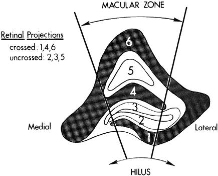
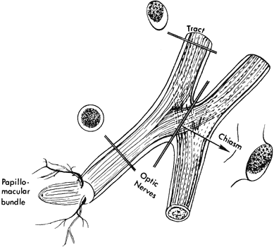
optic tracts. J Neurol Neurosurg Psychiatry 32:259, 1969

82. Weiskrantz L: Blindsight: A Case Study in Implications. Oxford Psychological Series #10. Oxford, Clarendon Press, 1986

83. Sadun AA: The afferent visual system. Anatomy and physiology. In M Yanoff, JS Duker, eds: Ophthalmology. London, Mosby, 1999

84. Hoyt WF, Luis O: Visual fiber anatomy in the infrageniculate pathway of the primate: Uncrossed and crossed retinal quadrant fiber projections studied with Nauta silver stain. Arch Ophthalmol 6:94, 1962

85. Hoyt WF, Luis O: The primate chiasm: Details of visual fiber organization studies by silver impregnation techniques. Arch Ophthalmol 70:69, 1963

86. Hoyt WF, Tudor RC: The course of parapapillary temporal retinal axons through the anterior optic nerve: A Nauta degeneration study in the primate. Arch Ophthalmol 69:503, 1963

87. Potts AM, Hodges D, Shelman CB et al: Morphology of the primate optic nerve: I-III. Invest Ophthalmol Vis Sci 11:980, 1972

88. Spalding JMK: Wounds of the visual pathway: I. The visual radiation. J Neurol Neurosurg Psychiatry 15:99, 1952

89. Van Buren JM, Baldwin M: The architecture of the optic radiation in the temporal lobe of man. Brain 81:15, 1958

90. Holmes G: A contribution to the cortical representation of vision. Brain 54:470, 1931

91. Spalding JMK: Wounds of the visual pathway: II. The striate cortex. J Neurol Neurosurg Psychiatry 15:169, 1952

92. Stensaas MA, Eddington DK, Dobelle WH: The topography and variability of the primary visual cortex in man.J Neurosurg 40:747, 1974

93. Horton JC, Hoyt WF: The representation of the visual field in human striate cortex. Arch Ophthalmol 109:816, 1991

94. Brindley GS: Sensory effects of electrical stimulation of the visual and 
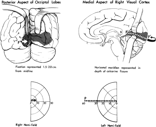
paravisual cortex in man. In June R, ed: Handbook of Sensory Physiology, Vol. VII/3B, pp 583–594. Berlin, Springer-Verlag, 1973

95. Karten HJ, Keyser KT, Brecha NC: Biochemical and morphological heterogeneity of retinal ganglion cells. In Cohen B, Bodis-Wollner I, eds: Vision and the Brain. New York, Raven Press, 1984

96. Ehrlich D, Keyser A, Karten HJ: Distribution of substance P-like immunoreactive retinal ganglion cells and their pattern of termination in the optic tectum of the chick (Gallus gallus). J Comp Neurol 266:220, 1987

97. Zucker CL, Dowling JE: Centrifugal fibers synapse on interplexiform cells in the teleost retina. Nature 330:166, 1987

98. Schiller PH: The on and of channels of the visual system. In Cohen B, Bodis-Wollner I, eds: Vision and the Brain. New York, Raven Press, 1990

99. Reiter HO, Stryker MP: Neural plasticity without postsynaptic action potentials: Less active inputs become dominant when kitten visual cortex cells are pharmacologically inhibited. Proc Natl Acad Sci USA 85:3623, 1988

100. Livingstone M: Segregation of form, color, movement, and depth processing in the visual system: Anatomy, physiology, art and illusion. In Cohen B, Bodis-Wollner I, eds: Vision and the Brain. New York, Raven Press, 1990

101. Mikami A, Newsom WT, Wurtz RH: Motion selectivity in macaque visual cortex: I. Mechanisms of direction and speed selectivity in extrastriate area MT. J Neurophysiol 55:1308, 1986

102. Sadun AA, Bassi C: Optic nerve damage in Alzheimer's disease. Ophthalmology 97:1, 1990

103. Fox PT, Fox JM, Raichle ME, Burde RM: The role of cerebral cortex in the generation of voluntary saccades: A positron emission tomographic study. J Neurophysiol 54:348, 1985

104. Fitzke FW: Imaging the optic nerve and ganglion cell layer. Eye 14:450, 2000

105. Chen W, Kato T, Zhu XH et al: Mapping of lateral geniculate nucleus activation during visual stimulation in human brain using fMRI. Magn Reson Med 39:89, 1998

106. Dupont P, Vogels R, Vandenberghe R et al: Regions in the human brain activated by simultaneous orientation discrimination: a study with positron emission tomography. Eur J Neurosci 10:3689, 1998

107. Bergland RM, Ray BS: The arterial supply of the human optic chiasm. J Neurosurg 31:327, 1969

108. Smith CG, Richardson WFG: The course and distribution of the arteries supplying the visual (striate) cortex. Am J Ophthalmol 61:1391, 1966

Back to Top