|
|
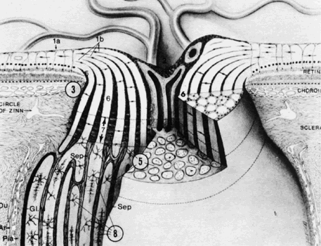
| Fig. 3. Schematic structure of optic disc and nerve. 1a, internal limiting membrane of retina; 1b, nerve fiber layer; 2, optic cup, lined by astrogial cells, and central retinal vessels; 3, ophthalmoscopically visible disc edge; 5, glial and connective tissue columns; 6, nerve fiber fascicles; 7, major portion of lamina cribrosa; 8, oligodendrocytes; Du, dura; Ar, arachnoid; Gl. M, glial mantle; Sep, pial septum. (Modified from Anderson DR, Hoyt WF: Ultrastructure of intraorbital portion of human and monkey optic nerve. Arch Ophthalmol 82;506, 1969) |