Chapter 39
Vitreous Pathobiology
J. SEBAG
Main Menu   Table Of Contents

Search

DEVELOPMENTAL AND INHERITED DISORDERS
VITREOUS AGING AND POSTERIOR VITREOUS DETACHMENT
ANOMALOUS POSTERIOR VITREOUS DETACHMENT
DISRUPTION OF VITREOUS IN ANOMALOUS POSTERIOR VITREOUS DETACHMENT
MISCELLANEOUS CONDITIONS
REFERENCES

DEVELOPMENTAL AND INHERITED DISORDERS

HYALOID VASCULAR SYSTEM

The embryonic vascular system of the vitreous (vasa hyaloidea propria) and lens (tunica vasculosa lentis) attains its maximum prominence during the ninth week of gestation or 40-mm stage.1 Atrophy of the vessels begins posteriorly with dropout of the vasa hyaloidea propria, followed by the tunica vasculosa lentis. Recent studies have detected the onset of apoptosis in the endothelial cells of the tunica vaculosa lentis as early as day 17.5 in the mouse embryo.2 At the 240-mm stage (seventh month) in the human, blood flow in the hyaloid artery ceases.3 Regression of the vessel itself begins with glycogen and lipid deposition in the endothelial cells and pericytes of the hyaloid vessels.3 Endothelial cell processes then fill the lumen, and macrophages form a plug that occludes the vessel. The cells in the vessel wall then undergo necrosis and are phagocytized by mononuclear phagocytes.4 Gloor5 claimed that macrophages are not involved in vessel regression within the embryonic vitreous but that autolytic vacuoles form in the cells of the vessel walls, perhaps in response to hyperoxia. Interestingly, the sequence of cell disappearance from the primary vitreous begins with endothelial and smooth muscle cells of the vessel walls, followed by adventitial fibroblasts and lastly phagocytes,6 consistent with a gradient of decreasing oxygen tension. Recent studies have suggested that the vasa hyaloidea propria and the tunica vasculosa lentis regress via apoptosis.7 These studies concluded that macrophages are important in this process. Subsequent studies by a different group confirmed the importance of macrophages in promoting regression of the fetal vitreous vasculature and further characterized these macrophages as hyalocytes.8

It is not known what stimulates regression of the hyaloid vascular system, but studies have identified a protein native to the vitreous that inhibits angiogenesis in various experimental models.9–11 Activation of this protein and its effect on the primary vitreous may be responsible for the regression of the embryonic hyaloid vascular system as well as the inhibition of pathologic neovascularization in the adult. Mitchell and colleagues12 point out that the first event in hyaloid vasculature regression is endothelial cell apoptosis and propose that lens development separates the fetal vasculature from vascular endothelial growth factor (VEGF)-producing cells, decreasing the levels of this survival factor for vascular endothelium, inducing apoptosis. Following endothelial cell apoptosis, there is loss of capillary integrity, leakage of erythrocytes into the vitreous, and phagocytosis of apoptotic endothelium by hyalocytes. Meeson and colleagues13 proposed that there are actually two forms of apoptosis that are important in regression of the fetal vitreous vasculature. The first (“initiating apoptosis”) results from macrophage induction of apoptosis in a single endothelial cell of an otherwise healthy capillary segment with normal blood flow. The isolated dying endothelial cells project into the capillary lumen and interfere with blood flow. This stimulates synchronous apoptosis of downstream endothelial cells (“secondary apoptosis”) and ultimately obliteration of the vasculature. Removal of the apoptotic vessels is achieved by hyalocytes.

Pathology

Regression of the hyaloid artery usually occurs completely and without complications. Persistence of the hyaloid vascular system occurs in 3% of full-term infants but in 95% of premature infants14 and can be associated with prepapillary hemorrhage.15 Anomalies involving incomplete regression of the embryonic hyaloid vascular system occur in more than 90% of infants born earlier than 36 weeks of gestation and in over 95% of infants weighing less than 5 lb at birth.16 There is a spectrum of disorders resulting from persistence of the fetal vasculature.17

Mittendorf's dot is a remnant of the anterior fetal vascular system located at the former site of anastomosis of the hyaloid artery and tunica vasculosa lentis. It is usually inferonasal to the posterior pole of the lens and is not associated with any known dysfunction.

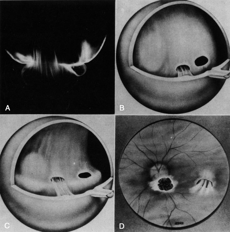
Bergmeister's papilla is the occluded remnant of the posterior portion of the hyaloid artery with associated glial tissue. It appears as a gray, linear structure anterior to the optic disc and adjacent retina and does not cause any known functional disorders. Exaggerated forms can present as prepapillary veils.

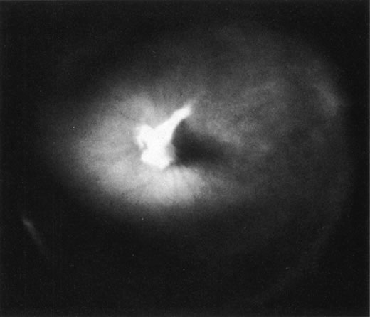
Vitreous cysts are generally benign lesions that are found in eyes with abnormal regression of the anterior18 or posterior19 hyaloid vascular system; otherwise normal eyes20,21; and eyes with coexisting ocular disease, such as retinitis pigmentosa22 and uveitis.23 Some vitreous cysts contain remnants of the hyaloid vascular system,24 supporting the concept that the cysts result from abnormal regression of these embryonic vessels.25 However, one histologic analysis of aspirated material from a vitreous cyst purportedly revealed cells from the retinal pigment epithelium.26 Vitreous cysts are generally not symptomatic and thus do not require surgical intervention. However, argon laser photocoagulation has been employed, and a recent report27 described the use of neodymium:yttrium-aluminum-garnet (Nd:YAG) laser therapy to rupture a free-floating posterior vitreous cyst.

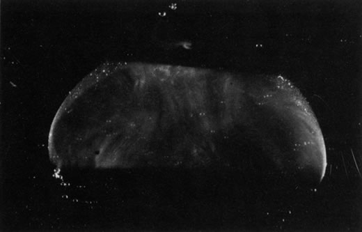
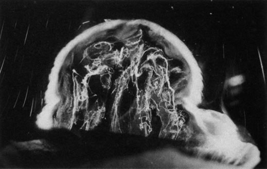
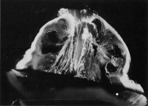
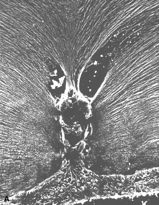
Persistent hyperplastic primary vitreous (PHPV) was first described by Reese28 as a congenital malformation of the anterior portion of the primary vitreous appearing as a plaque of retrolental fibrovascular connective tissue. This tissue is adherent to the posterior lens capsule and extends laterally to attach to the ciliary processes, which are elongated and displaced centrally. Although 90% of cases are unilateral, many of the fellow eyes have Mittendorf's dot or other anomaly of anterior vitreous development.29 A persistent hyaloid artery, often still perfused with blood, arises from the posterior aspect of the retrolental plaque in the affected eye. In severe forms, there is microphthalmos with anterior displacement of the lens-iris diaphragm, shallowing of the anterior chamber, and secondary glaucoma. PHPV is believed to arise from abnormal regression and hyperplasia of the primary vitreous.28 Experimental data suggest that the abnormality begins at the 17-mm stage of embryonic development.30 The hyperplastic features result from generalized hyperplasia of retinal astrocytes and a separate component of glial hyperplasia arising from the optic nerve head.31 The fibrous component of the PHPV membrane is presumably synthesized by these astrocytes and glial cells.32 A recent case report with clinicopathologic correlation found that collagen fibrils in this fibrous tissue had diameters of 40 to 50 nm with a cross-striation periodicity of 65 nm. The investigators concluded that the collagen fibrils differed from those of the primary vitreous and suggested that they arose either from a different population of cells or were the result of abnormal metabolism by the same cells that synthesize vitreous collagen.33

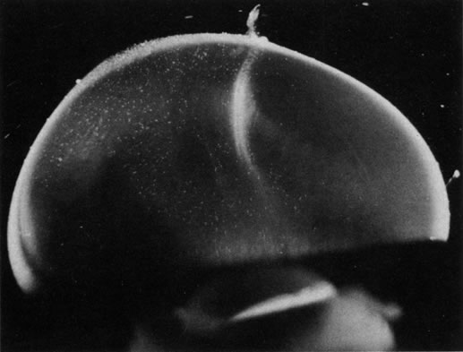
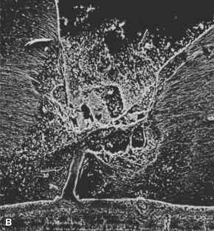
The retina is usually not involved in anterior PHPV. Indeed, previous studies have suggested that the anterior form is due to a primary defect in lens development and that vitreous changes are all secondary.34 This postulate has never been substantiated. There are rare instances of posterior PHPV in which opaque connective tissue arises from Bergmeister's papilla and persistent hyaloid vessels (Fig. 1).32,35 These can cause congenital falciform folds of the retina and, if severe, can cause tentlike retinal folds, leading on rare occasions to tractional and/or rhegmatogenous retinal detachment. Font and investigators36 demonstrated the presence of adipose tissue, smooth muscle, and cartilage within the retrolental plaque and suggested that PHPV arises from metaplasia of mesenchymal elements in the primary vitreous.

Fig. 1. Persistent hyperplastic primary vitreous (PHPV). A wide-angle fundus photograph of the left eye in a 10-year-old boy with the posterior form of PHPV demonstrates a fibrous stalk arising from the optic disc and extending into the central vitreous. (Sebag J: The Vitreous: Structure, Function and Pathobiology, p 99. New York, Springer-Verlag, 1989.)

DOMINANT EXUDATIVE VITREORETINOPATHY

Dominant exudative vitreoretinopathy was first described in 1969 by Criswick and Schepens37 as a bilateral, slowly progressive abnormality of the vitreous and retina that resembles retinopathy of prematurity but with no history of prematurity or postnatal oxygen administration. Gow and Oliver38 identified this disorder as an autosomal dominant condition with complete penetrance. They characterized the course of this disease in stages ranging from posterior vitreous detachment with snowflake opacities (stage I), to thickened vitreous membranes and elevated fibrovascular scars (stage II), and vitreous fibrosis with subretinal and intraretinal exudates, ultimately developing retinal detachment due to fibrovascular proliferation arising from neovascularization in the temporal periphery (stage III). Plager and coworkers39 recently reported the same findings in four generations of three families, but found X-linked inheritance. Van Nouhuys40–42 studied 101 affected members in 16 Dutch pedigrees and five patients with sporadic manifestations. He found that the incidence of retinal detachment was 21%, all but one case occurring prior to the age of 30. These were all tractional or combined traction/rhegmatogenous detachments, and there were no cases of exudative retinal detachment. Van Nouhuys42 concluded that the etiology of dominant exudative vitreoretinopathy lies in premature arrest of development in the retinal vasculature, since the earliest findings in these patients were nonperfusion of the peripheral temporal retina with stretched retinal blood vessels and shunting with vascular leakage. Thus, Van Nouhuys considers dominant exudative vitreoretinopathy as a retinopathy with secondary vitreous involvement. However, Brockhurst and colleagues43 described that vitreous membrane formation begins just posterior to the ora serrata and that this precedes retinal vessel abnormalities, suggesting a vitreous origin to this disorder. Others suggested that there may be a combined etiology involving anomalies of the hyaloid vascular system and primary vitreous as well as retinovascular dysgenesis.44

RETINOPATHY OF PREMATURITY

Retinopathy of premature infants was first described in 1942 by Terry45 as “retrolental fibroplasia.” That term is no longer appropriate since it is actually a descriptive term of the pathology in advanced (stage V) cases of cicatricial retinopathy of prematurity (ROP). We now identify acute stages of the disease that are not retrolental and do not have much fibroplasia.46

The pathogenesis of ROP begins with birth, prior to complete maturation and development of the peripheral retina, followed by postnatal oxygen administration, triggering retinal vasoconstriction with endothelial cell necrosis and vaso-obliteration in response to hyperoxia.47 After the discontinuation of supplemental oxygen, arterial pO2 levels return to normal and the obliterated (or at best, highly constricted) vessels are not adequately reperfused, causing the peripheral retina they subserve to become ischemic and release neovascular growth factors. An alternative hypothesis of pathogenesis proposes that spindle cells in the immature peripheral retina are stimulated by excessive amounts of reactive oxygen species, whether related to oxygen therapy and subsequent relative hypoxia or other metabolic circumstances, to release angiogenic growth factors.48 In either case, the result is migration and proliferation of capillary endothelial cells that form new blood vessels at the posterior ridge of tissue between the vascularized and avascular retina. This results in neovascularization arising from the ridge that demarcates the developed posterior retina from the immature peripheral retina. The new vessels grow into the vitreous body, similar to neovascularization in diabetic retinopathy,49 although they grow farther anteriorly and more exuberantly. This is perhaps because of the participation of cells of the ocular fetal vasculature, whose apoptosis has been retarded or arrested by the presence of high levels of VEGF.50

There are no clearly identified vitreous changes during stages I and II of acute ROP, although this may simply be due to our present inability to detect such abnormalities.51 Indeed, the abundance of reactive oxygen species in the retina and vitreous of premature infants could induce widespread vitreous liquefaction.52 There are also likely to be localized areas of liquid vitreous, particularly at the periphery. At surgery for stage IV-A ROP with retinal detachment, there is a “trough” in the periphery.53 This structure is most likely the consequence of underlying retinal immaturity in the periphery, with consequent lack of typical gel vitreous synthesis, normally a Müller cell function, overlying the immature retina. The liquid vitreous trough is probably present early in the natural history of disease but goes undetected by present vitreous imaging techniques.54 Such disruption of normal vitreous composition and structure probably alters a number of physiologic processes within the vitreous, including the ability of vitreous to inhibit cellular and vascular invasion.9–11 Furthermore, the interface between posterior gel vitreous and peripheral liquid vitreous at the ridge causes vitreous traction to be exerted at the retinal ridge.

In stage III ROP, new blood vessels extend from the inner retina into the vitreous cortex. The cortex, overlying the rear guard of differentiated capillary endothelial cells, becomes opaque and contains linear, fibrous structures adjacent to a large pocket of liquid vitreous.55 In advancing from stage III to stage IV, the neovascular tissue arising from the rear guard grows through the vitreous body toward Wiegert's ligament on the posterior lens capsule.56 This configuration of neovascularization is probably the result of cell migration and proliferation along the walls of the future Cloquet's canal or the tractus hyaloideus of Eisner. Cells of the primary vitreous likely contribute to the formation of the dense central vitreous stalk and retrolental membrane seen in the cicatricial stage, since these cells could also undergo migratory and proliferative responses to intraocular angiogenic stimuli.

VITREORETINAL DYSTROPHY OF GOLDMANN-FAVRE

The autosomal recessive condition vitreoretinal dystrophy of Goldmann-Favre was first described in 1957 by Goldmann57 as consisting of vitreous abnormalities with peripheral retinoschisis and chorioretinal atrophy. Francois58 characterized the vitreous changes as syneresis (collapse), fibrillar degeneration with white strand formation, and punctate deposits. The retinal changes are pigmentary degeneration (with a different appearance from the bone spicules of retinitis pigmentosa), attenuated blood vessels, and microcystic degeneration of the macula and retinal periphery. Night blindness is an important feature, and the electroretinogram is markedly abnormal. Fishman and collaborators59 used fluorescein angiography to demonstrate cystoid macular edema (CME) and vascular occlusion with leakage in the peripheral retina corresponding to the areas of schisis. Schepens60 described that there is vitreous organization attached to the areas of schisis. Histopathologic studies by Peyman and coworkers61 showed attenuation of the outer nuclear layer and absence of photoreceptors. Based on the presence of normal retinal pigment epithelium and choriocapillaris, these investigators suggested that the etiology was a primary retinal degeneration. In this regard, Müller cell dysfunction could lead to the vitreous abnormalities as well as peripheral retinoschisis. It follows that in these cases, CME results from peripheral and central vitreous changes that occur in the absence of posterior vitreous detachment, thus inducing traction on the macula, as has been noted in cases of peripheral anterior vitritis with CME62,63 and in some cases of aphakic macular edema.64–66

VITREOUS COLLAGEN DISORDERS

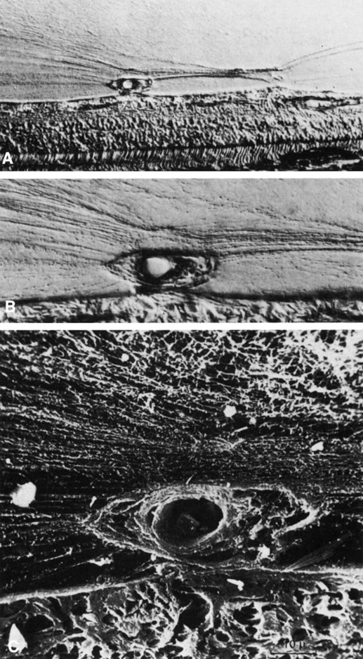
Vitreous is one of many connective tissues in the body. Collagen is one of the most important structural molecules in all connective tissues. Thus, it is of interest to consider parallel phenomena occurring in the vitreous and connective tissues elsewhere, especially as related to collagen. For example, Gartner67 pointed out the similarities between the intervertebral disk and the vitreous, in which age-related changes with herniation of the nucleus pulposus was associated with presenile vitreous degeneration in 40% of cases. He proposed that a generalized connective tissue disorder resulted in disk herniation and presenile vitreous degeneration in these cases. Based on these findings, Gartner likened herniation of the nucleus pulposus in the disk to prolapse of vitreous into the retrohyaloid space by way of the posterior vitreous cortex following posterior vitreous detachment (see Fig. 9).

Fig. 9. Vitreomacular traction. Vitreous can remain attached to the macula even in the presence of posterior vitreous detachment. In such cases, vitreous can extrude through the premacular vitreous cortex and fibers can insert into the macula. B, C, and D are an artist's rendition of this phenomenon. A demonstrates vitreous extrusion into the retrohyaloid space in a specimen from a middle-aged human. (Adapted from Jaffe NS: The Vitreous in Clinical Ophthalmology. St. Louis, CV Mosby, 1969; and Jaffe NS: Vitreous traction at the posterior pole of the fundus due to alterations in the posterior vitreous. Trans Am Acad Ophthalmol Otolarynogol 71:642, 1967.)

Maumenee68 identified several different disorders with single-gene autosomal dominant inheritance in which dysplastic connective tissue primarily involves joint cartilage. In these conditions, there is associated vitreous liquefaction, collagen condensation, and vitreous syneresis (collapse). Since type II collagen is common to cartilage and vitreous, Maumenee suggested that the various arthro-ophthalmopathies may result from different mutations, perhaps of the same or neighboring genes, on the chromosome involved with type II collagen metabolism. In these disorders, probably including such conditions as Wagner's disease,69 the fundamental problem in the posterior segment of the eye is that the vitreous is liquefied and unstable, tending to syneresis at an early age. However, there is no dehiscence at the vitreoretinal interface in concert with the changes inside the vitreous body, perhaps owing to the fact that the internal limiting lamina of the retina is composed of type IV collagen. Thus, in these cases, abnormal type II collagen metabolism causes destabilization of the vitreous and results in traction on the retina that can lead to large posterior tears and difficult retinal detachments.

In Marfan's syndrome, an autosomal dominant disorder featuring poor musculature, lax joints, aortic aneurysms, and arachnodactyly, there is lens subluxation, thin sclera, peripheral fundus pigmentary changes, and vitreous liquefaction at an early age. The combination of myopia, vitreous syneresis, and abnormal vitreoretinal adhesions at the equator accounts for the frequency of rhegmatogenous retinal detachment due to equatorial or posterior horseshoe tears.60

Ehlers-Danlos syndrome has some similarities to Marfan's syndrome, most notably joint laxity, aortic aneurysms, and an autosomal dominant pattern of inheritance. However, there are as many as six types of Ehlers-Danlos patients, and the genetics of this condition are probably more heterogeneous than in Marfan's syndrome. A further distinction from Marfan's patients is the hyperelastic skin and poor wound healing of all connective tissues, including cornea and sclera, that is seen in Ehlers-Danlos patients. Ocular manifestations include lens subluxation, angioid streaks, thin sclera, and high myopia due to posterior staphyloma. Vitreous liquefaction and syneresis occur at a young age. Vitreous traction causes vitreous hemorrhage, perhaps also due to blood vessel wall fragility, and retinal tears with rolled edges, often causing bilateral retinal detachments.60

In 1965, Stickler and associates70 described a condition in five generations of a family that was found to be autosomal dominant with complete penetrance and variable expressivity. The features were a marfanoid skeletal habitus and orofacial and ocular abnormalities. Subsequent studies identified subgroups with short stature and a Weill-Marchesani habitus. The skeletal abnormalities now accepted as characteristic of Stickler's syndrome are radiographic evidence of flat epiphyses, broad metaphyses, and especially spondyloepiphyseal dysplasia.71 Ocular abnormalities are high myopia, greater than –10 diopters in 72% of cases,72 and vitreoretinal changes characterized by vitreous liquefaction, fibrillar collagen condensation, and a perivascular lattice-like degeneration in the peripheral retina believed to be the cause of a high incidence (greater than 50%) of retinal detachment.71 More recent studies correlated specific gene defects with particular phenotypes, thereby enabling the classification of Stickler syndrome patients into four subgroups.73 Patients with abnormalities in the genes coding for type II procollagen and type V/XI procollagen are the ones who have severe vitreous abnormalities.

Knobloch74 described a syndrome similar to Stickler's syndrome with hypotonia, relative muscular hypoplasia, and mild to moderate spondyloepiphysealdysplasia causing hyperextensible joints. The vitreoretinopathy is characterized by vitreous liquefaction, veils of vitreous collagen condensation, and perivascular lattice-like changes in the peripheral retina.

It is presently unknown whether myopia unrelated to the aforementioned arthro-ophthalmopathies should be considered a form of vitreous collagen disease as well. The extensive liquefaction of vitreous (myopic vitreopathy) and propensity for retinal detachment due to peripheral retinal traction and myopic peripheral retinal degeneration suggest that this postulate may deserve closer scrutiny.

Back to Top
VITREOUS AGING AND POSTERIOR VITREOUS DETACHMENT

BIOCHEMICAL CHANGES

Using slit lamp biomicroscopy in a clinical setting, Busacca75 and Goldmann76 observed that after the ages of 45 to 50, there is a decrease in the gel volume and an increase in the liquid volume of human vitreous. Eisner77 qualitatively confirmed these clinical findings in his postmortem studies of dissected human vitreous and observed that liquefaction begins in the central vitreous. In a large autopsy study of formalin-fixed human eyes, O'Malley78 provided quantitative evidence to support these observations. He found that more than half of the vitreous body was liquefied in 25% of persons age 40 to 49 and that this increased to 62% of those age 80 to 89. Oksala79 used ultrasonography in vivo to detect echoes from gel-liquid interfaces in 444 normal human eyes and found progressive liquefaction with age. Vitreous liquefaction actually begins much earlier than the ages at which clinical examination or ultrasonography detects changes. Balazs and colleagues80,81 found evidence of liquid vitreous after age 4 and observed that by the time the human eye reaches its adult size (ages 14 to 18) 10% to 12.5% of the total vitreous volume consists of liquid vitreous (Fig. 2). In these postmortem studies of 610 fresh, unfixed human eyes, it was observed that after age 40, there is a steady increase in liquid vitreous that occurs simultaneously with a decrease in gel volume. By the ages of 80 to 90 years more than half the vitreous body is liquid. The finding that the central posterior vitreous is where liquefaction begins77 and where fibers are first observed82–84 is consistent with the concept that dissolution of the hyaluronic acid (HA)–collagen complex results in the simultaneous formation of liquid vitreous and aggregation of collagen fibrils into bundles of parallel fibrils seen as large fibers.83 In the posterior vitreous, such age-related changes often form large pockets of liquid vitreous.85

Fig. 2. Liquefaction of human vitreous. The volumes of gel and liquid vitreous in 610 human eyes were measured. The results are plotted versus the age of the donor. Liquid vitreous appears by the age of 5 and increases throughout life until it constitutes more than 50% of the volume of the vitreous during the tenth decade. Gel vitreous volume increases during the first decade while the eye is growing in size. The volume of gel vitreous then remains stable until about age 40, when it begins to decrease simultaneous with the increase in liquid vitreous. (Balazs EA, Denlinger JL: Aging changes in the vitreous. In: Aging and Human Visual Function, pp 45–57. New York, Alan R. Liss, 1982.)

Age-related rheologic changes in the vitreous may result from an alteration in HA-collagen interaction. Chakrabarti and Park86 claimed that the interaction between collagen and HA is dependent on the conformational state of each macromolecule and that a change in the conformation of HA molecules could result in vitreous liquefaction and aggregation or cross-linking of collagen molecules. Armand and Chakrabarti87 detected differences in the structure of the HA molecules present in gel vitreous and those in liquid vitreous, suggesting that such conformational changes occurred during liquefaction. Whether these changes are cause or effect is not known. However, Andley and Chapman88 demonstrated that singlet oxygen can induce conformational changes in the tertiary structure of HA molecules. Ueno and associates52 suggested that free radicals generated by metabolic and photosensitized reactions could alter HA and/or collagen structure and trigger a dissociation of collagen and HA molecules, ultimately leading to liquefaction. This is plausible because the cumulative effects of a lifetime of daily exposure to light may influence the structure and interaction of collagen and HA molecules by the proposed free radical mechanism(s). Enzymes, such as endogenous metalloproteinases, may also be involved in liquefying the gel vitreous. The importance of vitreous liquefaction in the pathogenesis of posterior vitreous detachment is discussed below.

Total vitreous collagen content does not change after the ages of 20 to 30 (Fig. 3). However, the collagen concentration in the gel vitreous at the ages of 70 to 90 (approximately 0.1 mg/mL) was found to be significantly greater than at the ages of 50 to 60 (slightly more than 0.05 mg/mL; p < .05). Because the total collagen content does not change, this finding is most likely due to the decrease in the volume of gel vitreous that occurs with aging (see Fig. 2) and a consequent increase in the concentration of the collagen remaining in the gel. The collagen fibrils in this gel become packed into bundles of parallel fibrils,83 perhaps with cross-links between them. Aging of collagen throughout the body is associated with increased cross-linking as manifested by decreased solubility,89 increased collagen “stiffness,”90 and increased resistance to enzymatic degradation.91 There is also an increase in a reducible lysine-carbohydrate condensation product with increasing age.92 Although similar investigations have not been performed on vitreous collagen, studies by Snowden and colleagues93 demonstrated that with aging, there is a decrease in the quantity of bovine vitreous collagen solubilized by heat alone. This could result from an increase in collagen cross-linking or, as suggested by the authors, from changes in the surrounding glycoproteins and proteoglycans. Abnormal collagen cross-links have been identified in diabetic human vitreous,94 suggesting “precocious senescence” of vitreous collagen as has been described for other organs and tissues in diabetic patients.95–97

Fig. 3. Age-related changes in human vitreous collagen levels. Collagen content (mg; right ordinate) is indicated by solid dots (means) and darkly hatched boxes in the upper portion of the graph (height = standard error). Collagen concentration (mg/mL; left ordinate) is indicated by asterisks (means) and lightly hatched boxes in the lower portion of the graph (height = standard error). Vitreous collagen concentration decreases during the first decade of life because there is no net synthesis of collagen during this period of active growth of the eye. There are no significant changes in collagen content following age 20, consistent with the lifelong “stability” of this molecule. However, collagen concentration in the gel vitreous increases after ages 40 to 50. This is due to the decrease in gel vitreous that occurs during this time (see Fig. 2), concentrating the remaining collagen in an ever-decreasing volume of gel vitreous. The increase in gel vitreous collagen concentration between ages 50 to 60 and 80 to 90 is statistically significant (p < .05). (Balazs EA, Denlinger JL: Aging changes in the vitreous. In: Aging and Human Visual Function, pp 45–57. New York, Alan R. Liss, 1982.)

Vitreous HA concentration increases until about the age of 20, when adult levels are attained (Fig. 4A). HA concentration does not change in either the liquid or gel vitreous from ages 20 to 60 (Fig. 4).81,98 This necessarily means that there is an increase in the HA content of liquid vitreous and a concomitant decrease in the HA content of gel vitreous, since the amount of liquid vitreous increases and the amount of gel vitreous decreases with age (see Fig. 2). This is consistent with the concept that vitreous liquefaction is associated with a “redistribution” of HA from the gel to the liquid vitreous. With advanced liquefaction, there is a marked increase in the concentration of HA in liquid vitreous at ages 70 to 90 (Fig. 4B).

Fig. 4. Age-related changes in human vitreous hyaluronic acid (HA) concentration. The dots represent the means of the samples. The vertical height of the boxes represents the standard error of the means. The horizontal width of the boxes represents the age ranges in the sample group. A. HA concentration in the gel vitreous. There is a fourfold increase in the concentration of HA during the first two decades of life. Considering that this is also a period of active growth of the eye and substantial increase in vitreous volume, there must be prolific synthesis of HA to increase concentrations so dramatically. After age 20, HA concentrations in the gel vitreous are stable. Since this is a period of decreasing amounts of gel vitreous (see Fig. 2), there must be a net decrease in the HA content of the gel to result in no substantial changes in concentration. B. HA concentration in the liquid vitreous. There are no data for the first 4 years since there is no liquid vitreous during this time. From the ages of 5 to 50 to 60 there is a 50% increase in the concentration of HA in the liquid vitreous. After this time, there is a substantial increase in liquid vitreous HA concentration. The magnitude of this accumulation of HA in the liquid component of the vitreous is even greater when one considers that this occurs during the time when the volume of liquid vitreous is increasing by twofold (see Fig. 2). (Balazs EA, Denlinger JL: Aging changes in the vitreous. In: Aging and Human Visual Function, pp 45–57. New York, Alan R. Liss, 1982.)

Vitreous soluble protein concentrations also increase from 0.5 to 0.6 mg/mL at ages 13 to 50, to 0.7 to 0.9 mg/mL at ages 50 to 80, and to 1.0 mg/mL above the age of 80.80,81,98 This increase may result from an age-related breakdown in the blood–ocular barriers of the retinal vasculature, retinal pigment epithelium, and ciliary body epithelium.

STRUCTURAL CHANGES

Basal Laminae and Vitreous Base

The vitreoretinal interface is the site of many blinding vitreoretinal disorders.99 The basal laminae surrounding the vitreous thicken with age,100 a phenomenon that occurs in basal laminae throughout the body.101 Hogan and associates102 claimed that the thickening of the internal limiting lamina of the retina occurs during life as a result of synthesis by subjacent Müller's cells. This phenomenon may play a role in weakening vitreoretinal adhesion, thus contributing to the development of posterior vitreous detachment.

Teng and Chi103 found that the vitreous base posterior to the ora serrata varies in width depending on the age of the person. More than half of eyes from people older than age 70 had a posterior vitreous base wider than 1 mm. The width increased with advancing age to nearly 3 mm, bringing the posterior border of the vitreous base closer to the equator. This posterior migration of the vitreous base probably plays an important role in the pathogenesis of peripheral retinal breaks and rhegmatogenous retinal detachment, since this is the area of strongest vitreoretinal adhesion. The phenomenon of posterior migration of the posterior border of the vitreous base was recently confirmed and an explanation for increased vitreoretinal adhesion in this location was offered.104 It appears that there is intraretinal synthesis of collagen fibrils that penetrate the internal limiting lamina of the retina and ‘splice’ with vitreous collagen fibrils. Within the vitreous base, Gartner105 found no differences in the thickness of collagen fibrils when comparing five eyes from humans ages 9 months, 29, 39, 61, and 71. He did, however, note that there was “lateral aggregation” of collagen fibrils in the eyes from older persons, similar to aging changes within the central vitreous.82–84 All these aging changes at the vitreous base very likely contribute to increased traction on the peripheral retina and the development of retinal tears and detachment.

Central Vitreous

During postnatal development, there is a transition from a dense, highly light-scattering structure (Fig. 5), because at birth vitreous is mostly collagen, to homogeneous transparency that results when HA “swells” and spreads collagen fibrils apart (Fig. 6). The redistribution of HA and collagen during maturation to the adult results in the aggregation of collagen fibrils into packed bundles of parallel fibrils that appear as fibers. During the latter decades of life, these fine parallel fibers in the central vitreous become thickened and tortuous (Fig. 7). Immediately adjacent to these coarse structures are areas with little or no light-scattering properties that are filled with “liquid” vitreous. When advanced, this vitreous degeneration forms large pools of liquid vitreous identified clinically as “lacunae” (Fig. 8). When the posterior vitreous detaches from the retina, there is an overall reduction in the size of the vitreous body due to the collapse (syneresis) that occurs when liquid vitreous enters the space posterior to the vitreous cortex and anterior to the retina. This displacement of liquid vitreous occurs by way of the prepapillary “hole” and possibly the premacular vitreous cortex, and is important in the pathogenesis of posterior vitreous detachment.

Fig. 5. Vitreous structure in a human embryo at 33 weeks of gestation. The posterior aspect of the lens is seen below. The vitreous body is enclosed by the dense, highly light-scattering cortex. Parapapillary glial tissue was torn away during dissection and hangs from the prepapillary vitreous cortex. Within the vitreous body, Cloquet's canal arcs from the prepapillary vitreous toward the lens. Since its course undulates through the vitreous body, not all of Cloquet's canal can be visualized in a single horizontal section. (Sebag J: Age-related changes in human vitreous structure. Graefes Arch Clin Exp Ophthalmol 22:89, 1987. Specimens courtesy of the New England Eye Bank, Boston, MA.)

Fig. 6. Human vitreous structure during childhood. This view of the posterior and central vitreous from a 6-year-old child demonstrates a dense vitreous cortex with hyalocytes. There is vitreous extrusion into the retrohyaloid space through the premacular vitreous cortex. However, no fibers are present. (Sebag J: The Vitreous: Structure, Function and Pathobiology, p 79. New York, Springer-Verlag, 1989.)

Fig. 7. Fibrous structure of human vitreous during old age. The vitreous body of an 88-year-old woman has undergone substantial degeneration in the fibrous structure. Fibers are thickened and tortuous. The entire vitreous body appears to have undergone dissolution with empty spaces adjacent to the thickened fibers. (Sebag J: Age-related changes in human vitreous structure. Graefes Arch Clin Exp Ophthalmol 22:89, 1987. Specimens courtesy of the New England Eye Bank, Boston, MA.)

Fig. 8. Human vitreous lacunae in old age. The central vitreous has thickened, tortuous fibers. The peripheral vitreous has pockets devoid of any structure. These contain liquid vitreous and correspond to lacunae as seen clinically on biomicroscopy. (Sebag J, Balazs EA: Human vitreous fibres and vitreoretinal disease. Trans Ophthalmol Soc UK 104:123, 1985.)

POSTERIOR VITREOUS DETACHMENT

The most common age-related event in the vitreous is posterior vitreous detachment (PVD). True PVD can be defined as a separation between the posterior vitreous cortex and the internal limiting lamina (ILL) of the retina.106 PVD can be localized, partial, or total (up to the posterior border of the vitreous base). PVD should be distinguished from other forms of vitreoretinal separation that clinically may be mistaken for PVD. One of these forms involves separation of the ILL and some of the inner retina along with the detached posterior vitreous cortex. This can follow severe tractional events in the young, in whom the posterior vitreous cortex/ILL adhesion is strong.107

Another form of vitreoretinal separation that can mimic true PVD features forward displacement of the anterior portion of the posterior vitreous cortex, leaving part of the posterior layer of the vitreous cortex still attached to the retina. Balazs4 has coined the term “vitreoschisis” to denote prominent liquefaction with cavitation in the posterior vitreous but persistent attachment of the outermost layers of the posterior vitreous cortex to the ILL. Clinically, this is often mistaken as true PVD and occurs in cases with prominent liquefaction and no PVD, such as advanced axial myopia. The skilled examiner can distinguish this from PVD, since true PVD displays a characteristic ascension/descension movement of the posterior vitreous cortex on vertical saccades. The term vitreoschisis should be reserved for cases in which an actual split has occurred in the posterior vitreous cortex. Green and Byrne108 recently reported that splitting of the posterior vitreous cortex may be present in eyes with proliferative diabetic retinopathy (PDR) and vitreous hemorrhage. It is possible that in these cases, vitreoschisis cavities in the posterior vitreous cortex result from dissection of blood. However, a split in the vitreous cortex can also cause bleeding, since neovascular complexes grow into the vitreous cortex.109 Chu and associates110 studied 140 patients with diabetic vitreous hemorrhage and found echographic evidence of vitreoschisis in 20% of cases.

Lindner111 and Jaffe112 described that in some cases of PVD, there is herniation of vitreous through the vitreous cortex of the posterior pole. As previously mentioned, Gartner67 drew an analogy between this phenomenon and the herniation of the nucleus pulposus in the intervertebral disks of the spine. When a PVD involves herniation of the vitreous into the retrovitreal space by way of the premacular vitreous cortex, there can be persistent attachment to the macula and traction (Fig. 9).113

Epidemiology

In clinical studies, the incidence of purported PVD was found to be 53% in persons older than age 50, and 65% in those over age 65.111 Autopsy studies revealed an incidence of 27% to 51% in the seventh decade and 63% in the eighth decade.114 However, these figures may be overestimates due to the methods employed in these postmortem studies. PVD is more common in myopia, occurring 10 years earlier than in emmetropia and hyperopia.111,112 This likely results from effects of myopia on the structure of the vitreous. Cataract extraction in myopic patients introduces additional effects, causing PVD to develop in all but one of 103 myopic (greater than –6 D) eyes that were studied following cataract extraction (presumably intracapsular).115

Several studies114,116 found a higher incidence of PVD in women than in men, a finding that may be due to hormonal changes following menopause. This hypothesis is supported by studies in the vitreous117 as well as other organs and tissues.118,119

Pathogenesis

PVD results from rheologic changes within the vitreous that lead to synchysis (liquefaction), in conjunction with weakening of the vitreous cortex/ILL adhesion. O'Malley78 suggested that PVD was strongly correlated with synchysis since both are correlated with age, although PVD had a later onset than synchysis. Foos and Wheeler114 studied 4492 autopsy eyes and found a statistically significant correlation between the degree of synchysis and the incidence of PVD. Larsson and Osterlin120 studied 61 human eyes post mortem and correlated the degree of vitreous liquefaction with the extent of PVD. Once “liquid” vitreous forms (see previous section, Biochemical Changes) and the collagen network is destabilized, owing to a loss of the stabilizing effect of HA molecules and cross-linking due to the loss of type IX collagen on the surface of collagen fibrils, collapse (syneresis) of the vitreous body can occur. It is likely that dissolution of the posterior vitreous cortex/ILL adhesion at the posterior pole allows liquid vitreous to enter the retrocortical space by way of the prepapillary hole and perhaps the premacular vitreous cortex as well.84,111,113,121 With rotational eye movements, liquid vitreous can dissect a plane between the vitreous cortex and the ILL, leading to true PVD. This volume displacement from the central vitreous to the preretinal space causes the observed collapse of the vitreous body (syneresis). It is recognized114,121 that PVD begins at the posterior pole. Vitreoretinal dehiscence at the macula may result from a predisposition or an increased stimulus for vitreous degeneration in the premacular region. Indeed, it is here that studies85 have identified a large pocket of liquid vitreous related to aging. Foos and Wheeler114 proposed that liquefaction in the posterior pole results from light toxicity to the premacular vitreous, since this is where the eye focuses incident light. There can also be a contribution of toxicity caused by metabolic waste products resulting from the high density of metabolically active neurons in the macula. Both light irradiation and metabolic processes can generate free radicals, which could alter HA and/or collagen structure and disrupt the HA-collagen association, causing liquefaction.52 These phenomena could also influence the extracellular matrix components binding the vitreous cortex to the ILL, contributing to the pathogenesis of PVD.

Sequelae

In youth, the vitreous is transparent (see Fig. 6) and has little or no effect on glare sensitivity.122 In old age, the aggregation of vitreous collagen fibrils into thick, irregular, visible fibers (see Fig. 7) can induce glare sensitivity, which may be subjectively bothersome. Furthermore, the high incidence of PVD in old age may also induce glare due to scattering of light by the dense collagen fibril network in the posterior vitreous cortex. One group of individuals in whom glare discomfort is a common complaint are patients who have undergone scleral buckle surgery for rhegmatogenous retinal detachment. The complaint of glare appears to be due to postoperative vitreous turbidity and not a change in the threshold sensitivity of retinal receptors.123 Because vitreous biochemical and structural changes likely predispose these patients to rhegmatogenous retinal detachment, prominent vitreous changes are probably already present preoperatively. Scleral buckle surgery adds to the preexisting vitreous inhomogeneity by breaking down the normal blood–vitreous barriers and mechanisms that maintain vitreous clarity.124

“Floaters” are the most common complaint of patients with PVD. These usually result from entoptic phenomena caused by condensed vitreous fibers, glial tissue of epipapillary origin that adheres to the posterior vitreous cortex, and/or intravitreal blood.125 Floaters move with vitreous displacement during eye movement and scatter incident light, casting a shadow on the retina that is perceived as a gray hairlike or flylike structure. Although not considered a disease in the classical sense, this condition can be extremely bothersome, prompting some patients to undergo vitrectomy. While this form of treatment is considered by most ophthalmologists to be too aggressive for this problem, it is conceivable that the future will see the injection of pharmacologic agents to dissolve vitreous opacities and eliminate this problem more safely.126,127 In an autopsy series of 320 cases with complete PVD, 57% had glial tissue on the posterior vitreous cortex.128 Vogt's or Weiss' ring are the terms applied when peripapillary tissue is torn away during PVD and forms a ring around the prepapillary hole in the posterior vitreous cortex. Murakami and coworkers129 studied 148 cases of floaters and detected glial tissue on the posterior vitreous cortex in 83%. They claimed that patients complaining of multiple small floaters usually have minimal vitreous hemorrhage, frequently associated with retinal tears. Lindner111 found that minimal vitreous hemorrhage occurred in 13% to 19% of cases with PVD.

In 1935, Moore130 described that “light flashes” are sometimes a complaint resulting from PVD. Wise131 noted that light flashes occurred in 50% of cases at the time of PVD and were usually vertical and temporally located. These flashes are generally thought to result from vitreoretinal traction and thus are considered by most to signify a higher risk of retinal tears. However, Voerhoeff132 suggested that the light flashes are actually caused by the detached vitreous cortex impacting on the retina during eye movement. Once PVD occurs, there are differences in the characteristics of the detached vitreous that influence, for example, vitreous movement following displacement by ocular excursions. Differences in the “stiffness” of the detached vitreous have been identified using ultrasonic Doppler techniques.133 These probably relate to the biochemical composition and organization of the vitreous and are likely to change with time, depending on the effects of vitreous detachment on retinal and vitreous metabolism.

Back to Top
ANOMALOUS POSTERIOR VITREOUS DETACHMENT
When vitreous liquefaction occurs concurrently and in tandem with weakening at the vitreoretinal interface, an “innocuous” PVD occurs, resulting in the aforementioned phenomena. When there is discordance between the rates and extents of these two processes, anomalous PVD results. This is most often due to advanced vitreous liquefaction without concomitant dehiscence at the vitreoretinal interface. Anomalous PVD exerts traction at the vitreoretinal interface with a variety of potential sequelae. This unifying concept in vitreoretinal diseases is a useful way to conceptualize all vitreoretinal disorders.

In 1904, Best134 emphasized that every movement of the eye results in movement of the vitreous body and that this causes traction at any point of strong vitreoretinal adhesion. Normally strong adhesion occurs at the vitreous base, disc, and macula; over retinal vessels; and often as a sheetlike adhesion in the posterior pole of young persons.135 The following describes how anomalous PVD results in traction and the various vitreoretinal pathologies observed in humans. The spectrum of disorders can be categorized into two large groups: disruptions of retina and disruptions of vitreous.

RETINAL DISRUPTION IN ANOMALOUS PVD

Peripheral Traction

Schepens136 first described the clinical appearance of peripheral vitreoretinal traction as “white with or without pressure.” White with pressure refers to the appearance of the peripheral fundus when examined during scleral depression, and white without pressure is the appearance without scleral depression. Daicker137 proposed that these appearances result from “collagenic” formations in the peripheral retina, whereas Gartner138 suggested that they are due to irregularities of the internal limiting lamina. Green139 described that the appearance of these lesions results from incident light (from the ophthalmoscope) that is tangential to dense bundles of vitreous collagen. There are those who consider that these findings predispose to peripheral retinal tears.137 However, Byer140 does not believe that this ophthalmoscopic appearance has any diagnostic or prognostic significance. Watzke141 performed clinicopathologic correlation of a case with this finding and described that the lesion was due to portions of the vitreous cortex that remained attached to the retina following PVD. This would therefore represent a variant of vitreoschisis (splitting of the vitreous cortex), in this instance, at the peripheral fundus.

Peripheral Retinal Tears

Because the vitreous base is the site of strongest vitreoretinal adhesion, it is here, usually at the posterior border, that vitreoretinal traction causes peripheral retinal tears. Green139 reviewed many clinical and postmortem studies on the prevalence of peripheral retinal tears. Clinical studies found retinal tears in 0.59% to 7.2%, whereas autopsy studies showed a prevalence from 3.3% to 8.8%. Other studies have focused on the relationship between PVD and peripheral retinal tears. Autopsy studies found that PVD is associated with retinal breaks in 14.3% of all cases.142 Clinical studies found retinal tears in 8% to 15% of eyes with acute PVD.143–145 Lindner111 and Jaffe112 found that retinal tears occurred in the superior fundus in over 94% of cases. In the presence of high myopia (greater than –6 D), PVD is associated with peripheral retinal breaks in 11.1%.143 In high myopes who underwent uncomplicated cataract extraction (presumably by intracapsular techniques), the prevalence of retinal breaks following PVD was as high as 16.2%.144 Clearly, in all these cases, the PVD was anomalous.

In 1957, Teng and Chi145 claimed that “irregularities” of the vitreous base are responsible for the vitreoretinal traction that results in peripheral retinal tears. It is known that at the vitreous base, there are collagen fibrils oriented perpendicular to the wall of the eye,138,139,146 with insertions anterior and posterior to the ora serrata.82,147 Gartner100 found that with aging, there is aggregation of the collagen fibrils of the vitreous base similar to the aggregation of collagen fibrils seen within the central vitreous.84 Various conditions of the peripheral retina that are associated with increased vitreoretinal traction include meridional folds, peripheral pigment clumps, retinal rosettes, granular patches, and progressive lattice-like degeneration.144–152 Recently, two structures have been identified (Fig. 10) that are probably remnants of the fetal vasculature in the primary vitreous and are likely to play a role in transmitting traction to the peripheral retina.153

Fig. 10. A. Cystic retinal tuft. The tuft is a cystoid formation of fibers, similar to those of the nerve fiber layer, and cells similar to those found in the inner plexiform layer of the retina. The tuft is connected to the internal limiting lamina of the retina. This scanning electron micrograph shows the insertion of the vitreous collagen fibers on the tuft's apical surface. Their orientation changes toward the tuft's surface. B. Verruca. The verruca has a structure similar to that of a tree. Its “roots” are embedded in the inner layers of the retina. Cellular elements resembling cells of the inner plexiform layer can be seen near the retinal surface. The “trunk” of this structure extends from the retina to the middle parts of the vitreous cortex. The “branches” of the verruca are intertwined with interrupted vitreous collagen fibers. Local condensation of collagen fibers exists as well as local collagen destruction (arrows) and interruption of the internal limiting lamina of the retina. (Photographs courtesy of Dr. Stephan Dunker.153)

LATTICE DEGENERATION

Discrete oval areas of retinal thinning are associated with localized vitreous liquefaction, separation of the overlying vitreous, and increased vitreoretinal adhesion at the margins of “lattice” lesions.146 Retinal breaks are found at these margins and posterior to the areas of lattice degeneration. Retinal tears resulting from vitreous traction are relatively infrequent as compared with atrophic retinal holes. Clinical studies found retinal tears in only 1% of eyes with lattice degeneration, wheras 16.3% to 18.2% of lattice lesions had atrophic retinal holes.146,147 The risk of retinal tears is greater when the area of lattice degeneration is located juxtabasal or extrabasal relative to the vitreous base.147,148 The retinal tears are believed to result from aggregated collagen fibrils inducing traction up the retina. Obliterative fibrosis of the blood vessels in areas of lattice degeneration is present in only 11.9% of lesions146 and is seen as a “lattice-wicker” of white lines, for which the condition is named. The presence of this vascular anomaly has led to the hypothesis that retinal circulatory abnormalities are the primary cause of this condition. According to this theory, the vitreous changes are secondary and only important in the subsequent development of retinal tears as a local phenomenon. Vitreous liquefaction149 and PVD150 may not be important contributing factors in this local traction, but may be important in the subsequent development of retinal detachment.151

Rhegmatogenous Retinal Detachment

In a study of 100 patients with bilateral surgical aphakia (presumably intracapsular) and rhegmatogenous retinal detachment in one eye, Hovland154 found that 26% eventually developed peripheral tears and retinal detachments in the fellow eye. In this study, the absence of PVD in the fellow eye at the time of retinal detachment in the first eye was the poorest prognostic sign for the fellow eye. Indeed, Bradford and coworkers155 found that in rhegmatogenous retinal detachments that developed within 6 months after cataract surgery, equatorial tears were significantly more common than in detachments that occurred 2 or more years after cataract extraction. Because this is not the typical profile of an “aphakic” retinal detachment, the authors hypothesized that these retinal tears occurred at the time of PVD and were due to the presence of anomalous vitreoretinal adhesions. Furthermore, since this appearance is no different from phakic rhegmatogenous retinal detachments, cataract surgery was probably not an important factor in these cases, although it may have precipitated the PVD as a result of biochemical changes within the vitreous. The authors concluded that the small anterior tears that cause most retinal detachments long after cataract extraction result from chronic vitreoretinal traction at the vitreous base, rather than acute (anomalous) PVD.

The relationship of a retinal tear to the vitreous base is an important prognostic feature that determines the risk of a peripheral retinal tear resulting in a retinal detachment.147 Juxtabasal tears are the most dangerous because of the degree of peripheral vitreous traction associated with these retinal tears; thus, they should be treated. Eyes with retinal breaks that occur at the time of symptomatic PVD are also considered appropriate candidates for prophylaxis, since treatment of retinal tears with continuing vitreous traction significantly reduces the risk of retinal detachment. How to best evaluate the presence or absence of vitreous traction and how to quantitate the degree of vitreous traction are presently not known.

Syneresis and PVD are important factors in the development of peripheral traction and retinal detachment. O'Malley156 found that in 23 eyes with peripheral breaks, all had more syneresis than did age-matched controls and 21 had extensive PVD. Pischel157 noted syneresis and PVD in 90% of cases with retinal detachment. This is consistent with the concept that excess liquefaction without concomitant vitreoretinal dehiscence is important in the pathogenesis of anomalous PVD. After Best,134 Lindner158 emphasized the importance of eye movements in the pathogenesis of retinal tears and detachment. Rosengren and Osterlin159 provided experimental evidence in support of the concept that PVD initiates the following sequence of events: After PVD, the retrocortical space is occupied by liquid vitreous. During eye movements, liquid vitreous, which is lighter than gel, is set in motion before gel vitreous. Because all eye movements are rotational, the liquid acts as a wedge in the retrocortical space, further separating detached vitreous from retina. Where the vitreous is firmly attached to the peripheral retina (at the vitreous base as well as at anomalous sites), traction will be exerted by the force of the liquid vitreous moving in the retrocortical space and pressing against the vitreous cortex. Furthermore, when an ocular saccade stops, it usually does so suddenly. The heavier gel vitreous continues moving because of inertia. It is believed that substantial traction is thus placed upon sites of firm vitreoretinal adhesion resulting in peripheral tears of the retina.60 Furthermore, once a retinal break develops, continued displacement of the vitreous cortex attached to a retinal flap can lift a tear, allowing the liquid vitreous behind the posterior vitreous cortex to enter the subretinal space, resulting in retinal detachment.

Retinoschisis

In “senile” or degenerative retinoschisis, there is a split in the outer nuclear layer of the neural retina. Studies have shown that in 85% of cases of senile retinoschisis, there is PVD but that the vitreous cortex remains attached to the inner layer of the schisis cavity.160 Schepens60 stated that liquefaction and PVD are frequent findings in senile retinoschisis and that a prominent fibrous structure in the gel adjacent to the schisis cavity may be responsible for peripheral retinal traction, contributing to schisis formation. This was confirmed by Caspers-Velu and colleagues,161 who found significant vitreous traction at surgery in 13 patients with “progressive degenerative retinoschisis.” This disorder clearly illustrates the principle of anomalous PVD.

TRACTION ON RETINAL BLOOD VESSELS

During PVD, traction on retinal blood vessels can cause retinal and/or vitreous hemorrhage. Retinal hemorrhages resulting from PVD are most often located along the vitreous base.125,145 Peripapillary125 and macular162 hemorrhages have also been reported, corresponding to those locations known to have strong vitreoretinal adhesion. In addition to anatomic sources of increased vitreoretinal adhesion over retinal blood vessels,163 histopathologic studies in humans164 have shown the presence of paravascular retinal rarefaction, which was hypothesized to be associated with abnormal vitreous attachments. Foos165 noted a thinning of the ILL of the retina over blood vessels and hypothesized that this results in breaks through which vitreous fibers attach and/or glial cells migrate to form small epiretinal membranes, resulting in abnormal vitreoretinal adhesions. Furthermore, the absence of the stabilizing influence of Müller cell processes in these regions may make the retinal blood vessels more susceptible to the effects of vitreous traction.

Lindner111 found that vitreous hemorrhage was present in 13% to 19% of cases with PVD. Jaffe112 noted that in the presence of vitreous hemorrhage, PVD was associated with retinal breaks in 69% of cases. Spencer and Foos164 claimed that the strong vitreoretinal adhesion over blood vessels accounted for the presence of paravascular retinal tears in 11% of 252 eyes studied at autopsy. In eyes with PVD, 29% had full-thickness paravascular tears and 46% had full- or partial-thickness tears. These partial-thickness tears are sometimes referred to as “retinal pits.”

Several authors described a syndrome of recurrent vitreous hemorrhage due to avulsion of retinal vessels with persistent attachment of the intact vessel to an operculum overlying a retinal break.166,167 Lincoff and associates168 treated nine cases of avulsed vessels and noted that six were associated with retinal veins and three with arteries. There were retinal breaks in six cases and retinal detachments in five of these six cases.

POSTERIOR POLE TRACTION

Vitreomacular Traction

Vitreous traction at the macula has been implicated in contributing to the development of aphakic CME.64–66 The high prevalence of an attached posterior vitreous in youth is believed to contribute to the high incidence of CME in peripheral uveitis or peripheral anterior vitritis.63 Jaffe169 and Maumenee170 reported a vitreomacular traction syndrome in phakic patients that results in macular edema that does not have a petalloid pattern on fluorescein angiography. Jaffe169 described that in some patients with PVD, there is persistent attachment of vitreous to the macula with visible vitreous strands that traverse the retrocortical space and insert into the macula (see Fig. 9). He further described that in many cases, symptoms disappear and vision returns when the posterior vitreous completely detaches. Smiddy and colleagues171 found that all patients with this vitreomacular traction syndrome had PVD with persistent vitreous attachment to the macula. Cystic changes in the macula were present in 12 out of 16 cases, supporting the hypothesis that vitreous traction can cause, or at least contribute to, formation of CME. The authors reported favorable results following vitrectomy for macular traction.

Juvenile Retinoschisis

Juvenile X-linked retinoschisis is a bilateral condition that features splitting of the retina in the macula and sometimes the inferotemporal quadrant. In 50% of cases, there is no peripheral schisis cavity.172 The macular schisis cavity does not progress if the overlying vitreous is detached and is progressive in areas of persistent vitreous attachment.60 Yanoff and colleagues173 studied the histopathology of this condition and postulated that the causative defect may lie in Müller's cell. Selective b-wave reduction on electroretinography supports this postulate. Furthermore, this is consistent with the hypothesis that there is a localized failure of development of the secondary vitreous that enables the schisis cavity to form, since Müller's cells are at least in part responsible for local vitreous formation.

A recent study found that in 104 eyes of 52 patients with X-linked congenital retinoschisis, the electro-oculogram was normal, whereas the electroretinogram had a normal a wave, reduced b-wave amplitude, prolonged b-wave latencies and implicit times, and a reduced 30-Hz flicker response.174 The most frequent untoward events were retinal detachment (11%) and vitreous hemorrhage (4%). The recurrence rate following scleral buckle surgery was an astounding 40%, further supporting the concept that there are profound abnormalities in the vitreous, perhaps specifically at the vitreous cortex, that render the normally curative mechanisms of scleral buckle surgery ineffective.

Back to Top
DISRUPTION OF VITREOUS IN ANOMALOUS POSTERIOR VITREOUS DETACHMENT
When there is sufficient vitreous liquefaction to destabilize the vitreous body and insufficient weakening at the vitreoretinal interface, there can be disruptions within the posterior vitreous cortex and secondary untoward effects upon the retina. In some of these conditions, the role of anomalous PVD is evident. In others, its role is putative, supported mostly by the efficacy of vitrectomy with membrane dissection and removal in curing the disease.

Macular Hole

There has recently been considerable discussion on the role of vitreous traction in macular hole formation and the nature of such traction. In the formation of spontaneous macular holes, sometimes called senile or idiopathic, there is an operculum lying immediately in front of the macular hole and no PVD in approximately 25% of cases.172 In these patients, vitreous traction is localized at the fovea due either to “tangential” contraction of the prefoveal vitreous cortex175 or to sagittal traction transmitted to this site by intravitreal fibers.64 In the latter case, vitreous fibers are sometimes seen attached to an operculum after the macular hole is formed.174–178

Gass was the first to propose that “tangential” prefoveal vitreous traction is the primary cause of spontaneous macular holes.172,175 Johnson and colleagues179 recently refined this concept by proposing that the cause of this traction is perifoveal vitreous detachment, localizing onto the fovea the dynamic vitreous traction associated with ocular saccades. According to Gass, this traction, by whatever mechanism, causes a retinal detachment localized to the fovea with radiating retinal folds (stage 1), whereas others believe that there is central cyst at this stage.180 Indeed the Paris group has proposed that foveal pseudocyst is the first step in macular hole formation.181,182 As a result of these changes, there is greater visibility of xanthophyll pigment, accounting for a central yellow spot (stage 1A) or ring (stage 1B). There is also a negative Watzke's sign at this stage. Several weeks later, a single or several minute holes develop near the center of the fovea (stage 2). These holes continue to enlarge, at times in a “can-opener” fashion, over the ensuing weeks and months until an operculated full-thickness hole develops (stage 3). If a PVD occurs, an operculum can be found attached to the posterior vitreous cortex in the midvitreous (stage 4). There are reports that this is actually a pseudo-operculum, but recent studies in London with optical coherence tomography support the possibility that, at least in some cases, a significant amount of retinal tissue is torn from the foveal area during macular hole formation.183 In 10% of cases, a true PVD occurs before stage 2 develops and patients experience spontaneous resolution of symptoms. In the majority of cases, however, the PVD is anomalous, as substantiated by the intraoperative finding of a preretinal membrane composed mostly of posterior vitreous cortex. An explanation for the claim that there is PVD preoperatively even though preretinal membranes are found at surgery is that there is a split in the posterior vitreous cortex with persistent attachment of the outer layer of the split vitreous cortex to the macula and anterior displacement of the anterior wall of this vitreoschisis cavity. In this disorder, anomalous PVD results from excess liquefaction of vitreous without concomitant weakening at the vitreoretinal interface and the consequent traction causes a split in the posterior vitreous cortex, known as vitreoschisis. This phenomenon has been described in proliferative diabetic vitreoretinopathy184 and may well be a factor in the pathogenesis of macular holes.

An alternate hypothesis describes that PVD can actually contribute to the pathogenesis of macular holes.185 Deficient choroidal blood flow and ischemia to the retinal pigment epithelium and fovea (step 1) initiate spontaneous macular hole formation. Cystic changes subsequently develop in response to this ischemia (step 2). The development of atrophic changes in the fovea (step 3) then results in “involutional macular thinning.” In step 4, PVD is believed to pull on this susceptible, thinned fovea, causing a macular hole. To support this hypothesis, the authors cited the finding that in their study, 10 out of 12 eyes (83%) with involutional macular thinning that developed PVD experienced a full-thickness macular hole. This was also noted by McDonnel and colleagues,186 who found that 11 out of 22 eyes (50%) with macular cysts developed a macular hole following PVD. The results reported by Frangieh and associates177 support the concept of a role for PVD in the development of macular holes. In their series of 35 cases with lamellar or full-thickness holes, partial or complete PVD was present in all 35 (100%) of the eyes. Vitreous fibers were found inserting to the macula or onto a detached operculum in eight out of 35 eyes (22.8%). Margherio and Schepens176 found evidence of vitreous traction in 36 out of 56 eyes (64.2%) with macular holes.

Since macular hole surgery was first devised by Kelly and Wendel,187 many patients have benefited from this innovation. Recent modifications of their original approach include attempts to dissect away the ILL of the retina. There are reports that this increases surgical success rates,188 which is understandable because by removing the ILL, one assures that the posterior vitreous cortex no longer exerts untoward effects on the central macula. Due to the tenacious attachment of Müller cells to the ILL, it is inconceivable that if the entire ILL were being dissected off the retina, the postoperative vision would be undisturbed. Thus, it is far more likely that only the inner lamina (lamina rara interna) or, at the most, the inner and central (lamina densa) laminae are being dissected away, leaving the lamina rara externa and intact Müller cells behind.

PREMACULAR MEMBRANES

Premacular membranes arise from cell proliferation at or near the optic disc that extends into the macula. Vision is affected because of disruption of macular anatomy. However, this does not occur in all cases, since evidence of abnormal proliferation at the optic disc has been found in about one-half of normal human autopsy eyes.189 The high prevalence of cell proliferation at this site may be due to the absence of a true basal lamina at the optic disc (see Chapter 38 in this volume). Rather, the membrane of Elschnig and meniscus of Kuhnt replace the thicker ILL of the retina at the optic nerve head.190 It is likely that these thin structures have diminished barrier properties compared with a true basal lamina, such as the ILL. Furthermore, the absence of the posterior vitreous cortex in the prepapillary region would also diminish the inhibitory effects of the vitreous cortex on cell migration and proliferation.

Clinically, the prevalence of premacular membranes is 3.5%189 and the condition is unilateral in 80% of cases.191 Premacular membranes are usually only recognized clinically when the macula has an irregular surface and there are traction lines in the inner retina. With increasing severity, the retinal vessels become dilated and tortuous.191 Small white spots can be seen, as well as yellow exudates, blot hemorrhages, and microaneurysms.191 Gass172 classified premacular membranes according to the severity of retinal distortion, concurrent biomicroscopic changes, and associated ocular disorders, as a spectrum ranging from “cellophane maculopathy,” which can advance to a “surface-wrinkling retinopathy,” and then “macular pucker.” Vision may be reduced because of macular edema induced by incomplete PVD and vitreous traction on the macula. In a study of 250 cases with premacular membranes, there was a significantly higher prevalence of poor vision, cystoid macular edema, or angiographic macular edema in cases with a partial PVD.192 When there was no PVD or complete PVD, there were very few cases of such maculopathy. In certain cases, progression from partial to complete PVD can dissect the membrane off the macula and symptoms resolve.193

Histopathologic studies of premacular membranes show fibrocellular sheets with varying degrees of cellularity.194 Identifying the exact cell type(s) is difficult because astrocytes, hyalocytes, fibrocytes, macrophages, and retinal pigment epithelial cells can all transform into cells with similar appearances on light and electron microscopy.195 Many of these cell types also have the ability to develop the myofibroblast characteristics intrinsic to the pathophysiology of this disorder.196 Recent studies using immunofluorescent binding to cell surface antigens have begun to characterize these cells more precisely.

The pathogenesis of premacular membrane formation is poorly understood. PVD is present in 80% to 95% of cases.189,191 In a prospective study of 34 eyes with acute PVD, only 9% had evidence of epiretinal membranes at presentation.197 Follow-up 18 months later found that epiretinal membranes had formed in 41% of the eyes, suggesting that once the vitreous detaches away from the retina, there is a loss of some inhibitory influence. Gass172 pointed out that premacular membranes are often likely to develop following transient vitreomacular traction during PVD. There are two sequelae of PVD that could possibly explain this observation. First, during PVD with transient vitreoretinal traction, dehiscences could arise at the optic nerve head and along retinal blood vessels (where vitreoretinal adhesion is strong) that would allow migration and proliferation of fibrous astrocytes onto the anterior aspect of the ILL. Second, during PVD, vitreoretinal separation does not occur cleanly between the vitreous cortex and the ILL of the retina, but splits the vitreous cortex (vitreoschisis), leaving cortical remnants and hyalocytes adherent to the ILL.4 Indeed, an autopsy series of normal human eyes with spontaneous PVD found that 26 out of 59 (44%) had cortical vitreous remnants at the fovea.198 On scanning electron microscopy, the adherent vitreous cortex appeared either disc-shaped, ringlike, or cystic. It is likely that these vitreous cortex remnants also contained hyalocytes, although in this study, the use of the scanning electron microscope made it impossible to analyze this feature. Proliferation, fibrous metaplasia, and contraction of hyalocytes can result in epiretinal membranes that are confined to the central macula172 and are hypocellular on histopathology.194 In these cases, surgical membrane peeling procedures are technically easier and far less traumatic to the underlying retina. Membranes that arise following true PVD and proliferation of fibrous astrocytes from the retina are more difficult to dissect because their origin from the retina maintains firm connections between the membrane and the retina. Thus, there are probably subcategories of epiretinal membranes whose etiologies and responses to surgical intervention differ from one another. Preoperative identification of the more favorable cases would enable better case selection and improved surgical results.

PROLIFERATIVE VITREORETINOPATHY

Proliferative vitreoretinopathy (PVR) usually occurs following retinal tears and retinal detachment, especially after therapeutic interventions for these conditions, and is a major cause of failed retinal detachment surgery. Clinically, an increase in the number and size of pigmented cells in the vitreous (see Chapter 38 in this volume) of patients with retinal detachment (preoperatively or postoperatively) heralds the development of massive PVR.199 Early in the course of PVR, membranes are very cellular, whereas later, there is an increase in extracellular collagen.200 Experimental and clinical evidence suggests that this collagen is predominantly type I.201 Scheiffarth and coworkers202 found type II collagen in pathologic vitreous membranes of only six of 19 patients with PVR, whereas 12 of 13 patients with PDR had type II collagen. It is therefore likely that the collagen in PVR membranes is newly synthesized by cells that do not normally synthesize vitreous collagen. The finding of type II collagen in almost one-third of cases in Scheiffarth's series probably resulted from the fact that PVR membranes are often intimately associated with the posterior vitreous cortex.

The cells of PVR membranes are fibroblast-like but have several progenitors, particularly astrocytes194,203 and retinal pigment epithelial (RPE) cells.199,204 The prominence of RPE cells in PVR probably results from the access to the vitreous afforded RPE cells by the retinal break, the dispersion of viable RPE cells into the vitreous during cryopexy treatment of retinal tears,205 and their migration into the vitreous in response to chemoattractants.206 Because one of the functions of vitreous is to inhibit cell invasion of the vitreous cavity,124 it is not clear how the vitreous is altered to permit cell migration and proliferation in PVR. Campochiaro and coworkers207 found that vitreous from cases of PVR had much greater stimulatory activity of RPE migration than vitreous from patients with premacular membranes and uncomplicated retinal detachment. The presence of macrophages within the vitreous in PVR could stimulate RPE cell migration206 by means of chemoattractants such as platelet-derived growth factor208 and fibronectin, a chemoattractant for fibroblasts.209 Not surprisingly, macrophages are not a prominent cell type in premacular membranes, explaining why this condition does not feature aggressive cell proliferation. RPE cells could also migrate and proliferate in response to increased stimulatory factors endogenous to the vitreous.210 In addition to stimulatory factors, PVR may result from a decrease in the properties of the vitreous that normally inhibit cell migration and proliferation.9–11,124 There may be individual patient variations in the levels of these inhibitory properties, which, when suboptimal, predispose to the development of PVR. Disruption of the blood–vitreous barrier following retinal detachment and surgery may further reduce these inhibitory effects. Vitrectomy surgery could also result in a reduction of the antiproliferative, inhibitory properties of the vitreous. Indeed, Hsu and colleagues211 found that in a rabbit model, vitrectomy aggravated intraocular fibroblast proliferation and traction retinal detachment. Thus, although vitrectomy is indispensable in the management of PVR, it should be reserved for cases of established PVR. In the absence of appropriate indications, vitrectomy probably should not be performed as prophylaxis for PVR.

The consequence of traction by PVR membranes is not simply wrinkling of the retina, as is the case in premacular membranes, but actual retinal detachment. This may relate to the contractility of the cells in PVR212 and their linking to one another by gap junctions, resulting in compact sheets.213 Studies of human vitrectomy specimens have demonstrated the presence of myofibroblasts associated with extracellular collagen fibrils of about 20 nm diameter that are entwined with normal vitreous collagen and are thus able to transmit any contractile forces from the cells to the surrounding vitreous and, in turn, to the retina, wherever the posterior vitreous cortex is still attached.214

INFLAMMATION

Cells can be found within the vitreous in association with severe chorioretinal or ciliary body inflammation. In such conditions, the presence of inflammatory cells within the vitreous is secondary to active inflammation at an adjacent site. Histologic evidence of cell infiltration of the vitreous base was found in eyes with iris or corneal inflammation.106 Hogan215 described three stages in the effects of inflammation on the vitreous: liquefaction and shrinkage; formation of a coarse membrane with further vitreous shrinkage and marked central liquefaction; and scar formation, which may occur along the retinal surface, at the disc, and/or at the ciliary body, where a cyclitic membrane may arise. Scarring in the posterior pole can result in macular traction if the vitreous cortex is still attached posteriorly.

Inflammatory cells within the vitreous tend to aggregate along the course of collagen fibrils.71 The presence of inflammatory cells within the vitreous may be associated with an aggregation of collagen fibrils into linear structures, originally described in 1932 by Koby as “elongated cylindroids.” Schlaegel216 believed these structures were primarily associated with ocular toxoplasmosis. However, as Roizenblatt and colleagues217 pointed out, these structures have also been reported in cases of uveitis, high myopia, retinal detachment, pars planitis, and tumors seeding the vitreous body. When such cases were studied histopathologically,217 the “cylinders” were found to be condensations of vitreous collagen fibrils coated by cells of different types, depending on the etiology of the infiltrate. These structures inserted into the vitreous base,217 following the course of fibers seen in the normal adult vitreous (see Chapter 38 in this volume).

PROLIFERATIVE DIABETIC VITREORETINOPATHY

Various disorders result in the outgrowth of blood vessels from the optic disc and retina into the vitreous. A factor common to many of these disorders is the presence of ocular, particularly retinal, ischemia,49,218 which is hypothesized to release an angiogenic factor or factors that stimulate neovascularization.49,219 It is important to note that the current concept of the phenomenon of intraocular neovascularization considers that in addition to the stimulatory factors promoting the growth of new blood vessels, there is a breakdown in the mechanism(s) normally involved in preventing neovascularization, similar to the situation encountered in PVR.

In ischemic retinopathies, new vessels arise from the optic disc to invade the vitreous. The absence of a prepapillary vitreous cortex and the thinness and biochemical composition of the membrane of Elschnig and the meniscus of Kuhnt at the optic disc (see Chapter 38 in this volume) probably diminish the inhibitory activity of the barriers at this site, thus predisposing the blood vessels of the optic disc to preferentially respond to stimuli that promote neovascularization. For new vessels to grow, they need a surface for cell migration and proliferation. Histologic studies have shown that in humans with PDR, new blood vessels grow into as well as on the posterior vitreous cortex (Fig. 11).109 As the disease progresses, these vessels continue to grow out of the retinal plane and along and/or into the posterior vitreous cortex. Any displacement of the vitreous due to eye movement, trauma, vitreous detachment, or osmotic fluxes would transmit traction to the new blood vessels. Experimental studies have shown that such traction can induce further growth of new vessels.220 These data explain the findings of Jalkh and associates,221 who noted that diabetic patients with a totally detached posterior vitreous had the lowest risk of progression in the severity of their retinopathy. This was confirmed by Wong and co-workers,222 who found “aborted neovascular outgrowths” at diabetic vitrectomy surgery in areas where the vitreous was detached. In the previously mentioned clinical study,221 patients with a partially detached vitreous had the highest risk of progressing to more severe proliferative diabetic retinopathy, possibly due to traction inducing further stimulation of the neovascular response and hemorrhage. Furthermore, prospective clinical studies have shown that panretinal laser photocoagulation induces posterior vitreous detachment.223 This may explain part of the therapeutic effect of laser therapy, since any subsequent neovascularization will not have a posterior vitreous cortex to grow into, and thus these new vessels will be “abortive” and have a better prognosis. Biochemical changes in human vitreous collagen224,225 are believed to result in the precocious senescence of vitreous structure in diabetic patients94 that contributes to vitreous liquefaction and partial detachment with traction on new vessels in these patients.226 Clinical studies have shown the presence of funnel-shaped vitreous attachment to the disc in PDR and/or focally to the walls of new blood vessels.227 Traction upon the fragile walls of these new vessels results in vitreous hemorrhage. Diabetic patients with vitreous attachment to neovascular fronds are at particular risk for traction on these vessels because of contraction and expansion of the diabetic vitreous following significant fluctuations in serum osmolarity.

Fig. 11. A–C. Proliferative diabetic vitreoretinopathy. Neovascularization from the disc and retina involves vascular endothelial cell migration and proliferation onto and into the posterior vitreous cortex. These photomicrographs demonstrate the growth of neovascular complexes into the posterior vitreous cortex of a human eye (bar = 10 μm). (Faulborn J, Bowald S: Microproliferations in proliferative diabetic retinopathy and their relation to the vitreous: Corresponding light and electron microscopic studies. Graefes Arch Clin Exp Ophthalmol 223:130, 1985.)

Back to Top
MISCELLANEOUS CONDITIONS

RETINITIS PIGMENTOSA

In 1949, Berliner228 made very detailed descriptions of the vitreous changes in retinitis pigmentosa as seen by slit lamp biomicroscopy. The appearance of particulate structures is believed to result from melanin pigment granules embedded in the matrix of vitreous collagen fibrils.229 Cells that are found in retinitis pigmentosa vitreous include uveal melanocytes230 and macrophages derived from blood monocytes that gain access by way of abnormal retinal vessels, migrated retinal pigment epithelial cells, or hyalocytes. In favor of the hyalocyte origin are the histologic features of invaginated nuclei and long, slender pseudopodia.229 Newsome and Michels231 recently employed monoclonal antibody techniques to identify the cells in retinitis pigmentosa vitreous and found T helper and suppressor cells, activated T cells, and some B cells.

SYNCHYSIS SCINTILLANS

Synchysis scintillans, a condition of vitreous opacification, is related to any pathologic process that results in chronic vitreous hemorrhage. The vitreous opacities are noted when frank hemorrhage is no longer present and appear as flat, golden brown, refractile bodies that are freely mobile. They are associated with liquid vitreous, so they settle to the most dependent portion of the vitreous body when eye movement stops. The vitreous surrounding these opacities is degenerated and liquefied, leading to collagen displacement peripherally.71 Chemical studies have demonstrated the presence of cholesterol crystals in these opacities, and the condition is sometimes referred to as cholesterolosis bulbi.232 Free hemoglobin spherules in the vitreous have also been reported.233

ASTEROID HYALOSIS

Asteroid hyalosis is a generally benign condition characterized by small white or yellow-white spheric or disc-shaped opacities throughout the vitreous body. The prevalence of this condition in the general population is about 0.042% to 0.5%, affecting all races, but with a male-to-female ratio of 2:1. Curiously, asteroid hyalosis is unilateral in over 75% of cases. Asteroid bodies are intimately associated with the vitreous gel and move with typical vitreous displacement during eye movement. This fact led Rodman and colleagues234 to suggest that there was a relationship to vitreous fibril degeneration. However, posterior vitreous detachment, either complete or partial, occurs less frequently in asteroid hyalosis than in age-matched controls.235

Histologic studies demonstrated a crystalline appearance to asteroid bodies, with a positive staining pattern to fat and acid mucopolysaccharide stains that was unaffected by hyaluronidase pretreatment.234 Electron diffraction studies showed the presence of calcium oxalate monohydrate and calcium hydroxyphosphate.236 Streeten237 performed ultrastructural studies and found intertwined ribbons of multilaminar membranes with a 6-nm periodicity that she interpreted as characteristic of complex lipids, especially phospholipids, lying in a homogeneous background matrix. In these investigations, energy-dispersive x-ray analysis showed calcium and phosphorus to be the main elements in asteroid bodies. Electron diffraction structural analysis demonstrated calcium hydroxyapatite and possibly other forms of calcium phosphate crystals.

The etiology of asteroid hyalosis is not clearly understood. There have been reports suggesting an association between asteroid hyalosis and diabetes mellitus.238,239 Others dispute such an association.240,241 Asteroid hyalosis appears to be associated with certain pigmentary retinal degenerations as reported by Sebag and coworkers,242 although it is not known whether this is related to the presence of diabetes mellitus in these patients. Yu and Blumenthal243 proposed that asteroid hyalosis resulted from aging collagen, whereas other studies suggested that asteroid formation is preceded by depolymerization of hyaluronic acid.244

AMYLOIDOSIS

Amyloidosis may result in the deposition of opacities in the vitreous of one or both eyes. Bilateral involvement may be an early manifestation of the dominant form of familial amyloidosis, although rare cases of vitreous involvement in nonfamilial forms have been reported.245 The opacities first appear in the vitreous cortex adjacent to retinal blood vessels and later appear in the anterior vitreous.246 Initially, the opacities are granular, with wispy fringes, and later take on a “glass wool” appearance.71 When the opacities form strands, they appear to attach to the retina and the posterior aspect of the lens by thick footplates.237 Following PVD, the posterior vitreous cortex is observed to have thick, linear opacities that follow the course of the retinal vessels. The opacities seem to aggregate by “seeding” on vitreous fibrils and along the posterior vitreous cortex.237

When visually significant, the opacified vitreous can be safely removed surgically. Specimens that have been studied histopathologically have contained starlike structures with dense, fibrillar centers. The amyloid fibrils are 5 to 10 nm in diameter and are distinguished from the 10- to 15-nm vitreous fibrils by stains for amyloid and by the fact that the vitreous fibrils are very straight and long.237 Electron microscopic studies confirmed the presence of amyloid, and immunocytochemical studies247 identified the major amyloid constituent as a protein resembling prealbumin. Streeten237 considered that hyalocytes could perform the role of macrophage processing of the amyloid protein before its polymerization. This may further explain why the opacities initially appear at the posterior vitreous cortex, where hyalocytes reside (see Chapter 38 in this volume).

NEOPLASIA

Cell invasion of the vitreous may occur as a result of intraocular neoplasia as well as ocular involvement in extraocular neoplasia. In retinoblastoma, especially the endophytic form, cells traverse the vitreous and adhere to basal laminae at various sites. Occasionally, clumps of retinoblastoma cells can pass through the vitreous body and form aggregates in the anterior chamber.

Choroidal melanoma cells can also enter the vitreous and are associated with intraocular hemorrhage and melanomalytic glaucoma.248

Reticulum cell sarcoma is a malignant histiocytic lymphoma in which cell invasion of the vitreous is often the only manifestation of the disease. Ocular disease may antedate neurologic symptoms by 3 months to 8 years.249 Cells arise from the optic nerve and retina and are initially localized to the vitreous cortex, where they form fluffy, nondiscrete opacities. Later in the course of the disease, cells may spread throughout the vitreous body. Histologic diagnosis can be achieved by vitreous biopsy,250 and immunologic identification of cell surface markers in such biopsies can influence the choice of treatment modalities.251

Back to Top
REFERENCES

1. Mann I: The vitreous and suspensory ligament of the lens. In: The Development of the Human Eye, p 150. New York, Grune & Stratton, 1964

2. Mitchell CA, Risau W, Drexler HC: Regression of vessels in the tunica vasculosa lentis is initiated by coordinated endothelial apoptosis: A role for vascular endothelial growth factor as a survival factor for endothelium. Dev Dyn 213:322, 1998

3. Jack RL: Regression of the hyaloid artery system: An ultrastructural analysis. Am J Ophthalmol 74:261, 1972

4. Balazs EA: Fine structure of the developing vitreous. Int Ophthalmol Clin 15:53, 1973

5. Gloor BP: Zur entwicklung des glaskorpers und der Zonula. III. Henkunft, Lebenszeit und ersatz der glaskorpezellen beim kaninchen. Graefes Arch Clin Exp Ophthalmol 187:21, 1973

6. Balazs EA, Toth LZ, Ozanics V: Cytological studies on the developing vitreous as related to the hyaloid vessel system. Graefes Arch Clin Exp Ophthalmol 213:71, 1980

7. Ito M, Yoshioka M: Regression of the hyaloid vessels and papillary membrane of the mouse. Anat Embryol (Berl) 200:403, 1999

8. McMenamin PG, Djano J, Wealthall R, Griffin BJ: Characterization of the macrophages associated with the tunica vasculosa lentis of the rat eye. Invest Ophthalmol Vis Sci 43:2076, 2002

9. Raymond L, Jacobson B: Isolation and identification of stimulatory and inhibiting growth factors in bovine vitreous. Exp Eye Res 34:267, 1982

10. Lutty GA, Mello RJ, Chandler C, et al: Regulation of cell growth by vitreous humour. J Cell Sci 76:53, 1985

11. Jacobson B, Dorfman T, Basu PK, et al: Inhibition of vascular endothelial cell growth and trypsin activity by vitreous. Exp Eye Res 41:581, 1985

12. Mitchell CA, Risau W, Drexler HC: Regression of vessels in the tunica vasculosa lentis is initiated by coordinated endothelial apoptosis: a role for vascular endothelial growth factor as a survival factor for endothelium. Dev Dyn 213:322, 1998

13. Meeson A, Palmer M, Calfon M, Lang R: A relationship between apoptosis and flow during programmed capillary regression is revealed by vital analysis. Development 122:3929, 1996

14. Jones H: Hyaloid remnants in the eyes of premature babies. Br J Ophthalmol 47:39, 1963

15. Delaney WV: Prepapillary hemorrhage and persistent hyaloid artery. Am J Ophthalmol 90:419, 1980

16. Renz B, Vygantas C: Hyaloid vascular remnants in human neonates. Ann Ophthalmol 9:179, 1977

17. Goldberg M: Persistent fetal vasculature: An integrated interpretation of signs and symptoms associated with PHPV. Am J Ophthalmol 124:587, 1997

18. Lisch W, Rochels R: Zur pathogenese kongenitaler Glaskorperzysten. Klin Monatsbl Augenheilkd 195:375, 1989

19. Steinmetz RL, Straatsma BR, Rubin ML: Posterior vitreous cyst. Am J Ophthalmol 109:295, 1990

20. Bullock JD: Developmental vitreous cysts. Arch Ophthalmol 91:83, 1974

21. Fewan SM, Straatsma BR: Cyst of the posterior vitreous. Arch Ophthalmol 91:328, 1974

22. Perera P: Bilateral cyst of the vitreous: Report of a case. Arch Ophthalmol 16:1015, 1936

23. Brewerton EW: Cysts in the vitreous. Trans Ophthalmol Soc UK 33:93, 1913

24. Francois J: Pre-papillary cyst developed from remnant of the hyaloid artery. Br J Ophthalmol 34:365, 1950

25. Duke-Elder S: Anomalies in the vitreous body. In Duke-Elder S (ed): System of Ophthalmology, pp 763–770. Vol 3, part 2. London, Henry Klimpton, 1964

26. Orellana J, O'Malley RE, McPherson AR, et al: Pigmented free floating vitreous cysts in two young adults: Electron microscopic observations. Ophthalmology 92:297, 1985

27. Ruby AJ, Jampol LM: Nd:YAG treatment of a posterior vitreous cyst. Am J Ophthalmol 110:428, 1990

28. Reese AB: Persistent hyperplastic primary vitreous. Am J Ophthalmol 40:317, 1955

29. Awan KJ, Thumayam M: Changes in the contralateral eye in uncomplicated persistent hyperplastic primary vitreous. Am J Ophthalmol 99:122, 1985

30. Boeve MH, Stades FC: Glaucom big hond und Kat. Overzicht en retrospective evaluatie van 421 patienten. I. Pathobiologische achtergronden, indehing en raspredisposities. Tijdschr Diergeneeskd 110:219, 1985

31. Wolter JR, Flaherty NW: Persistent hyperplastic vitreous. Am J Ophthalmol 47:491, 1959

32. Manschot WA: Persistent hyperplastic primary vitreous. Arch Ophthalmol 59:188, 1958

33. Akiya S, Uemura Y, Tsuchiya S, et al: Electron microscopic study of the developing human vitreous collagen fibrils. Ophthalmol Res 18:199, 1986.

34. Spitznas M, Koch F, Phols P: Ultrastructural pathology of anterior persistent hyperplastic primary vitreous. Graefes Arch Clin Exp Ophthalmol 228:487, 1990

35. Pruett RC, Schepens CL: Posterior hyperplastic primary vitreous. Am J Ophthalmol 69:535, 1970

36. Font RL, Yanoff M, Zimmerman LE: Intraocular adipose tissue and persistent hyperplastic primary vitreous. Arch Ophthalmol 82:43, 1969

37. Criswick VG, Schepens CL: Familial exudative vitreo-retinopathy. Am J Ophthalmol 68:578, 1969

38. Gow J, Oliver GL: Familial exudative vitreoretinopathy. Arch Ophthalmol 86:150, 1971

39. Plager DA, Orgel IK, Ellis FD, et al: X-linked recessive familial exudative vitreoretinopathy. Am J Ophthalmol 114:145, 1992

40. Von Nouhuys CE: Juvenile retinal detachment as a complication of familial exudative vitreoretinopathy. Fortschr Ophthalmol 86:221, 1989

41. Van Nouhuys CE: Dominant exudative vitreoretinopathy and other vascular developmental disorders of the peripheral retina. Doc Ophthalmol 54:1, 1982

42. Van Nouhuys CE: Signs, complications, and platelet aggregation in familial exudative vitreoretinopathy. Am J Ophthalmol 111:34, 1991

43. Brockhurst RJ, Albert DM, Zakov ZN: Pathologic findings in familial exudative vitreoretinopathy. Arch Ophthalmol 99:2143, 1981

44. Miyakubo H, Inohara N, Hashimoto K: Retinal involvement in familial exudative vitreoretinopathy. Ophthalmologica 185:125, 1982

45. Terry TL: Extreme prematurity and fibroblastic overgrowth of persistent vascular sheath behind each crystalline lens. Am J Ophthalmol 25:203, 1942

46. Committee for the Classification of Retinopathy of Prematurity: An international classification of retinopathy of prematurity. Arch Ophthalmol 102:1130, 1984

47. Patz A: Clinical and experimental studies on retinal neovascularization. Am J Ophthalmol 94:715, 1984

48. Kretzer FL, Hittner M: Spindle cells and retinopathy of prematurity. Birth Defects 24:147, 1988

49. Sebag J, McMeel JW: Diabetic retinopathy: Pathogenesis and role of retina-derived growth factor in angiogenesis. Surv Ophthalmol 30:377, 1986

50. Alon T, Hemo I, Itin A, et al: Vascular endothelial growth factor acts as a survival factor for newly formed retinal vessels and has implications for retinopathy of prematurity. Nat Med 1:1024, 1995

51. Sebag J: Imaging vitreous. Eye 16:429, 2002

52. Ueno N, Sebag J, Hirokawa H, et al: Effects of visible-light irradiation on vitreous structure in the presence of a photosensitizer. Exp Eye Res 44:863, 1987

53. Machemer R: Description and pathogenesis of late stages of retinopathy of prematurity. Ophthalmology 92:1000, 1985

54. Sebag J: Imaging vitreous. Eye 16:429, 2002

55. Foos RY: Chronic retinopathy of prematurity. Ophthalmology 92:563, 1985

56. Hirose T, Sang DA: Vitreous changes in retinopathy of prematurity. In Schepens CL, Neetens A (eds): The Vitreous and Vitreo-Retinal Interface, pp 165–177. New York, Springer-Verlag, 1987

57. Goldmann H: Biomicroscopie du corps vitre et du fond de 1'oeil. Bull Mem Soc Fr Ophtalmol 70:265, 1957

58. Francois J: Degenerescence hyaloideotapeto-retinenne de Goldmann-Favre. Ophthalmologica 168:81, 1974

59. Fishman GA, Jampol LM, Goldberg MF: Diagnostic features of the Favre-Goldmann syndrome. Br J Ophthalmol 60:345, 1976

60. Schepens CL: Retinal Detachment and Allied Diseases. Philadelphia, WB Saunders, 1983

61. Peyman GA, Fishman GA, Sanders DR, et al: Histopathology of Goldmann-Favre syndrome obtained by full-thickness eye-wall biopsy. Ann Ophthalmol 9:479, 1977

62. Sebag J: The Vitreous: Structure, Function and Pathobiology, pp 116–120. New York, Springer-Verlag, 1989

63. Hirokawa H, Takahashi M, Trempe CL: Vitreous changes in peripheral uveitis. Arch Ophthalmol 103:1704, 1985

64. Schepens CL: Clinical aspects of pathologic changes in the vitreous body. Am J Ophthalmol 38:8, 1954

65. Sebag J, Balazs EA: Pathogenesis of CME: Anatomic consideration of vitreo-retinal adhesions. Surv Ophthalmol 28(suppl):493, 1984

66. Schepens CL, Avila MP, Jalkh AE, et al: Role of the vitreous in cystoid macular edema. Surv Ophthalmol 28(suppl):499, 1984

67. Gartner J: Photoelastic and ultrasonic studies on the structure and senile changes of the intervertebral disc and of the vitreous body. Mod Probl Ophthalmol 8:136, 1969

68. Maumenee IH: Vitreoretinal degeneration as a sign of generalized connective tissue diseases. Am J Ophthalmol 88:432, 1979

69. Maumenee IH, Stoll HU, Meta MB: The Wagner syndrome versus hereditary arthroophthalmopathy. Trans Am Ophthalmol Soc 81:349, 1982

70. Stickler GB, Belau PG, Farrell FJ, et al: Hereditary progressive arthroophthalmopathy. Mayo Clin Proc 40:443, 1965

71. Spencer WH: Vitreous. In Spencer WH ed : Ophthalmic Pathology: An Atlas and Text, pp 548–588. Vol2. Philadelphia, WB Saunders, 1985

72. Hermann J, France TO, Spranger JW, et al: The Stickler syndrome (hereditary arthro-ophthalmopathy). Birth Defects 11:76, 1978

73. Snead MP, Yates JRW: Clinical and molecular genetics of Stickler syndrome. J Med Genet 36:353, 1999

74. Knobloch WH: Inherited hyaloideoretinopathy and skeletal dysplasia. Trans Am Ophthalmol Soc 73:417, 1975

75. Busacca A: La structure biomicroscopique du corps vitre normal. Annales d'Oculiste 91:477, 1958

76. Goldmann H: Senescenz des Glaskorpers. Ophthalmologica 143:253, 1962

77. Eisner G: Zur anatomie des Glaskorpers. Graefes Arch Clin Exp Ophthalmol 193:33, 1975

78. O'Malley P: The pattern of vitreous syneresis: A study of 800 autopsy eyes. In Irvine AR, O'Malley P eds: Advances in Vitreous Surgery, pp 17–33. Springfield, IL,Charles C Thomas Publisher, 1976

79. Oksala A: Ultrasonic findings in the vitreous body at various ages. Graefes Arch Clin Exp Ophthalmol 207:275, 1978

80. Balazs EA, Flood MT: Age-related changes in the physical and chemical structure of human vitreous. Proceedings of the Third International Congress for Eye Research, Osaka, Japan, 1978

81. Balazs EA, Denlinger JL: Aging changes in the vitreous. In: Aging and Human Visual Function, pp 45–57. New York, Alan R. Liss, 1982

82. Sebag J, Balazs EA: Human vitreous fibres and vitreoretinal disease. Trans Ophthalmol Soc UK 104:123, 1985

83. Sebag J, Balazs EA: Morphology and ultrastructure of human vitreous fibers. Invest Ophthalmol Vis Sci 30:187, 1989

84. Sebag J: Age-related changes in human vitreous structure. Graefes Arch Clin Exp Ophthalmol 225:89, 1987

85. Kishi S, Shimizu K: Posterior precortical vitreous pocket. Arch Ophthalmol 108:979, 1990

86. Chakrabarti B, Park JW: Glycosaminoglycans: Structure and interaction. CRC Crit Rev Biochem 8:255, 1980

87. Armand G, Chakrabarti B: Conformational differences between hyaluronates of gel and liquid human vitreous: Fractionation and circular dichroism studies. Curr Eye Res 6:445, 1987

88. Andley UP, Chapman SF: Effect of oxidation on the conformation of hyaluronic acid [ARVO abstract]. Invest Ophthalmol Vis Sci 25:318, 1984

89. Schnider SL, Kohn RR: Effects of age and diabetes mellitus on the solubility of collagen from human skin, tracheal cartilage, and dura mater. Exp Gerontol 17:185, 1982

90. Kohn RR, Rollerson E: Relationship of age to swelling properties of human diaphragm tendon in acid and alkaline solutions. J Gerontol 13:241, 1958

91. Hamlin CR, Kohn RR: Evidence for progressive age-related structural changes in post mature human collagen. Biochem Biophys Acta 236:458, 1971

92. Robins SP, Bailey AJ: Age-related changes in collagen: The identification of reducible lysine-carbohydrate condensation products. Biochem Biophys Res Commun 48:76, 1972

93. Snowden JM, Eyre DR, Swann DA: Age-related changes in the thermal stability and crosslinks of vitreous, articular cartilage and tendon collagens. Biochim Biophys Acta 706:153, 1982

94. Sebag J, Buckingham B, Charles MA, Reiser KA: Biochemical abnormalities in vitreous of humans with proliferative diabetic retinopathy. Arch Ophthalmol 110:1472, 1992

95. Rosenbloom AL, Silverstein JM, Lezotte DC, et al: Limited joint mobility in childhood diabetes mellitus indicates increased risk for microvascular disease. N Engl J Med 305:191, 1981

96. Monnier VM, Vasaroth Vishwamath BA, Frank KE, et al: Relation between complications of type 1 diabetes mellitus and collagen-linked fluorescence. N Engl J Med 314:403, 1986

97. Buckingham B, Perejda AJ, Sandborg C, et al: Skin, joint, and pulmonary changes in type I diabetes mellitus. Am J Dis Child 140:420, 1986

98. Berman ER, Michaelson IC: The chemical composition of the human vitreous body as related to age and myopia. Exp Eye Res 3:9, 1964

99. Sebag J, Hageman GS: Interfaces [Guest editorial]. Eur J Ophthalmol 10:1, 2000

100. Gartner J: Electron microscopic observations on the cilio-zonular border of the human eye with particular reference to the aging changes. Z Anat Entwickl Gesch 131:263, 1970

101. Caulfield JB: Medical progress in the application of the electron microscope to renal diseases. N Engl J Med 270:183, 1964

102. Hogan MJ, Alvarado JA, Weddel JE: Histology of the Human Eye: An Atlas and Textbook, p 607. Philadelphia, WB Saunders, 1971

103. Teng CC, Chi HH: Vitreous changes and the mechanism of retinal detachment. Am J Ophthalmol 44:335, 1957

104. Wang J, McLeod D, Henson DB, Bishop PN: Age-dependent changes in the basal retinovitreal adhesion. Invest Ophthalmol Vis Sci 44:1793, 2003

105. Gartner J: Electronmicroscopic study on the fibrillar network and fibrocyte-collagen interactions in the vitreous cortex at the ora serrata of human eyes with special regard to the role of disintegrating cells. Exp Eye Res 42:21, 1986

106. Hogan MJ: The vitreous: Its structure in relation to the ciliary body and retina. Invest Ophthalmol 2:418, 1963

107. Sebag J: Age-related differences in the human vitreo-retinal interface. Arch Ophthalmol 109:966, 1991

108. Green RL, Byrne SF: Diagnostic ophthalmic ultrasound. In Ryan SJ (ed): Retinal Disease, chap 17. St. Louis, CV Mosby, 1989

109. Faulborn J, Bowald S: Microproliferations in proliferative diabetic retinopathy and their relation to the vitreous: Corresponding light and electron microscopic studies. Graefes Arch Clin Exp Ophthalmol 223:130, 1985

110. Chu TG, Green RL, Cano MR, et al: Schisis of the posterior vitreous cortex: An ultrasonographic finding in diabetic retinopathy [ARVO abstract]. Invest Ophthalmol Vis Sci 32:1028, 1991

111. Lindner B: Acute posterior vitreous detachment and its retinal complications. Acta Ophthalmol 87(suppl):l, 1966

112. Jaffe NS: Vitreous traction at the posterior pole of the fundus due to alterations in the posterior vitreous. Trans Am Acad Ophthalmol Otolaryngol 71:642, 1967

113. Sebag J: Vitreo-retinal interface and the role of vitreous in macular disease. In Brancato R, Coscas G, Lumbroso (eds): Proceedings of the Retina Workshop, pp 3–6. Amsterdam, Kugler & Ghedini, 1987

114. Foos RY, Wheeler NC: Vitreoretinal juncture. Synchysis senilis and posterior vitreous detachment. Ophthalmology 89:1502, 1982

115. Hyams SW, Neumann E, Friedman Z: Myopia-aphakia. II. Vitreous and peripheral retina. Br J Ophthalmol 59:483, 1975

116. Novak MA, Welch RB: Complications of acute symptomatic posterior vitreous detachment. Am J Ophthalmol 97:308, 1984

117. Larsen G: The hyaluronic acid in the rabbit vitreous: Variations following hormonal treatment. AMA Arch Ophthalmol 60:815, 1971

118. Smith TJ: Dexamethasone regulation of glycosaminoglycan synthesis in cultured human skin fibroblasts: Similar effects of glucocorticoid and thyroid hormones. J Clin Invest 74:2157, 1984

119. Sirek OV, Sirek A, Fikar K: The effect of sex hormones on glycosaminoglycan content of canine aorta and coronary arteries. Atherosclerosis 27:227, 1977

120. Larsson L, Osterlin S: Posterior vitreous detachment: A combined clinical and physiochemical study. Graefes Arch Clin Exp Ophthalmol 223:92, 1985

121. Eisner G: Biomicroscopy of the Peripheral Fundus. New York, Springer-Verlag, 1973

122. Wolf E, Gardiner JS: Studies on the scatter of light in the dioptric media of the eye as a basis of visual glare. Arch Ophthalmol 74:338, 1965

123. Deiter P, Wolf E, Geer S: Glare and the scatter of light in the vitreous: Effect in postoperative retinal detachment patients. Arch Ophthalmol 87:12, 1972

124. Sebag J: The Vitreous: Structure, Function and Pathobiology, pp 60–61. New York, Springer-Verlag, 1989

125. Cibis GE, Watzke RC, Chua J: Retinal hemorrhages: Posterior vitreous detachment. Am J Ophthalmol 79:358, 1975

126. Sebag J: Pharmacologic vitreolysis. Retina 18:1, 1998

127. Sebag J: Is pharmacologic vitreolysis brewing? [Guest editorial] Retina 22:1, 2002

128. Foos RY, Roth AM: Surface structure of the optic nerve head. 2. Vitreopapillary attachments and posterior vitreous detachment. Am J Ophthalmol 76:662, 1973

129. Murakami K, Jalkh AE, Avila MD, et al: Vitreous floaters. Ophthalmology 90:1271, 1983

130. Moore RF: Subjective “lightning streak’. Br J Ophthalmol 19:545, 1935

131. Wise GN: Relationship of idiopathic preretinal macular fibrosis to posterior vitreous detachment. Am J Ophthalmol 79:358, 1975

132. Voerhoeff FH: Are Moore's lightning streaks of serious portent? Am J Ophthalmol 41:837, 1956

133. Wong D, Restori M: Ultrasonic Doppler studies of the vitreous. Eye 2:87, 1988

134. Best F: Der glaskoerper bei Augen bewegangen. Klin Monatsbl Augenheilkd 42:538, 1904

135. Sebag J: Age-related differences in the human vitreo-retinal interface. Arch Ophthalmol 109:966, 1991

136. Schepens CL: Subclinical retinal detachments. Arch Ophthalmol 47:593, 1952

137. Daicker B: Sind die symptome “Weiss mil Druck” und “Weiss ohne Druck” durch die peripherie netz-shautsklerose bedingt? Mod Probl Ophthalmol 15:82, 1975

138. Gartner J: New research on the aetiology and surgery of retinal detachment. Mod Probl Ophthalmol 15:112, 1975

139. Green WR: Vitreoretinal juncture. In Ryan SJ (ed): Retinal Disease, pp 13–69. St. Louis, CV Mosby, 1989

140. Byer NE: The Peripheral Retina in Profile: A Steroscopic Atlas. Torrance, CA, Criterion Press, 1982

141. Watzke RC: The ophthalmoscopic sign “white with pressure”: A clinicopathologic correlation. Arch Ophthalmol 66:812, 1961

142. Foos RY: Posterior vitreous detachment. Trans Am Acad Ophthalmol Otolaryngol 76:480, 1972

143. Tasman WS: Posterior vitreous detachment and peripheral retinal breaks. Trans Am Acad Ophthalmol Otolaryngol 72:217, 1968

144. Hyams SW, Neumann E: Peripheral retina in myopia with particular reference to retinal breaks. Br J Ophthalmol 53:300, 1969

145. Teng CC, Chi HH: Vitreous changes and the mechanism of retinal detachment. Am J Ophthalmol 44:335, 1957

146. Byer NE: Lattice degeneration of the retina. Surv Ophthalmol 23:213, 1979

147. Sigelman J: Vitreous base classification of retinal tears: Clinical application. Surv Ophthalmol 25:59, 1980

148. Straatsma BR, Alien RA: Lattice degeneration of the retina. Trans Am Acad Ophthalmol Otolaryngol 66:600, 1962

149. Sato K, Tsunakawa N, Inaba K, et al: Fluorescein angiography on retinal detachment and lattice degenerations. I. Equatorial degeneration with idiopathic retinal detachment. Acta Soc Ophthalmol Jpn 75:635, 1971

150. Foos RY, Simons KB: Vitreous in lattice degeneration of retina. Ophthalmology 91:452, 1984

151. Byer NE: Discussion of “Vitreous in lattice degeneration of retina.” Ophthalmology 91:457, 1984

152. Teng CC, Katzin HM: An anatomic study of the peripheral retina. III. Congenital retinal rosettes. Am J Ophthalmol 36:169, 1953

153. Dunker S, Glinz J, Faulborn J: Morphologic studies of the peripheral vitreoretinal interface reveal structures implicated in the pathogenesis of retinal tears. Retina 17:124, 1997

154. Hovland KR: Vitreous findings in fellow eyes of aphakic retinal detachment. Am J Ophthalmol 86:350, 1978

155. Bradford JD, Wilkinson CP, Fransen SR: Pseudophakic retinal detachments: The relationships between retinal tears and the time following cataract surgery at which they occur. Retina 9:181, 1989

156. O'Malley P: The pattern of vitreous syneresis: A study of 800 autopsy eyes. In Irvine AR, O'Malley P (eds): Advances in Vitreous Surgery, pp 17–33. Springfield, IL, Charles C Thomas Publisher, 1976

157. Pischel KD: Detachment of the vitreous as seen by slit lamp examination. Am J Ophthalmol 36:1497, 1953

158. Lindner K: Zur Klinik des Glaskorpers. III. Glaskorper und netzhautabhebung. Albecht Von Graefes Arch Ophthalmol 137:157, 1937

159. Rosengren B, Osterlin S: Hydrodynamic events in the vitreous space accompanying eye movements: significance for the pathogenesis of retinal detachment. Ophthalmologica 173:513, 1976

160. Boisdequin D, Croughs P, Regnier P, et al: Vitre et retinoschisis. Bull Soc Belge Ophtalmol 192:75, 1981

161. Caspers-Velu LE, Libert J, Pokorny KS: Vitreous changes in progressive symptomatic retinoschisis. Invest Ophthalmol Vis Sci 32:915, 1991

162. Schachat AP, Sommer A: Macular hemorrhages associated with posterior vitreous detachment. Am J Ophthalmol 102:647, 1986

163. Sebag J: The Vitreous: Structure, Function and Pathobiology, pp 47–55. New York, Springer-Verlag, 1989

164. Spencer LM, Foos RY: Paravascular vitreoretinal attachments: Role in retinal tears. Arch Ophthalmol 84:557, 1970

165. Foos RY: Vitreoretinal juncture, epiretinal membranes and vitreous. Invest Ophthalmol Vis Sci 16:416, 1977

166. Robertson DM, Curtin VT, Norton EWD: Avulsed retinal vessels with retinal breaks: A cause of recurrent vitreous hemorrhage. Arch Ophthalmol 85:669, 1971

167. Theodossiadis OP, Koutsandrea CN: Avulsed retinal vessels with and without retinal breaks. Trans Ophthalmol Soc UK 104:887, 1985

168. Lincoff H, Kreissig I, Richard G: Treating avulsed vessels with a temporary balloon buckle. Am J Ophthalmol 101:90, 1986

169. Jaffe NS: Macular retinopathy after separation of vitreoretinal adherence. Arch Ophthalmol 78:585, 1967

170. Maumenee AE: Further advances in the study of the macula. Arch Ophthalmol 78:151, 1976

171. Smiddy WE, Michels RG, Glaser BM, et al: Vitrectomy for macular traction caused by incomplete vitreous separation. Arch Ophthalmol 106:624, 1988

172. Gass JDM: Vitreous maculopathies. In Gass JDM (ed): Steroscopic Atlas of Macular Diseases, pp 676–713. St. Louis, CV Mosby, 1987

173. Yanoff M, Rahn ED, Zimmerman LE: Histopathology of juvenile retinoschisis. Arch Ophthalmol 79:49, 1968

174. Kellner U, Brummer S, Foerster MH, et al: X-linked congenital retinoschisis. Graefes Arch Clin Exp Ophthalmol 228:432, 1990

175. Johnson RN, Gass JDM: Idiopathic macular holes: Observations, stages of formation and implications for surgical intervention. Ophthalmology 95:917, 1988

176. Margherio RR, Schepens CL: Macular breaks. I. Diagnosis, etiology, and observations. Am J Ophthalmol 74:219, 1972

177. Frangieh GT, Green WR, Engel HM: A histopathologic study of macular cysts and holes. Retina 1:311, 1981

178. Avila MP, Jalkh AE, Murakami K, et al: Biomicroscopic study of the vitreous in macular breaks. Ophthalmology 90:1277, 1983

179. Johnson MW, Van Newkirk MR, Meyer KA: Perifoveal vitreous detachment is the primary pathogenic event in idiopathic macular hole formation. Arch Ophthalmol 119:215, 2001

180. Kokame GT: Clinical correlation of ultrasonographic findings in macular holes. Am J Ophthalmol 119:441–451, 1995

181. Gaudric A, Haouchine B, Massin P, et al: Macular hole formation: New data provided by OCT. Arch Ophthalmol 117:744, 1999

182. Haouchine B, Massin P, Gaudric A: Foveal pseudocyst as the first step in macular hole formation: A prospective study by optical coherence tomography. Ophthalmology 108:15, 2001

183. Tanner V, Chauhan DS, Jackson TL, Williamson TH: Optical coherence tomography of the vitreo-retinal interface in macular hole formation. Br J Ophthalmol 85:1092, 2001

184. Chu T, Lopez PF, Cano MR, Green RL: Posterior vitreoschisis—an echographic finding in proliferative diabetic retinopathy. Ophthalmology 103:315, 1996

185. Morgan CM, Schatz H: Involutional macular thinning: A pre-macular hole condition. Ophthalmology 93:153, 1986

186. McDonnel PJ, Patel A, Green WR: Comparison of intracapsular and extracapsular cataract surgery: Histopathologic study of eyes obtained postmortem. Ophthalmology 92:1208, 1985

187. Kelly NE, Wendel RT: Vitreous surgery for idiopathic macular holes. Results of a pilot study. Arch Ophthalmol 109:654, 1991

188. Haritoglu C, Gass CA, Schaumburgere M, et al: Long-term follow-up after macular hole surgery with internal limiting membrane. Am J Ophthalmol 134:661, 2002

189. Roth AM, Foos RY: Surface structure of the optic nerve head. I. Epipapillary membranes. Am J Ophthalmol 74:977, 1972

190. Heegaard S, Jensen OA, Prause JU: Structure of the vitread face of the monkey optic disc—SEM on frozen resin-cracked optic nerve heads supplemented by TEM and immunohistochemistry. Graefes Arch Clin Exp Ophthalmol 226:377, 1988

191. Wise G: Clinical features of idiopathic preretinal macular fibrosis. Am J Ophthalmol 79:349, 1975

192. Hirokawa H, Jalkh AE, Takahashi M: Role of vitreous in idiopathic preretinal macular fibrosis. Am J Ophthalmol 101:166, 1986

193. Messner KH: Spontaneous separation of preretinal macular fibrosis. Am J Ophthalmol 83:9, 1977

194. Clarkson JG, Green WR, Massof D: A histopathologic review of 168 cases of preretinal membranes. Am J Ophthalmol 84:1, 1977

195. Kampik A, Kenyon KR, Michels RG: Epiretinal and vitreous membranes: A comparative study of 56 cases. Arch Ophthalmol 99:445, 1981

196. Wallow IHL, Stevens TS, Greaser ML, et al: Actin filaments in contracting preretinal membranes. Arch Ophthalmol 102:1370, 1984

197. Wiznia RA: Posterior vitreous detachment and idiopathic preretinal macular gliosis. Am J Ophthalmol 102:196, 1986

198. Kishi S, Demaria C, Shimizu K: Vitreous cortex remnants at the fovea after spontaneous vitreous detachment. Int Ophthalmol 9:253, 1986

199. Laqua H, Machemer R: Clinicopathologic correlation in massive preretinal proliferation. Am J Ophthalmol 80:913, 1975

200. Constable IJ, Tolentino Fl, Donovan RH, et al: Clinicopathologic correlation of vitreous membranes. In Pruett RC, Regan CDJ (eds): Retina Congress, p 245. New York, Appleton-Century-Crofts, 1974

201. Burke JM, Kower HS: Collagen synthesis by rabbit neural retina in vitro and in vivo. Exp Eye Res 31:213, 1980

202. Scheiffarth OF, Kampik A, Gunther H, et al: Proteins of the extracellular matrix in vitreo-retinal membranes. Graefes Arch Clin Exp Ophthalmol 226:357, 1988

203. Laqua H, Machemer R: Glial cell proliferation in retinal detachment (massive preretinal proliferation). Am J Ophthalmol 80:602, 1975

204. Wallow IHL, Tso MOM: Proliferation of the retinal pigment epithelium over malignant choroidal tumors. Am J Ophthalmol 73:914, 1972

205. Singh AK, Glaser BM, Lemor M, et al: Gravity-dependent distribution of retinal pigment epithelial cells dispersed into the vitreous cavity. Retina 6:77, 1986

206. Burke JM, Twining S: Vitreous macrophage elicitation—generation of stimulants for pigment epithelium in vitro. Invest Ophthalmol Vis Sci 28:1100, 1987

207. Campochiaro PA, Jerdan JA, Glaser BM, et al: Vitreous aspirates from patients with proliferative vitreoretinopathy stimulate retinal pigment epithelial cell migration. Arch Ophthalmol 103:1403, 1985

208. Shimakado K, Raines E, Madtex D, et al: A significant part of macrophage-derived growth factor consists of at least two forms of KPDGF. Cell 43:277, 1985

209. Tsukamoto Y, Heisel W, Wahl S: Macrophage production of fibronectin, a chemoattractant for fibroblasts. J Immumol 127:673, 1981

210. Wiedemann P, Ryan SJ, Novak P, et al: Vitreous stimulates proliferation of fibroblasts and retinal pigment epithelial cells. Exp Eye Res 41:619, 1985

211. Hsu T, Dorey CK, Sorgente N, et al: Surgical removal of vitreous: Its effects on intraocular fibroblast proliferation in the rabbit. Arch Ophthalmol 102:605, 1984

212. Sugita G, Taus Y, Machemer R, et al: Intravitreal autotransplantation of fibroblasts. Am J Ophthalmol 89:121, 1980

213. Grierson I, Rahi AHS: Structural basis of contraction in viterous fibrous membranes. Br J Ophthalmol 65:737, 1981

214. Engel HA, Green WR, Michels RG: Diagnostic vitrectomy. Retina 1:121, 1981

215. Hogan MJ: Inflammation and its effect upon the vitreous. Trans Ophthalmol Soc UK 95:378, 1975

216. Schlaegel TFJr : Wet snow. Arch Ophthalmol 89:169, 1973

217. Roizenblatt J, Grant S, Foos RY: Vitreous cylinders. Arch Ophthalmol 98:734, 1980

218. Michaelson IC: The mode of development of the vascular system of the retina with some observations on its significance for certain retinal diseases. Trans Ophthalmol Soc UK 68:137, 1948

219. Glaser BM: Extracellular modulating factors and the control of intraocular neovascularization. Arch Ophthalmol 106:603, 1988

220. Yasmashita H, Hori S: A new model of neovascularization caused by tractional force [ARVO abstract]. Invest Ophthalmol Vis Sci 29:177, 1988

221. Jalkh A, Takahashi M, Topilow HW, et al: Prognostic value of vitreous findings in diabetic retinopathy. Arch Ophthalmol 100:432, 1982

222. Wong HC, Schmiks *, McLeod D: Abortive neovascular outgrowths discovered during vitrectomy for diabetic vitreous haemorrhage. Graefes Arch Clin Exp Ophthalmol 227:237, 1989

223. Sebag J, Buzney SM, Belyea DA, et al: Posterior vitreous detachment following panretinal laser photocoagulation. Graefes Arch Clin Exp Ophthalmol 228:5, 1990

224. Sebag J, Buckingham B, Reiser KA, et al: Non-enzymatic glycosylation of human vitreous collagen in proliferative diabetic retinopathy. Invest Ophthalmol Vis Sci 31:127, 1990.

225. Sebag J, Reiser KA, Nie S, et al: Raman spectroscopy of molecular changes in vitreous from humans with proliferative diabetic retinopathy (ARVO). Invest Ophthalmol Vis Sci 32:1258, 1991

226. Tagawa H, McMeel JW, Furukawa H, et al: Role of the vitreous in diabetic retinopathy. I. Vitreous changes in diabetic retinopathy and in physiologic aging. Ophthalmology 93:596, 1986

227. Davis MD: Vitreous contraction in proliferative diabetic retinopathy. Arch Ophthalmol 75:741, 1965

228. Berliner ML: Biomicroscopy of the Eye, p 1435. Vol 2. New York, Hoeber, 1949

229. Albert DM, Pruett RD, Craft JL: Transmission electron microscopic observations of vitreous abnormalities in retinitis pigmentosa. Am J Ophthalmol 101:665, 1986

230. Kolb H, Gouras P: Electron microscopic observations of human retinitis pigmentosa dominantly inherited. Invest Ophthalmol 13:487, 1974

231. Newsome DA, Michels RG: Retinitis pigmentosa vitreous contains immunocytes. Invest Ophthalmol Vis Sci 29:145, 1988

232. Andrews JS, Lynn C, Scobey JW, et al: Cholesterolosis bulbi: Case report with modern chemical identification of the ubiquitous crystals. Br J Ophthalmol 57:838, 1973

233. Grossniklaus HE, Frank KE, Farbi DC, et al: Hemoglobin spherulosis in the vitreous cavity. Arch Ophthalmol 106:961, 1988

234. Rodman HI, Johnson FB, Zimmerman LE: New histopathological and histochemical observations concerning asteroid hyalitis. Arch Ophthalmol 66:552, 1961

235. Wasano T, Hirokuwa H, Tagawa H, et al: Asteroid hyalosis: Posterior vitreous detachment and diabetic retinopathy. Am J Ophthalmol 19:255, 1987

236. March WF, Shoch D: Electron diffraction study of asteroid bodies. Invest Ophthalmol 14:399, 1975

237. Streeten BA: Disorders of the vitreous. In Garner A, Klintworth GK (eds): Pathobiology of Ocular Disease: A Dynamic Approach, Part B, pp 1381–1419. New York/Basel, Marcel Dekker, 1982

238. Smith JL: Asteroid hyalitis: Incidence of diabetes mellitus and hypercholesterolemia. JAMA 168:891, 1958

239. Cockburn DM: Are vitreous asteroid bodies associated with diabetes mellitus? Am J Optom Physiol Opt 62:40, 1985

240. Hatfield RE, Gastineau CF, Rucke CW: Asteroid bodies in the vitreous: Relationship to diabetes and hypercholesterolemia. Mayo Clin Proc 37:513, 1962

241. Luxenberg M, Sime D: Relationship of asteroid hyalosis to diabetes mellitus and plasma lipid levels. Am J Ophthalmol 67:406, 1969

242. Sebag J, Albert DM, Craft JL: The Alstrom syndrome: Ocular histopathology and retinal ultrastructure. Br J Ophthalmol 68:494, 1984

243. Yu SY, Blumenthal HT: The calcification of elastic tissue. In Wagner BM, Smith DE (eds): The Connective Tissue, pp 17–49. Baltimore, Williams & Wilkins, 1967

244. Lamba PA, Shukla KM: Experimental asteroid hyalopathy. Br J Ophthalmol 55:279, 1971

245. Schwartz MF, Green WR, Michels RG, et al: An unusual case of ocular involvement in primary systemic non-familial amyloidosis. Ophthalmology 889:394, 1982

246. Wong VG, McFarlin DE: Primary familial amyloidosis. Arch Ophthalmol 78:208, 1967

247. Doft BH, Rubinow A, Cohen AS: Immunocytochemical demonstration of prealbumin in the vitreous in heredo-familial amyloidosis. Am J Ophthalmol 97:296, 1984

248. El Baba F, Hagler WS, De La Cruz A, et al: Choroidal melanoma with pigment dispersion in vitreous and melanomalytic glaucoma. Ophthalmology 95:370, 1988

249. Vogel MH, Font RL, Zimmerman LE, et al: Reticulum cell sarcoma of the retina and uvea. Am J Ophthalmol 66:205, 1968

250. Chess J, Sebag J, Tolentino FI, et al: Pathologic processing of vitrectomy specimens. Ophthalmology 90:1560, 1983

251. Kaplan HJ, Meredith TA, Aaberg TM, et al: Reclassification of intraocular reticulum cell sarcoma: Immunologic characterization of vitreous cells. Arch Ophthalmol 78:707, 1980

Back to Top