|
|
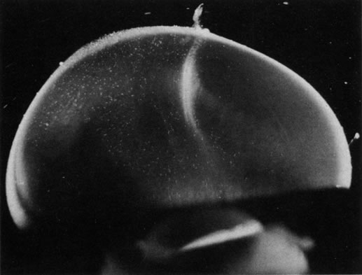
| Fig. 5. Vitreous structure in a human embryo at 33 weeks of gestation. The posterior aspect of the lens is seen below. The vitreous body is enclosed by the dense, highly light-scattering cortex. Parapapillary glial tissue was torn away during dissection and hangs from the prepapillary vitreous cortex. Within the vitreous body, Cloquet's canal arcs from the prepapillary vitreous toward the lens. Since its course undulates through the vitreous body, not all of Cloquet's canal can be visualized in a single horizontal section. (Sebag J: Age-related changes in human vitreous structure. Graefes Arch Clin Exp Ophthalmol 22:89, 1987. Specimens courtesy of the New England Eye Bank, Boston, MA.) |