Chapter 41
Neurogenic Tumors of the Orbit
THOMAS C. CANNON, KEITH D. CARTER and ROBERT FOLBERG
Main Menu   Table Of Contents

Search

NEUROFIBROMAS
SCHWANNOMAS
MALIGNANT PERIPHERAL NERVE SHEATH TUMORS
PARAGANGLIOMAS
PRIMITIVE NEUROECTODERMAL TUMORS
POSTAMPUTATION NEUROMAS
GRANULAR CELL TUMORS (GRANULAR CELL MYOBLASTOMAS)
REFERENCES

The orbit contains two types of nerves: the optic nerve (a tract of the central nervous system) and peripheral nerves (both branches of sensory nerves, such as the fifth nerve, and motor nerves, such as those that innervate the extraocular muscles). Oligodendrocytes produce and maintain the myelin of axons within the central nervous system. In contrast, the Schwann cells produce the myelin that surrounds the axons of nerves within the peripheral nervous system. This chapter is concerned primarily with tumors of neurogenic origin that have arisen from peripheral nerves. The complex diversity of tissues within the orbit is surpassed by no other anatomic site. Primary orbital tumors that are neurogenic range from the common orbital tumors, such as the neurofibroma and schwannoma, to the extremely rare orbital tumors, such as the malignant peripheral nerve sheath tumor, paraganglioma, and primitive neuroectodermal tumor.

To better understand the composition of neurogenic tumors of the orbit, we first must understand the basic histologic composition of a peripheral nerve. A peripheral nerve is composed of several bundles of axons referred to as fascicles. Within the peripheral nervous system, an axon is considered to be an extension of an individual nerve cell body. Each axon is invested with Schwann cells that provide the myelination for each individual axon of a fascicle. Between these individual longitudinally oriented axons is the endoneurium. The endoneurium is composed of amorphous extracellular matrix material and collagen fibrils, which provide the infrastructure for the support of the axons. The perineurium is the connective tissue that surrounds each fascicle (Fig. 1). The epineurium encompasses the entire nerve and contains the blood supply of the nerve.

Fig. 1. Histologic cross-sections of a peripheral nerve. m, myelinated fiber; pn, perineurium; a, axon; en, endoneurium; Schwann cell (s). (Toluidine blue; A, × 75; B, × 300.)

Back to Top
NEUROFIBROMAS
Neurofibromas are benign peripheral nerve tumors composed of axons, Schwann cells, and fibroblasts. Conversely, schwannomas are composed predominantly of Schwann cells. Neurofibromas represent from 0.5% to 3% of all orbital tumors.1 Neurofibromas may be classified into three major categories: isolated (localized) neurofibroma, diffuse neurofibroma, and plexiform neurofibroma. Some authors also categorize the amputation neuroma as a fourth category of neurofibroma, although these more appropriately may be considered a pseudotumor of nerve.2

ISOLATED NEUROFIBROMAS

The isolated (localized) neurofibroma is named because it generally grows as a circumscribed lesion. Although circumscribed, it usually is not encapsulated, as is the schwannoma. Isolated cutaneous neurofibromas may develop anywhere on the body and rarely produce symptoms. Isolated orbital neurofibromas tend to appear in the superior aspect of the orbit during the third to fifth decades (Fig. 2A). Decreased visual acuity is an uncommon symptom of localized neurofibroma confined to the anterior compartment of the orbit. When the posterior aspect of the orbit is affected, compression of the optic nerve may lead to decreased visual acuity and visual field defects and may produce a relevant afferent pupillary defect. Diplopia, which also may develop in isolated neurofibromas of the posterior aspect of the orbit, more likely is the result of mass effect rather than direct involvement of the innervating nerves.

Fig. 2. A. Isolated (localized) orbital neurofibroma with mild proptosis of the right eye. B. On MRI, the lesion appears to be circumscribed in the inferior aspect of the orbit. Despite the circumscribed radiologic appearance, these tumors are not encapsulated.

Radiographic studies often reflect the circumscribed nature of the tumor, although isolated neurofibromas are not encapsulated like schwannomas (see Fig. 2B). Some authors consider the classification of a neurofibroma as localized neurofibroma only in the absence of neurofibromatosis. Multiple localized orbital neurofibromas have been reported in patients who do not have neurofibromatosis.3,4 It is unclear whether these cases actually represent a forme fruste of neurofibromatosis.

Histologically, the localized neurofibroma is characterized by wavy cells with eosinophilic cytoplasm and comma-shaped nuclei (Fig. 3). Collagen can be present in variable quantities. Commonly, there is no palisading of the nuclei, in contrast to the Schwannoma. Ultrastructurally, the neurofibroma is composed of cells that more characteristically have features of the perineurial fibroblast. Conversely, schwannomas have long-spacing collagen and abundant basement membrane, seen on electron microscopic evaluation.

Fig. 3. Localized neurofibroma. Slender spindle cells with characteristic wavy nuclei are enmeshed in a fibrillar stroma. (Hematoxylin-eosin, × 75.)

Treatment of localized neurofibromas depends on whether the lesion is producing symptoms and its location. Most of these lesions are well defined by imaging studies such as computed tomography (CT) or magnetic resonance imaging (MRI). Surgical excision through an anterior or lateral orbitotomy usually is curative.

DIFFUSE AND PLEXIFORM NEUROFIBROMAS

Diffuse neurofibroma and plexiform neurofibroma both are associated with neurofibromatosis; the plexiform neurofibroma is pathognomonic of neurofibromatosis type 1. Both tumors tend to present within the first few decades of life. The diffuse neurofibroma is infiltrative and not circumscribed. Many diffuse neurofibromas are solid, but they also may have a gross myxoid or gelatinous appearance at the time of surgery. Because diffuse neurofibromas feature an infiltrative growth pattern, tumor cells often can be visualized, separating muscle fibers or infiltrating adipose tissue on histologic examination (Fig. 4). The radiographic appearance is infiltrative compared with the circumscribed nature of the localized neurofibroma and schwannoma. The plexiform neurofibroma also lacks encapsulation, and radiographic evaluation may not distinguish plexiform from diffuse neurofibroma.

Fig. 4. Diffuse neurofibroma with the characteristic wavy nuclei, as in Figure 3. This tumor dissects between fascicles of skeletal muscle. (Hematoxylin-eosin, × 75.)

The plexiform neurofibroma is the most common orbital manifestation of neurofibromatosis type 1. Features of malignancy are uncommon in plexiform neurofibromas, although patients with neurofibromatosis have a greater likelihood of developing a malignant peripheral nerve sheath tumor than individuals in the general population. Plexiform neurofibromas most commonly develop within large peripheral nerves. These tumors tend to distort large segments of nerve, most commonly in the extremities. This diffuse thickening experienced on palpation of these lesions has been referred to as a “bag of worms” (Fig. 5). Elephantiasis neuromatosa occurs when diffuse thickening of the nerve leads to enlargement and distortion of an entire extremity. A similar orbital presentation may be caused when plexiform neurofibroma invades the eyelids and surrounding facial nerves and tissues.

Fig. 5. Gross photograph of a plexiform neurofibroma of the cauda equina taken at postmortem examination. The patient died of malignant peripheral nerve sheath tumor (not illustrated). Plexiform neurofibromas and malignant peripheral nerve sheath tumors may involve the orbit.

Histologically, plexiform neurofibromas have several unique characteristics. Microscopically, they are composed of tortuous branches of expanded nerves that appear disorganized when cut in different planes of section. These tumors often appear to have an increased amount of myxoid extracellular matrix material, which produces the characteristic histologic appearance of the tumor (Fig. 6). Special stains, such as silver stains, often can be used to identify axons within these lesions. Neurofibromas, as opposed to Schwannomas, often invade the nerves, and entrapped axons can be identified with these staining methods. The major differentiating feature between the diffuse neurofibroma and the plexiform neurofibroma, however, is the clinical appearance. The plexiform neurofibroma causes a diffuse enlargement and distortion of the nerve and a characteristic eyelid deformity (Fig. 7).

Fig. 6. Histologic section of a plexiform neurofibroma with disorganization and expansion of the nerve fibers and extracellular myxoid matrix. (Hematoxylin-eosin, × 150.)

Fig. 7. Child with neurofibromatosis type 1 and orbital plexiform neurofibroma with extensive involvement of the eyelid.

Back to Top
SCHWANNOMAS
Schwannomas (neurilemomas) are benign peripheral nerve tumors that may arise de novo or within the setting of neurofibromatosis. Schwannomas in the orbit most frequently develop from the superior ciliary nerves, although they also may arise from smaller peripheral nerves. Orbital schwannomas appear to be encapsulated enlargements of the involved nerve. Foci of malignancy seldom are encountered in schwannomas.

Clinically, schwannomas represent approximately 1% of all orbital tumors and are found in 1.5% of all patients with neurofibromatosis type 1. They often are seen in young to middle-aged people and present with a slowly progressive proptosis (Fig. 8A). Often, the proptosis has evolved over decades or several years. Presenting complaints by patients can vary according to location of the tumor within the orbit. Tumors at the orbital apex may present earlier because their compressive effects are more likely to be symptomatic. In a literature review of symptomatic orbital schwannomas in 64 patients by Cantore et al,5 43 patients had exophthalmos and 49 patients had impairment of ocular motility. Conversely, deficits in visual acuity were seen in 28 patients, and optic disc abnormalities also were seen in only 28 patients. In nine of their own patients, Cantore et al5 reported a clinical history of the tumor being present for 1 year or longer. Encapsulation is a clinical feature distinguishing the schwannoma from neurofibroma (see Figs. 8B to D).

Fig. 8. A. Orbital schwannoma in a 16-year-old boy showing proptosis. B. CT scan of the orbit reveals an encapsulated schwannoma in the superior temporal aspect of the right orbit. The left eye is displaced downward (globe ptosis), and the eye does not appear to be the same size on this cut as the right eye. The discrepancy in the apparent size of the eye in this imaging plane is accounted for by the proptosis on the left side. C. Intraoperative photograph shows that the tumor is encapsulated. D. Surgically excised orbital schwannoma. A glistening capsule wraps around the tumor. The two dark red areas beneath the capsule represent zones of focal cystic degeneration.

Schwannomas often have distinctive histologic features that can facilitate their pathologic diagnosis. The main histologic components that are helpful, when present, include Antoni A type areas, Antoni B type areas, and the Verocay body. Antoni A refers to a histologic pattern of the tumor containing solid areas of tumor cells. The Antoni B pattern is textured more loosely, with cystic spaces present. The Verocay body is the name given to the palisading of the nuclei of schwannomas associated with acellular zones (Fig. 9). The Verocay body, when present, is a useful marker for schwannoma. Although the Verocay body may not be present in most schwannomas, palisading of the cells is a frequent finding (Fig. 10). Other ancillary histologic features can be useful in diagnosing schwannomas, such as hyalinization of vessel walls, hemosiderin-laden histiocytes, and foamy histiocytes. Encapsulation is manifest histologically by a band of eosinophilic fibrous tissue surrounding the tumor (Fig. 11). When the histopathologic examination cannot clearly differentiate between schwannoma and other tumors, such as meningioma, immunohistochemistry and electron microscopy can be performed. Immunohistochemical stains for S-100 frequently are positive in schwannoma and not in meningioma. By electron microscopy, Schwann cells have abundant basement membrane and long-spacing collagen (Luse bodies).

Fig. 9. Hematoxylin-eosin-stained histologic section of an orbital schwannoma with palisading of the tumor nuclei associated with acellular zones (Verocay body).

Fig. 10. Hematoxylin-eosin-stained histologic section of an orbital schwannoma with prominent palisading of the cells, a frequent finding in schwannomas.

Fig. 11. Orbital schwannoma. The tumor is encapsulated (arrows).

Treatment consists of complete surgical excision. The encapsulated nature of these tumors enables removal without significant orbital complications.

Back to Top
MALIGNANT PERIPHERAL NERVE SHEATH TUMORS
The rare tumor designated the malignant peripheral nerve sheath tumor (MPNST) has been referred to previously as malignant schwannoma, neurogenic sarcoma, neurofibrosarcoma, and malignant neurilemoma. Enzinger and Weiss6 indicate that the term malignant schwannoma connotes the false impression that these tumors arise from preexisting benign schwannomas, an event that occurs only extremely rarely. The histogenesis of these tumors remains elusive, despite advances in immunohistochemistry and electron microscopy. Although the Schwann cell and perineurial fibroblast have been implicated in the histogenesis of the malignant peripheral nerve sheath tumor, the exact cell of origin remains to be proven. These observations support the opinion that the preferable term for these neoplasms is malignant peripheral nerve sheath tumor.

MPNSTs may arise de novo or within the setting of neurofibromatosis. It has been reported that MPNST develops in 2% to 29% of patients with neurofibromatosis type 1.7 In general, approximately half of MPNSTs occur within neurofibromatosis and half arise de novo. These tumors most commonly occur in the large nerve trunks of the body, including the brachial plexus, the lumbar plexus, and the sciatic plexus. They therefore tend to occur around the spinal cord and often extend into the extremities. They often can extend far beyond the grossly defined tumor margins, and the performance of frozen sections to delineate surgical margins at the time of surgery is prudent if an MPNST is suspected. When arising in the orbit, they have a propensity to occur in the anterior supranasal orbit, arising from the supraorbital branch of the trigeminal nerve.

Jakobiec and associates8 reported a series of eight cases of orbital MPNST. All tumors in this series arose from a supraorbital branch of the trigeminal nerve. Clinically, all eight patients in this series had masses in the superior nasal aspect of the orbit immediately beneath the lid. A striking and worrisome feature of orbital MPNST is the frequent innocuous clinical appearance of the tumor at initial presentation.8 For example, despite clear histologic evidence of malignancy, no patient reported erosion of the overlying skin and in no patient was the malignant nature of the lesion suspected clinically. There was no gender predilection, and the right orbit was affected as commonly as the left orbit. Two of eight patients had neurofibromatosis in Jakobiec's study and five of eight patients died 1 to 5 years after resection of the tumor. Recurrent tumors tended to occur within 6 months after surgery. MPNSTs have been noted to extend long distances along nerves, and intracranial spread to the middle cranial fossa was reported in some patients.

The histopathologic appearance of MPNSTs is variable, and heterologous elements have been reported frequently. Classically, the tumor is markedly hypercellular, forming interlacing fascicles of tumor cells (Fig. 12). Mitotic figures often are conspicuous, although they may be represented variably within individual tumors. Often, densely cellular fascicles of tumor cells alternate with less densely cellular or hypocellular areas. In extraorbital locations, heterologous elements, such as elements of rhabdomyoblastic differentiation, may occur in up to 10% of tumors. MPNSTs containing zones of rhabdomyoblastic differentiation or triton tumors have not been reported in the orbit. Areas of cartilage, bone, and zones of glandular differentiation have been reported in extraorbital MPNSTs.

Fig. 12. Histologic sections of a malignant peripheral nerve sheath tumor (malignant schwannoma). A. Hypercellular interlacing fascicles of tumor cells can be seen. B. Marked hypercellularity and numerous spindle-shaped cells are visible. (Hematoxylin-eosin; A, × 75, B, × 320.)

MPNSTs are not particularly responsive to chemotherapy or radiation therapy; thus, surgical excision is the major treatment modality available. Large tumor size and the presence of necrosis have been associated with an aggressive clinical course.9 Because orbital occurrence is rare, it is not known whether the same prognostic indicators of MPNSTs elsewhere in the body are applicable to orbital MPNSTs.

Back to Top
PARAGANGLIOMAS
The paraganglia are groups of chromaffin cells distributed extensively throughout the body. They commonly are found along the aorta and are present within the carotid body, aortic arch, and the glomus jugulare, in the middle ear, and in the ganglion nodosum of the vagus nerve. The function of the paraganglia primarily is homeostatic. They function to sense fluctuations in blood pH or oxygen tension. Paragangliomas are tumors that arise from the paraganglia. They are histologically identical to the pheochromocytoma but are extraadrenal in location. Paragangliomas have a predilection for the head and neck region. Rarely, these tumor may occur in the orbit and are believed to arise from the ciliary ganglion. During a 47-year period, 73 paragangliomas were seen at Memorial Sloan Kettering, and only 1 of these arose within the orbit.10 Since then, occasional case reports and short case series have been reported in the literature.

Patients most commonly present with proptosis and decreased visual acuity. These tumors often are well vascularized and can be mistaken clinically for a vascular tumor, such as a hemangioma. They also may have a propensity to involve extraocular muscles.11 Recurrence rates of orbital paragangliomas are reportedly high, with 11 of 19 cases recurring in one study, and 7 of these cases resulting in death from the tumor.12 Histopathologically, they frequently can be mistaken as alveolar soft-part sarcoma if histologic preparation is poor. Goldstein and coworkers13 determined that 16 of an initial 29 reported cases of paraganglioma actually were alveolar soft-part sarcomas. Therefore, previous reports of recurrence rates may not represent accurately the prognosis after resection of this rare tumor.

The management of orbital paragangliomas is surgical. Diagnosis before surgery is difficult because of the rarity of the neoplasm and the vascular pattern of the tumor, demonstrable by ancillary studies. Exenteration may be a surgical option after recurrence, but it usually is not the first modality of treatment. Concerns for vision preservation usually prompt conservative management initially.

The histologic features of paragangliomas are distinctive. The cells usually are arranged in groups separated by fibrous septa. The histologic pattern is classically described as “Zellballen” (cell balls) because the cells usually are in spherical aggregates, separated by delicate vessels. Sustentacular cells, which are supportive cells with darkly basophilic nuclei and minimal cytoplasm, are present within paragangliomas and can be helpful in the histopathologic differential diagnosis. Electron microscopy of these tumors reveals large number of dense core neurosecretory granules that contain biogenic amines, indicative of the neural histogenesis of the tumor. Immunohistochemical studies also can be useful when the hematoxylin and eosin (H&E) stain does not demonstrate the classic histologic pattern associated with paraganglioma. Immunohistochemically, paragangliomas react with neural markers and markers for peptide hormones, including synaptophysin, neuron-specific enolase, chromogranin, neurofilaments, serotonin, somatostatin, and other peptide hormones.14

Back to Top
PRIMITIVE NEUROECTODERMAL TUMORS
The term primitive neuroectodermal tumor (PNET) originally was described as a collective term for a class of tumors that are composed of primitive cells of neuroectodermal origin. When they occur within the retina, they are referred to as retinoblastoma; in the pineal, they are pineoblastomas; and in the posterior fossa, they are referred to as medulloblastomas. When these tumors occur within the central nervous system but not in these traditional locations, they are referred to as primitive neuroectodermal tumors. When arising outside the central nervous system, they are referred to as “peripheral” primitive neuroectodermal tumors.15

PNETs of the orbit are extremely rare as primary tumors, although a few cases have been reported.16,17 Peripheral PNETs primarily affect children and adolescents. They occur most commonly in the chest wall but also can present in the abdomen, trunk, and pelvis. Clinically, these tumors present as other orbital masses with proptosis, which may affect ocular motility. The few reported cases of primary orbital PNETs most commonly involved children and adolescents. Orbital PNETs characteristically have the typical cytogenetic abnormality of t(11:22)(q24;q12). Many investigators believe that Ewing's sarcoma and peripheral PNETs are two primitive tumors that may have the same histogenesis.

Back to Top
POSTAMPUTATION NEUROMAS
The postamputation neuroma is a benign, reactive proliferation composed of Schwann cells, perineurial fibroblasts, and other fibroblasts and occurs after transection of a peripheral nerve. It is believed that the proliferation of these neural elements after transection is a reparative attempt by the nerve to reestablish the continuity of the nerve. Postamputation neuromas often are seen in amputations of the extremities, although they can arise anywhere in the body when a peripheral nerve is transected. The occurrence of the amputation neuroma in the orbit is uncommon. Theories proposed for the uncommon occurrence in this location include the relatively small size of the nerve and the lack of irritating factors to the nerve. Amputation neuromas in areas outside the orbit commonly present with pain. Those tumors presenting within the orbit are less likely to cause pain than proptosis. Rarely, these reactive proliferations can be so intense that the suspicion of recurrent tumor or other orbital tumors is raised. Rare cases have been reported in which amputation neuromas arose within or concurrent with recurrent choroidal melanoma.18

The postamputation neuroma is characterized histologically by a proliferation of bundles of nerves surrounded by dense collagenous septa. In patients who have undergone enucleation for malignant tumors, meticulous histologic examination of the tissue must be undertaken to exclude the recurrence of the malignancy within the amputation neuroma. Electron microscopic evaluation of amputation neuromas has revealed the intense metabolic activation of these tumors with prominent proliferating perineurial fibroblasts. The perineurial fibroblast has been postulated to modulate the proliferative activity of the axon after transection and is crucial to the regenerative attempts of the nerve.19

Treatment of orbital amputation neuroma is surgical excision of the neuroma. Reconstructive orbital surgery is dependent on the resultant defect.

Back to Top
GRANULAR CELL TUMORS (GRANULAR CELL MYOBLASTOMAS)
Recent studies indicate that granular cell tumors are of neural origin (not muscle, as formerly suspected) and that the histogenesis may involve a Schwann cell derivative. The granular cell tumor originally was described as being a tumor of muscle origin.20 The tumor originally was named granular cell myoblastoma, reflecting the histogenesis from muscle. With advances in immunohistochemistry and electron microscopy, the characteristic granular histologic appearance has been attributable to the presence of membrane-bound autophagic vacuoles that contain cellular debris, myelin, and fragments of other organelles, such as rough endoplasmic reticulum.

The granular cell tumor most commonly occurs in the fourth through sixth decades and is rare in children. The tongue is the most common location of the tumor; however, granular cell tumors have been reported in a variety of unusual locations, including the lung, the pituitary gland, and the gallbladder. Several cases have been reported within the orbit. Clinically, the granular cell tumor presents as poorly circumscribed nodules. These tumors may be composed of a single nodule or multiple nodules. Malignant granular cell tumors are extremely rare and no cases, to our knowledge, have been reported within the orbit.

Several granular cell tumors have been reported within the orbit.21–24 Granular cell tumors of the conjunctiva and eyebrow also have been reported.25–27 In a series of six orbital granular cell tumors reported by Jaeger and associates,21 the duration of signs and symptoms ranged from 2 weeks to 5 years. Orbital granular cell tumors most commonly may produce a mass lesion within the upper or lower eyelid. Diplopia may result when the tumors interfere with ocular motility.

Histologically, granular cell tumors have abundant eosinophilic granular cytoplasm with round uniform nuclei (Fig. 13). Classically, the nuclei are basophilic, with a small nucleolus. The tumor cells often are arranged in cords or clusters, with intervening fibrous tissue or skeletal muscle. Granular cell tumors commonly stain for S-100, neuron-specific enolase, laminin, and various myelin proteins. The cytoplasm also is periodic acid-Schiff-positive and diastase resistant. The cells do not stain for glial fibrillary acidic protein or neurofilament. Transmission electron microscopy reveals the previously described autophagic vacuoles.

Fig. 13. Histologic section of an orbital granular cell tumor with prominent eosinophilic granular cytoplasm and small, round nuclei having prominent nucleoli. (Hematoxylin-eosin, × 320.)

Back to Top
REFERENCES

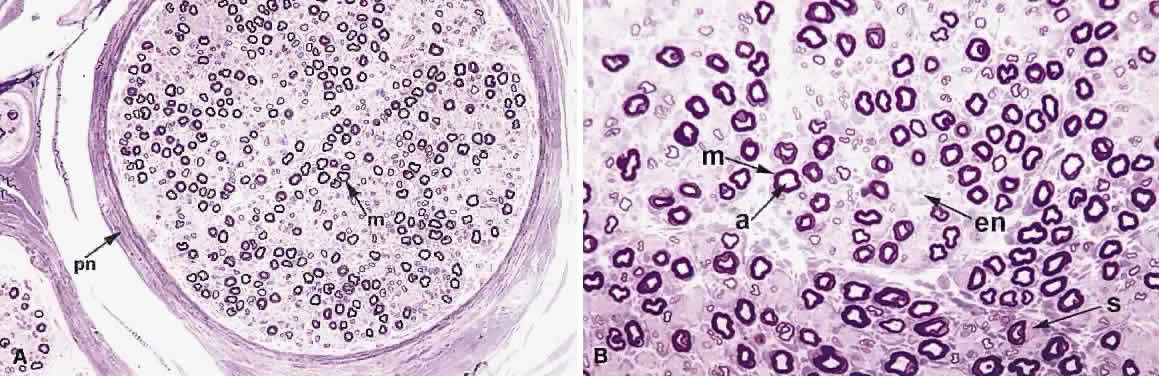
1. Levin LA, Jakobiec FA: Peripheral nerve sheath tumors of the orbit. In Albert DM, Jakobiec FA (eds): Principles and Practice of Ophthalmology: Clinical Practice, p 1978. Philadelphia: WB Saunders, 1994

2. Jakobiec FA, Bilyk JR, Font RL: Orbit. In Spencer WH (ed): Ophthalmic Pathology: An Atlas and Text, p 2438. 4th ed. Philadelphia: WB Saunders, 1996

3. Shields JA, Shields CL, Lieb WE: Multiple orbita neurofibromas unassociated with von Recklinghausen's disease. Arch Ophthalmol 108:80, 1990

4. Krohel GB, Rosenberg PN, Wright JE: Localized orbital neurofibromas. Am J Ophthalmol 100:458, 1985

5. Cantore G, Cipetta P, Raco A, et al: Orbital schwannomas: Report of nine cases and review of the literature. Neurosurgery 19:583, 1986

6. Enzinger FM, Weiss SW: Soft Tissue Tumors. 3rd ed. St. Louis: Mosby, 1995

7. Wanebo JE, Malik JM, Vandenberg SR et al: Malignant peripheral nerve sheath tumors. Cancer 71:1247, 1997

8. Jakobiec FA, Font RL, Zimmerman LE: Malignant peripheral nerve sheath tumors of the orbit: A clinicopathologic study of eight cases. Trans Am Ophthalmol Soc 83:332, 1985

9. Meis JM, Enzinger FM, Martz KL et al: Malignant peripheral nerve sheath tumors (malignant schwannomas) in children. Am J Surg Pathol 16:694, 1992

10. Lack E, Cubilla AL, Woodruff JM et al: Paragangliomas of the head and neck. Cancer 39:397, 1977

11. Netland PA, Font R, Jakobiec FA: Rare intraosseous and primary orbital tumors, in Albert DM, Jakobiec FA (eds): Principles and Practice of Ophthalmology: Clinical Practice, p 2051. Philadelphia: WB Saunders, 1994

12. Amemiya T, Kadoya M: Paraganglioma of the orbit. J Cancer Res Clin Oncol 96:169, 1980

13. Goldstein BG, Font RL, Alper MG: Granular cell tumor of the orbit: a case report including electron microscopic observations. Ann Ophthalmol 14:231, 1982

14. Rosai J: Adrenal gland and paraganglia. In Rosai J (ed): Ackerman's Surgical Pathology, p 1015. 8th ed. St. Louis: Mosby Year Book Inc, 1996

15. Dehner LP: Peripheral and central primitive neuroectodermal tumors: a nosologic concept seeking consensus. Arch Pathol Lab Med 110:997, 1986

16. Singh AD, Husson M, Shields CL: Primitive neuroectodermal tumor of the orbit. Arch Ophthalmol 112:217, 1994

17. Arora R, Sarkar C, Betharia SM: Primary orbital primitive neuroectodermal tumour with immunohistochemical and electron microscopy confirmation. Orbit 12:7, 1993

18. Folberg R, Bernardino VB, Aguilar GL et al: Amputation neuroma mistaken for recurrent melanoma in the orbit. Ophthalmic Surg 12:275, 1981

19. Katenkamp D, Sullivan TJ: Ultrastructure of perineurial cells during peripheral nerve regeneration: Electron microscopical investigations on the so-called amputation neuroma. Exp Pathol 16:5, 1978

20. Abrikossoff A: Uber myome ausgehend von der guergestreiften willkurlichen muskulatur. Virchows Arch A Pathol Anat Histopathol 260:215, 1926

21. Jaeger MJ, Green WR, Miller NR et al: Granular cell tumor of the orbit and ocular adnexa. Surv Ophthalmol 31:417, 1987

22. Karcioglu ZA, Hemphill GL, Wool BM: Granular cell tumor of the orbit: case report and review of the literature. Ophthalmic Surg 14:125, 1987

23. Morgan G: Granular cell myoblastoma: Report of a case. Arch Ophthalmol 94:2135, 1976

24. Drummond JW, Hall DL, Steen WH et al: Granular cell tumor (myoblastoma) of the orbit. Arch Ophthalmol 97:1492, 1979

25. Charles NC, Fox DM, Glasberg SS et al: Epibulbar granular cell tumor: report of a case and review of the literature. Ophthalmology 104:1454, 1997

26. Ferry AP: Granular cell tumor (myoblastoma) of the palpebral conjunctiva causing pseudoepitheliomatous hyperplasia of the conjunctival epithelium. Am J Ophthalmol 91:234, 1981

27. Pe'er J, Schwartzenberg T, Kopolovic J: Granular cell tumor (myoblastoma) of the eyebrow. Ophthalmology 194:1, 1987

Back to Top