|
|
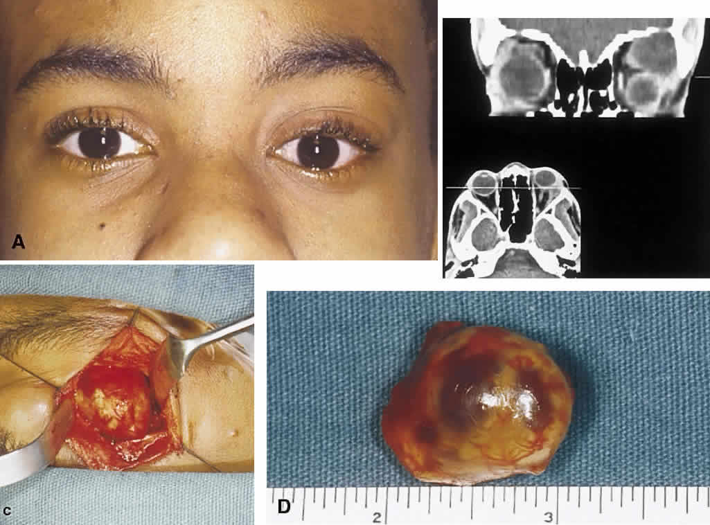
| Fig. 8. A. Orbital schwannoma in a 16-year-old boy showing proptosis. B. CT scan of the orbit reveals an encapsulated schwannoma in the superior temporal aspect of the right orbit. The left eye is displaced downward (globe ptosis), and the eye does not appear to be the same size on this cut as the right eye. The discrepancy in the apparent size of the eye in this imaging plane is accounted for by the proptosis on the left side. C. Intraoperative photograph shows that the tumor is encapsulated. D. Surgically excised orbital schwannoma. A glistening capsule wraps around the tumor. The two dark red areas beneath the capsule represent zones of focal cystic degeneration. |