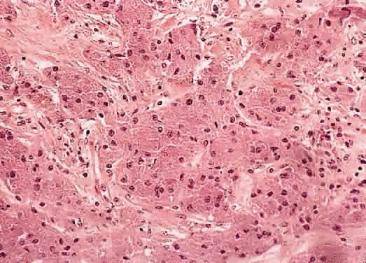
Fig. 13.
Histologic section of an orbital granular cell tumor with prominent eosinophilic granular cytoplasm and small, round nuclei having prominent nucleoli. (Hematoxylin-eosin, × 320.)