|
|
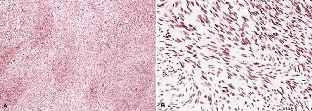
| Fig. 12. Histologic sections of a malignant peripheral nerve sheath tumor (malignant schwannoma). A. Hypercellular interlacing fascicles of tumor cells can be seen. B. Marked hypercellularity and numerous spindle-shaped cells are visible. (Hematoxylin-eosin; A, × 75, B, × 320.) |