Chapter 38
Nonsteroidal Anti-Inflammatory Drugs
ALLAN J. FLACH
Main Menu   Table Of Contents

Search

CHEMISTRY
PREPARATIONS
PHARMACOKINETICS
PHARMACODYNAMICS AND RATIONALE FOR USE
THERAPEUTIC POTENTIAL: REVIEW OF CLINICAL STUDIES
NONSTERIOIDAL ANTI-INFLAMMATORY DRUG TOXICITY AFTER ORAL ADMINISTRATION
REFERENCES

The term nonsteroidal anti-inflammatory drugs (NSAIDs) has been applied to clycooxygenase inhibitors (COIs) that are useful for the prevention and treatment of several commonly encountered ophthalmic syndromes and diseases. Topically effective preparations are commercially available as ophthalmic solutions that have been approved for use by the Food and Drug Administration (FDA) for more than two decades. Labeled indications include the inhibition of intraoperative miosis during cataract surgery, the treatment of ocular inflammation following cataract surgery, relief of ocular pruritis associated with seasonal allergic conjunctivitis and relief of pain and photophobia associated with refractive surgery. In addition, this group of drugs has been widely used by ophthalmologists throughout the world for the prevention and treatment of cystoid macular edema (CME) after cataract surgery. The purpose of this chapter is to summarize the status of topically applied NSAIDs. This effort will correlate the pharmacology and pharmacodynamics of NSAIDs with related advances in the medical sciences as reflected in laboratory and clinical studies, which is the goal and purpose of ophthalmic clinical pharmacology.1,2
Back to Top
CHEMISTRY
The NSAIDs currently available commercially represent a chemically heterogeneous group of compounds that can be divided into six different chemical classes: salicylates, fenamates, indoles, phenylalkanoic acids, phenylacetic acids, and pyrazolones. Individual members within respective groups are summarized in Table 1 and their structures are shown in Figure 1. NSAIDs is a term chosen to emphasize that drugs of this group have chemical structures that do not include a steroid nucleus derived biosynthetically from cholesterol. Aspirin, chemically known as acetylsalicylic acid, remains the most widely used NSAID throughout the world. However, it cannot be prepared in an aqueous delivery system because it is rapidly hydrolyzed to salicylic and acetic acids. Therefore, it is unstable in solution and is not available in other than solid dosage forms.3 Salicylic acid has anti-inflammatory activity, but it is too irritating for topical ophthalmic use. It is reserved as a keratolytic agent for dermatologic conditions such as psoriasis and warts. Similar to the salicylates, the fenamates and pyrazolones derivatives are considered too toxic for ocular application.4–6 Therefore, this chapter reviews the indoles, the phenylalkanoic acids, and the phenylacetic acids that are available as topical ophthalmic preparations.

TABLE 1. Nonsteroidal Anti-Inflammatory Drugs


Chemical classGeneric nameTrade name (manufacturer)
SalicylatesAspirin salicylic acid, diflunisalMany trade names (multiple)
IndolesIndomethacinIndocid Ophthalmic Suspension (Merck, Sharpe & Dohme, West Point, PA)
 SulindacClinoril (Merck, Sharpe & Dohme)
 TolmetinTolectin (Smith Nephew, Memphis, TN)
Phenylalkanoic acidsFenoprofenNalfon (Lily, Indianapolis, IN)
 Flurbiprofen*Ocufen Ophthalmic solution (Allergan, Irvine, CA)
 IbuprofenMotrin (Upjohn, New York, NY); Rufen (Boots, Inc.)
 KetoprofenAlrheumat; Orudis
 Ketorolac*Acular Ophthalmic solution (Allergan)
 NaproxenNaprosyn (Syntex); Anaprox (Syntex Labs, Palo Alto, CA)
 PiroxicamFeldene (Pfizer)
 Suprofen†Profenal Ophthalmic solution (Alcon, Ft. Worth, TX)
Phenylacetic acidsDiclofenac*Voltaren Ophthalmic solution (Ciba Vision Ophthalmics, Duluth, GA)
PyrazolonsOxyphenbutazoneTandearil (Geigy)
  Oxalid (VSV)
 PhenylbutazoneButazolidin (Geigy)
  Azolid (VSV)
 Antipyrine 
 Aminopyrine 
 AzapropazoneRheumox

*Commercially available and approved by the Food and Drug Administration (FDA) as an ophthalmic preparation in the United States.
†Not commercially available, but approved by FDA as an ophthalmic preparation in the United States.


Fig. 1. Structures of nonsteroidal anty-inflammatory drugs.

Most NSAIDs are weak acids with pKa values between 3 and 6. The unionized form of the molecule is associated with good lipid solubility providing good absorption from the stomach, upper gastrointestinal tract and permitting passage through the lipoidal corneal epithelium of the eye. Generally, pKa values are lower than the respective pH values of the extracellular environment, and, therefore, these drugs exist largely in their ionized forms. However, in a more acid environment, such as that produced by inflammation, the unionized form of drug is available to diffuse into cells. A pH change from 7.4 to 7.0 is accompanied by a 25% increase in the unionized form of the drug. Because the intracellular pH decreases less than the extracellular pH during an inflammatory process, the relatively alkaline environment provided inside the cells favors the ionized form of the drug trapping it within the cell. Thus, the pH difference favors increasing the NSAID intracellular concentration potentially enhancing therapeutic effects.7

The chemical structures of all of the NSAIDs incorporate at least one aromatic ring. The structures of indomethacin, diclofenac, and flurbiprofen include two aromatic rings that are twisted with respect to each other. A study of structure–activity relationships has helped determine the characteristics of at least one NSAID receptor.8 However, there are undoubtedly multiple receptors for NSAIDs with different structural requirements. Therefore, it is unlikely that the characteristics of any single proposed receptor as determined in vivo will explain the pharmacologic activity of this group of drugs.

Indomethacin is the first topically administered NSAID that found widespread use in ophthalmic clinical studies. However, it remains unapproved for use as an ophthalmic formulation in the United States. From a chemical viewpoint, it is the only indole derivative available. This drug is chemically known as 1-(4-chlorobenzoyl)-5-methoxy-2-methyl-1H-indole-3-acetic acid with the empirical formula of C19H16ClNO4 and a molecular weight of 357.80. Indomethacin is soluble in alcohol but insoluble in water. Initially, it was formulated in a solution of sesame seed oil.9 However, this eyedrop proved too irritating for patients. Therefore, the preparation currently available commercially in Europe and other parts of the world is a 1% indomethacin aqueous suspension (Indocid Ophthalmic Suspension; Merck, Sharpe & Dohme, West Point, PA).

The phenylalkanoic acids are water-soluble and, consequently, are easier to formulate as ophthalmic solutions. Flurbiprofen 0.03% (Ocufen Ophthalmic Solution; Allergan, Irvine, CA) and suprofen 1% (Profenal Ophthalmic Solution; Alcon, Ft. Worth, TX) are approved by the FDA for inhibition of intraoperative miosis. However, only flurbiprofen is commercially available. Flurbiprofen is chemically known as sodium (+/-)-2-fluoro-α methyl-4-biphenyl acetate dehydrate with a molecular weight of 302.3. Suprofen is α-methyl-4-(2-thienylcarbonly) benzene acetic acid with the empirical formula of C14H12O3S and a molecular weight of 260.3. It has not been available commercially for several years.

The third phenylalkanoic acid derivative, ketorolac tromethamine, is currently available as three different commercial preparations: the first approved 0.5% ophthalmic solution (Acular; Allergan), a preservative free 0.5% solution (Acular PF; Allergan) and a lower strength 0.4% ophthalmic solution (Acular LS; Allergan). Acular Ophthalmic Solution is approved for the treatment of postoperative inflammation in patients who have undergone cataract extraction and for the temporary relief of ocular itching caused by seasonal allergic conjunctivitis. Acular PF is indicated for the reduction of ocular pain and photophobia following incisional refractive surgery. Acular LS is approved for the reduction of ocular pain and burning/stinging following corneal refractive surgery. All of these preparations contain the same basic drug molecule ketorolac as a tromethamine salt and is known chemically as (+/-5-benzoyl-2,3-dihydro-) 1H-pyrrolizine-1-carboxylic acid with 2-amino-2-(hydroxymethyl)-1,3-propanediol(1:1). The molecular weight is 376.41 with the empirical formula of C19H24N2O6. It is formulated as tromethamine salt to provide ideal water solubility. Ketorolac exists as both S (−) and R (+) enantiomers and is marketed as the racemate. The S (−) enantiomer is approximately twice as active as an anti-inflammatory agent. Interestingly, and unlike other NSAIDs, the S enantiomer of ketorolac is levorotatory. Ketorolac's enantiomers exhibit stereoselective pharmacokinetics.10

Diclofenac sodium 0.1% (Voltaren; Novartis Ophthalmics, East Hanover, NJ) is a phenylacetic acid derivative that is approved for use by the FDA after cataract surgery to inhibit postoperative inflammation and for the relief of pain and photophobia following refractive surgery. It is designated chemically as 2-((2, 6-dichloro-phenyl) amino) benzene acetic acid, monosodium salt, with an empirical formula of C14H10Cl2NO2. Its molecular weight is 318.14. It is freely soluble in methanol and sparingly soluble in water. This ophthalmic solution has the faint odor of castor oil.

Back to Top
PREPARATIONS
The FDA approves flurbiprofen 0.03% and suprofen 1% for use as inhibitors of intraoperative miosis. However, suprofen 1% (Profenal Ophthalmic Solution 1%, Alcon) has not been commercially available since 1998. Although flurbiprofen was initially distributed in 2.5 mL, 5 mL and 10 mL volumes, the FDA requested that Allergan remove all but the 2.5 mL volume from the market in an attempt to discourage its use for unapproved indications. Therefore, flurbiprofen is only available as 2.5 mL within a 6-mL bottle. This preparation, called Ocufen, contains 0.03% flurbiprofen sodium, 1.4% polyvinyl alcohol, 0.005% thimerosal, edetate disodium, potassium chloride, sodium chloride, sodium citrate, citric acid, and purified water with an adjusted pH of 6.0 to 7.0 and an osmolality of 260 to 330 mOsm/kg. The manufacturer recommends a total of four drops of solution, administered one drop approximately every half hour beginning 2 hours before surgery, for the inhibition of intraoperative miosis during cataract surgery. The preparation is supplied in a plastic dropper bottle and stored at room temperature (59°F to 75°F or 15°C to 25°C). Flurbiprofen is also available in generic formulation (flurbiprofen 0.03% ophthalmic solution, Bausch & Lomb, Rochester, NY).

Voltaren Ophthalmic Solution is approved for use in inhibiting postoperative inflammation following cataract surgery and for the temporary relief of pain and photophobia after refractive surgery. It is available in 2.5-mL and 7.5-mL volumes dispensed within 5-mL and 10-mL bottles, respectively. This preparation includes diclofenac 0.1%, boric acid, edetate disodium, polyoxyl 35 castor oil, purified water, sorbic acid, and tromethamine adjusted to a pH of 7.2. This ophthalmic solution is supplied in a dropper-tip, plastic squeeze bottle that can be stored at room temperature (59°F to 86°F or 15°C to 30°C). The medication should be protected from light. The manufacturer suggests one drop of solution four times daily beginning 24 hours after cataract surgery and continuing throughout the first 2 weeks of the postoperative period. One drop can be given 1 hour before refractive surgery and 15 minutes after the procedure is completed. Thereafter, one drop is administered four times daily for as long as 3 days.

Ketorolac tromethamine 0.5% ophthalmic solution is available in three different formulations: Acular 0.5% Ophthalmic Solution, Acular LS Ophthalmic Solution, and Acular PF Ophthalmic Solution (Allergan). Acular 0.5% Ophthalmic Solution is available in 3 mL, 5 mL, and 10 mL volumes each within an opaque plastic bottle 5 mL, 10 mL, and 10 mL, respectively, each with a controlled dropper tip. In addition to ketorolac tromethamine 0.5%, these solutions contain benzalkonium chloride 0.01%, edetate disodium 0.1%, octoxynol 40, sodium chloride, and purified water with an adjusted pH of 7.4. This preparation is approved by the FDA for use following cataract surgery for the treatment of postoperative inflammation with treatment initiated 24 hours after surgery and given one drop four times daily for 2 weeks. It is also approved for the relief of ocular itching caused by seasonal allergic conjunctivitis and is recommended by the manufacturer as one drop four times daily.

Acular PF Ophthalmic Solution is available as a sterile solution supplied in single-use vials within a package containing twelve single use vials each containing 0.4 mL of solution. This preservative-free solution contains only ketorolac tromethamine 0.5%, sodium chloride, hydrochloric acid and sodium hydroxide to adjust pH to 7.4, and purified water. This preparation is indicated for the reduction of ocular pain and photophobia after incisional refractive surgery.

Acular LS Ophthalmic Solution is indicated for the reduction of ocular pain and burning/stinging after cataract surgery. It is supplied as a 5 mL volume within a 10-mL bottle. This ophthalmic preparation contains ketorolac tromethamine 0.4%, benzalkonium chloride 0.006%, sodium chloride, edetate disodium 0.015%, octoxynol 40, purified water and is adjusted to a pH of approximately 7.4. It is best stored at 15°C to 25°C or 59°F to 75°F and protected from light.

These ophthalmic NSAIDs are available throughout the world where they are approved for different ophthalmic indications including treatment of allergic conjunctivitis, inhibition of intraoperative miosis during cataract surgery, temporary relief of pain and photophobia after refractive surgery, prevention of postoperative inflammation after cataract surgery, and the prevention and treatment of cystoid macular edema after cataract surgery. In addition, Indomethacin 1% Ophthalmic Suspension is available outside of the United States in a 5-mL dropper bottle.

Pharmacists are trained to prepare NSAID ophthalmic eyedrops by dissolving crushed tablets or the contents of capsules in artificial tears or other vehicles in an aseptic or sterile procedure. However, if improperly prepared, the result can be a potentially dangerous ophthalmic product. For example, 12 hospitalizations in western Pennsylvania, eastern Ohio, and West Virginia for ocular infections were reported in newspapers throughout the United States during the 1990s. Two of these cases required enucleations (Associated Press, San Francisco Chronicle, February 1991). Therefore, a properly qualified pharmacist with a special interest in ophthalmic pharmacology and the compounding of ophthalmic preparations is a requirement for the extemporaneous preparation of NSAID ophthalmic dose forms.

Back to Top
PHARMACOKINETICS

SYSTEMIC ADMINISTRATION

The NSAIDs that are of greatest ophthalmic interest have anti-inflammatory, analgesic, and antipyretic pharmacologic properties that are similar to those of the salicylates.10–16 One can emphasize these common pharmacologic activities and refer to NSAIDs as aspirinlike drugs.1 These agents are well absorbed from the gastrointestinal tract after oral administration, thereafter reaching maximum serum levels in 1 to 3 hours in fasting patients.17–21 Subsequently, they are metabolized in the liver and excreted in the bile and urine. Plasma half-lives are variable, possibly related to enterohepatic cycling, which is known to occur with the NSAIDs. Age, gender, and endogenous and environmental factors can affect clearance and, ultimately, the half-life of elimination of these drugs. All these agents are highly bound to protein (90% to 99%) and tissues.22

The rate of absorption of NSAIDs may be decreased by administration with meals or by formulation as slow-release (SR) preparations. Although the efficacy and tolerance of most slow-release preparations are not significantly better than those of conventional-release formulations, there is some evidence that an indomethacin preparation with slow-release characteristics can minimize dizziness and diarrhea.23

Although the NSAIDs are cleared by hepatic metabolism, the activity of some of these agents (aspirin, phenylbutazone, fenbufen, benorylate, and sulindac) is dependent, in part, on the formation of active metabolites.24 Furthermore, there can be differences in the pharmacokinetic profiles of enantiomers of the same drug. However, although the potential importance of stereoselective disposition and metabolic activation of NSAIDs cannot be disputed, there is no evidence that these pharmacokinetic parameters are important clinically during treatment with the NSAIDs currently approved for ophthalmic use.

Orally administered NSAIDs are widely used in the treatment of rheumatic diseases. The marked intersubject variability in pharmacologic response and incidence of side effects remains unexplained. Differences in plasma pharmacokinetics have not been clearly associated with this variability, although studies of the relationship between efficacy and unbound concentrations of NSAIDs are clearly needed. During treatment, increasing the dosage of a given NSAID in a nonresponder seems to do little to enhance efficacy, and selection of a different NSAID is frequently attempted.7 The value of this approach (i.e., changing drugs rather than increasing dose) during ophthalmologic therapy remains to be proved.

Although the NSAIDs are weak acids, and therefore, their renal clearance is increased by alkalinization of the urine, the excretion of unmetabolized drug is not usually a major pathway of elimination. However, renal impairment is of concern because of potential retention of NSAID metabolites and because of an increased risk of renal toxicity. Insofar as renal function deteriorates with age, the elderly are at greater risk of adverse effects.25

TOPICAL ADMINISTRATION

NSAIDs have excellent biphasic solubility and therefore penetrate the eye better after topical application than after systemic administration. For example, the rabbit aqueous humor level of indomethacin was much greater after the administration of either a 1% indomethacin suspension or 1% indomethacin sesame seed oil solution than after the oral administration of indomethacin.26,27 These laboratory observations have been confirmed in human studies.28 Volunteers given oral indomethacin 25 mg four times daily beginning 24 hours before surgery and a 25-mg dose 2 hours preoperatively did not have detectable aqueous humor levels despite mean peak blood levels of 642 ng/mL. However, application of a topical 1% aqueous suspension or a 1% sesame oil solution, five drops applied over 24 hours the day before surgery and one drop 45 minutes before surgery, provided adequate mean peak aqueous levels of solution (429 ng/mL) and suspension (198 ng/mL). Furthermore, these aqueous levels were not associated with measurable blood levels.

The ocular instillation of NSAIDs provides ocular tissue levels, including aqueous humor, adequate to inhibit prostaglandin (PG) synthesis. Ocular absorption and distribution of topically administered flurbiprofen in normal and aphakic rabbit eyes has been studied.8,28 Ocular instillation of 50 μL of 0.03% flurbiprofen provides ocular tissue levels (cornea, conjunctiva, sclera, iris, choroid, retina, ciliary body, and aqueous humor) sufficient to provide 50% inhibition of PG synthetase in phakic eyes (more than 90 ng/mL). Higher concentrations of drug were found in the vitreous humor, choroid, and retina of aphakic eyes. Topical application of 0.5% ketorolac tromethamine solution to normal rabbit eyes provides drug to all the ocular tissues, including cornea, conjunctiva, sclera, iris, ciliary body, lens, choroid, retina, vitreous humor, and aqueous humor.29 Topical application of suprofen 0.5% results in significant ocular levels that are associated with the inhibition of the generation of PGE2, PGF2a, PGI2, and thromboxane B2 from inflamed rabbit corneas. This inhibition is more effective than that achieved with topical prednisolone 1%.15 A comparison of the ocular penetration and subsequent effects of diclofenac, flurbiprofen, and indomethacin on disruption of the blood–aqueous barrier (BAB) in rabbit eyes has been reported.30 The breakdown of the BAB after paracentesis of the anterior chamber is inhibited in a dose-dependent manner. The anti-inflammatory effects persisted 6 hours. Dose-response curves for the NSAIDs are comparable and are in the range of 3 × 10⁶ mol. These dose ranges are comparable to those that have been used for inhibition of PG synthesis in vitro in rabbit conjunctival tissue.

The therapeutic usefulness of the NSAIDs appears to derive, in part, from their ability to inhibit the synthesis of PGs and the associated antimiotic and anti-inflammatory activities.22 An increase in PGE and PGF in the aqueous humor after cataract extraction has been demonstrated.31 Topical indomethacin solution and suspension easily achieved the 60 ng/mL concentration of indomethacin that is the median infective dose (ID50) for in vitro inhibition of PG synthesis within the aqueous humor.32 Therefore, these clinical observations, coupled with the increased potential for undesirable systemic side effects associated with oral administration, suggest that topical administration of NSAIDs is preferred. However, it is too early to conclude that the topical administration of NSAIDs is not associated with significant systemic absorption or systemic toxicity. It appears that a significant percentage of topically applied NSAID can appear in the systemic circulation after absorption from the nasolacrimal outflow system and the vascular nasal mucosa. After topical application to the rabbit eye of 50 μL 0.5% ketorolac (250 μg), peak plasma concentrations of 0.2 μg/ml were measured within about 15 minutes.29,33 Furthermore, after topical administration of flurbiprofen solutions, 74% of the dose appeared to enter the systemic circulation.20 These data appear to conflict with the study previously described.28 However, the results of laboratory determinations can be influenced by the sensitivity and timing of the assay, species differences, and variations in absorption from the nasolacrimal system. Therefore, although systemic absorption of an NSAID is relatively small and variable after topical treatment, it is less than prudent to assume that the topical route lacks systemic toxicity. Eyelid closure and digital occlusion of the nasolacrimal outflow system enhance intraocular penetration of drug and minimize systemic absorption of the instilled medication.34,35 These techniques should help provide an optimum risk:benefit ratio for the patient.

Back to Top
PHARMACODYNAMICS AND RATIONALE FOR USE

PHARMACODYNAMICS

PG research began with Ambache's study of “irin” more than 60 years ago, but the chemical identification of this endogenous mediator occurred subsequently. Many of the major advances in understanding these autacoids have occurred in the past two decades.36 PGs produce many physiologic effects within the eye, including miosis, conjunctival hyperemia, increased permeability of the blood–ocular barriers, and changes in intraocular pressure.37–40 Furthermore, PGs have become recognized as potential chemical mediators of both cellular and humoral aspects of inflammation and allergy.41–43

Aspirin and other NSAIDs decrease the synthesis of PGs by inhibiting cyclooxygenase (COX), the enzyme that catalyzes the formation of endoperoxides from arachidonic acid. In addition, indomethacin has shown inhibitory activity against phospholipase A2 and C. There is little evidence that NSAIDs inhibit lipoxygenase, except at concentrations higher than those achieved therapeutically; therefore, NSAIDs have limited ability to inhibit the generation of leukotrienes, as shown in Figure 2.44–48 However, diclofenac may indirectly limit leukotriene production by sequestration of arachidonic acid into triglyceride pools.49 This would enhance its anti-inflammatory activity because leukotrienes possess chemotactic activity.

Fig. 2. Mediators of inflammation-potential inhibition.

Although it is tempting to summarize the pharmacodynamics of NSAIDs by thinking of them simply as COX inhibitors, it is unlikely that this single mechanism accounts for all their pharmacologic effects. Different hypotheses have been suggested. These agents, at higher concentrations, blunt the functions of inflammatory cells such as polymorphonuclear leukocytes. Investigators have described how NSAIDs inhibit the aggregation of polymorphonuclear leukocytes. These drugs inhibit calcium movements and enhance the poststimulation increase in intracellular cyclic adenosine monophosphate (cAMP); these actions could alter the early steps of neutrophil activation. NSAIDs have a free radical scavenger action. As stated previously, they inhibit peripheral blood monocyte phospholipase C activity. In addition, NSAIDs may indirectly reduce vascular permeability.7,22,47,50–54

NSAIDs are anionic, planar molecules that can partition into lipids, such as those found in cell membranes and particularly in the acid environments provided by inflammation. Many membrane effects are likely related to the ability of aspirin like drugs to insert into the plasma membrane's lipid bilayer where they can disrupt protein–protein interactions and normal signaling events. These drugs inhibit membrane-associated nicotinamide adenine dinucleotide phosphate (NADPH) oxidase activity, uptake of precursor arachidonate, transmembrane anion transport, and mitochondrial oxidative phosphorylation.47,55–57 Finally, at high doses; NSAIDs inhibit nonprostaglandin activities, such as proteoglycan synthesis by chondrocytes and chemoattractant binding.58–61

Despite these qualifications, the inhibition of COX activity does appear to correlate with the therapeutic usefulness of NSAIDs in ophthalmology (i.e., the inhibition of intraoperative miosis, the modification of postoperative inflammation, the prevention and treatment of CME after cataract surgery, the relief of excessive pain and photophobia after refractive surgery and the treatment of allergic conjunctivitis).

Most NSAIDs inhibit not only PGs at sites of inflammation but also PGs that serve to assist with important functions throughout the body. Such inhibition may be harmful as described within the toxicity section of this chapter. The tendency for NSAIDs to impair normal gastrointestinal mucosal function with associated mucosal damage can result in serious gastrointestinal complications. A prospective trial of more than 8,000 patients with rheumatoid arthritis who were taking NSAIDs found the rate of serious gastrointestinal complications to be 1.54% per year.62 NSAIDs are among the most widely used therapeutic agents with approximately $2 billion spent in the United States on prescription NSAIDs alone.63 This means thousands of patients are at risk each year.

CycloOxygenase Isoforms

A potentially useful approach to avoiding some of the complications of NSAID treatment became possible with the identification of two isoforms of COX: COX-1 and COX-2. COX-1, a constitutive enzyme, synthesizes PGs that regulate physiologic processes. It is present in most tissues and, therefore, normally expressed in the gastrointestinal tract, kidneys, platelets, and vascular endothelium when it plays a role in their normal physiologic function. COX-2 is an inducible enzyme that is expressed throughout the body primarily during inflammatory responses, and in association with pain or in relation to fever. However, COX-2 may be constitutive, to some degree, in the absence of inflammation, within body sites such as the brain and kidneys.64 The development of NSAIDs that preferentially inhibit COX-2 provides the potential for relieving pain and inflammation without the adverse effects of COX-1 blockade.65 The advantages of this approach have been questioned in that some believe the enzymatic inhibitory activities demonstrated in vitro may not reflect COX inhibition in human tissues and, therefore, there is some question whether the COX-1 sparing effect translates into prevention of untoward effects during treatment.66 Furthermore, the clinical studies most often quoted that conclude that patients have a 50% (VIGOR Study) to 70% (CLASS Study) decreased gastrointestinal toxicity while using COX-2 inhibitors report a reduction of complicated confirmed upper gastrointestinal events from 1.4% to 0.6% (VIGOR Study) and 1.2% to 0.44% (CLASS Study). Therefore, while the percentages discussed are impressive they reflect relatively small numbers of patients.67,68 Finally, the cardiovascular safety of COX-2 inhibitors has recently been questioned. After the demonstration of an increase by twofold of heart attacks and strokes in patients using rofecoxib (Vioxx, Merck & Co., Whitehouse Station, NJ) for longer than 18 months, this COX-2 inhibitor was removed from the market.69 It is not clear whether other COX-2 inhibitors (celecoxib, Celebrex, Pharmacia, New York, NY; valdecoxib, Bextra, Pharmacia) share this cardiovascular toxicity.70–74

In conclusion, COX-2 inhibitors do not inhibit platelet function, they appear to reduce but not eliminate the risk of gastrointestinal bleeds and perforations and their nephrotoxicity is equivalent to conventional NSAIDs. Finally, it remains to be proven whether inhibition of COX-3 in the brain and the spinal cord may be the mechanism of action of acetaminophen.75

RATIONALE FOR THERAPEUTIC USE

Intraoperative Inhibition of Miosis

Frequently, ophthalmologists are confronted with a miotic pupil during cataract surgery. This can significantly complicate the removal of the cataract and the insertion of an appropriate intraocular lens (IOL). PGs are recognized as one of the mediators of the ocular inflammatory response, including atropine-resistant pupillary miosis, that occurs after ocular trauma.76 After many different types of ocular trauma, including ophthalmic surgery, these mediators are released within the eye and induce miosis.77,78 Pretreatment of experimental animals and humans with indomethacin, and other NSAIDs, reduces the miotic effect of surgical trauma.79–81 Flurbiprofen and suprofen were approved for use by the FDA for inhibition of intraoperative miosis during cataract surgery. However, suprofen is no longer commercially available. The studies providing support for this therapeutic benefit from topically applied NSAIDs are discussed in a subsequent section.

Treatment of Postoperative Inflammation After Cataract Surgery

Postoperative inflammation can induce tissue injury after contemporary cataract surgery in spite of the advances in technique and instrumentation.82–84 An evaluation of the treatment of inflammation is complicated by the multiplicity of ocular tissues: conjunctiva, sclera, cornea, aqueous humor, iris, ciliary body, choroid, retina, vitreous humor, and optic nerve. Each tissue represents a different environment for the expression of the inflammatory response.85,86

In clinical practice, the ophthalmologist examines postoperative patients with the slit lamp biomicroscopy to look for evidence of excessive inflammation in the early postoperative period.87–89 The accumulation of cells and protein (recognized as flare) within the aqueous humor is accepted as the clinical sign of a surgically induced anterior uveitis.88 Some physicians have advocated the use of anterior segment fluorophotometry to permit greater objectivity, better reproducibility, and quantification of these observations of inflammation.90–96 This technique assumes that the breakdown of the BAB, as measured by fluorescein accumulation in the anterior chamber, correlates with the accumulation of cells and protein within the anterior chamber of the eye. The fluorophotometric technique has been used to follow acute anterior uveitis90 and extracapsular cataract surgery postoperative inflammation.96 The laser cell flare meter also provides accurate and sensitive instrumentation to quantify inflammatory cells and flare following an inflammatory insult. However, it is not yet in widespread use even in clinical research performed within the United Stares despite the frequent criticism of its absence in clinical studies.97

The pathophysiologic significance of many of the potent endogenous agents (bradykinin, histamine, serotonin, chemotactic factors, and arachidonic acid metabolites such as PGs, prostacyclin, thromboxane A2, and leukotrienes) liberated during and following ocular surgery is unclear. However, evidence has accumulated that PGs are one of the mediators of the ocular inflammatory response.39,98 Therefore, the ability of NSAIDs to inhibit the synthesis of PGs appears to be a major part of the mechanism by which they reduce the clinical signs and symptoms of postoperative inflammation. The FDA approves diclofenac 0.1% and ketorolac tromethamine 0.5% ophthalmic solutions for use in treating postoperative inflammation following cataract surgery in the United States. In addition, indomethacin 1% ophthalmic suspension is available in other parts of the world for the treatment of postoperative inflammation.

Topically applied glucocorticoids are commonly used by ophthalmologists to reduce postoperative inflammation.99 They are approved for use after cataract surgery and implantation of an IOL to prevent excessive inflammation: 1% rimexolone (Vexol 1%, Alcon) and 0.5% loteprednol (Lotemax 0.5%, Bausch & Lomb)100–102 The pharmacodynamics of the glucocorticoids and the NSAIDs overlap in that glucocorticoids inhibit phospholipase A2 and are, therefore, also inhibitors of PG synthesis.32 However, glucocorticoids have many other effects on inflammation, including those on white cells (inhibit migration of macrophages and neutrophils; induce lymphocytopenia, eosinopenia, and monocytopenia; and induce neutrophilic leukocytosis), blood vessels (reduce capillary permeability and suppress vasodilatation), and inflammatory chemical mediators (inhibit degranulation of neutrophils, mast cells, and basophils; stabilize lysosomes and suppress action of lymphokines).103,104 In addition, by inhibiting phospholipase A2, steroids not only decrease the synthesis of PGs but also inhibit the lipoxygenase pathway and decrease leukotriene formation (see Fig. 2). These differences in pharmacodynamic activity between steroids and NSAIDs may result in different therapeutic efficacies and toxicities. Furthermore, they may provide for synergism (additivity or potentiation) when NSAIDs are used with steroids. Historically, the concomitant use of corticosteroids during studies of postoperative inflammation has confused the critical evaluation of NSAID effects and has contributed to a delay in the FDA approval process in the United States. Glucocorticoids have only recently been approved for use as a postoperative anti-inflammatory agent and, therefore, prior to this approval the FDA considered them a placebo during studies of postoperative inflammation. This is discussed in a subsequent section of this chapter.

Prevention and Treatment of Cystoid Macular Edema After CataractSurgery

CME is one of the most common causes of reduced vision following cataract surgery.105 Inasmuch as the pathophysiology of this problem is not completely understood,106–110 any attempt to understand the therapeutic effect from a given medical treatment will be incomplete. However, inflammation appears to be a major component of the pathogenesis of CME.111–118 In support of this rationale are several observations and associations, including the signs and symptoms,112 the increased capillary dilatation and permeability observed with the fluorescein angiogram,119 and the retinal pericapillary cellular infiltration within pathology specimens taken from patients with CME at the time of their death.114 Increased leakage from the anterior uvea has been observed during iris angiography performed in the presence of CME.111,120–123 Finally, there is greater anterior ocular inflammation in the immediate postoperative period, as measured with slit lamp examination and anterior ocular fluorophotometry, in eyes that eventually develop angiographic CME 6 weeks after surgery, compared to eyes that do not.96

PGs appear to be part of the pathogenesis of CME. PGs produce inflammation,124–126 are present in ocular tissues,127 are released during ocular surgery,128 and can disrupt the BAB.39,111,120,121,123,129,130 An active transport system for PGs has been demonstrated in the ciliary body area,131 and a transvitreal path from the anterior segment to the retina has been identified.132 PGs' ocular pharmacologic effects are consistent with several of the anatomic changes recognized as part of CME, such as perifoveal capillary dilatation, increased capillary leakage, and breakdown of the BAB.39,133 Therefore, it is not surprising that the rationale for prevention and treatment of CME has focused on NSAIDs, with the goal of reducing the production of PGs.110 NSAIDs are approved for use in the treatment and prophylaxis of CME throughout the world but not in the United States. This is discussed in a later section of this chapter.

Treatment of Seasonal Allergic Conjunctivitis

Approximately 20% of the general population experience symptoms of allergy.134 The most common type of allergic conjunctivitis is seasonal rhinoconjunctivitis accounting for more than 50% of all cases of allergic conjunctivitis. The hallmark symptom is itching. Evidence exists that ketorolac has mast cell stabilizing ability.135 PGE1 lowers the threshold of human skin to histamine-evoked itching.136 PGD2 is the primary PG produced by the mast cell in type I hypersensitivity reactions.43 There is some evidence, including its identification in tears of patients with vernal conjunctivitis, that PGF may also be involved in allergic disease.137 Interestingly, some PGs may have anti-inflammatory activities possibly participating in negative feedback systems that result in the allergic response being self-limited.138

Ketorolac tromethamine 0.5% solution has been approved for use in relieving ocular pruritus associated with allergic seasonal conjunctivitis. Ketorolac solution given four times daily for 1 week was more effective than placebo in relieving itching and clinical signs and symptoms of allergic conjunctivitis, including erythema, edema, and mucous discharge.139,140

Relief of Pain and Photophobia Associated With Refractive Surgery

Patients experience pain after refractive surgical procedures. The role of PGs in the production of pain is unclear.141 The corneal epithelium and stroma have PG-synthesizing capability that is enhanced after injury and different endogenous mediators, including PGs, stimulate peripheral nerves and cause discomfort.142 Small concentrations of PGE2 and PGI2 produce sensitivity to touch. Investigators have reported that while PGs increase the sensitivity of pain receptors, they do not induce the pain itself.143 Autacoids, including PGs, increase the amounts of cyclic adenosine monophosphate and ionic calcium at the nociceptor membrane and decrease the activation threshold.144 A randomized, double-masked, placebo-controlled study of the effect of topical administration of indomethacin 1% suspension in patients with corneal scars, erosions, infiltrates, and edema reported significant improvements in symptoms such as photophobia, itching, burning, foreign-body sensation, tearing, and pain.145

At present, the FDA approves diclofenac 0.1% and ketorolac tromethamine 0.5% ophthalmic solutions for use after refractive surgery. Clinical studies supporting the use of these topically effective NSAIDs for this indication are summarized in a subsequent section of this chapter.

Back to Top
THERAPEUTIC POTENTIAL: REVIEW OF CLINICAL STUDIES

INHIBITION OF INTRAOPERATIVE MIOSIS

Adequate pupil dilatation is desirable during cataract extraction and IOL implantation. Decreasing pupil size is the only statistically significant risk factor for vitreous loss and zonular breaks in 1,000 extracapsular cataract extractions, as described in a prospective study of intraoperative complications.146 The incidence of vitreous loss with larger pupils (greater than 6 mm) averaged 2.8%, compared to 5.9% for smaller pupils (less than 6 mm). Flurbiprofen 0.03% (Ocufen Ophthalmic Solution) and suprofen 1% (Profenal Ophthalmic Solution) are approved by the FDA for use as intraoperative inhibitors of miosis.147,148 The published clinical studies that provide support for the claim that NSAIDs inhibit intraoperative miosis are summarized in Table 2. These studies show that NSAIDs have a statistically significant effect in maintaining mydriasis, compared with placebo, when administered preoperatively with concurrent topical sympathomimetics and parasympatholytics.

TABLE 2. Nonsterioidal Anti-Inflammatory Drugs and Inhibition of Intraoperative Miosis
Click Here to view Table 2.

The published data suggest that the pharmacologic effect of NSAIDs on pupil size is small.149,150 Furthermore, this small effect appears to be quite variable from one surgical practice to another. One well-designed clinical study that provides support for the efficacy of flurbiprofen is a double-masked, placebo-controlled, randomized study of 34 white patients who received one drop of flurbiprofen 0.03% solution or vehicle in the eye to be operated at 6:00 AM and 7:00 AM, the morning of surgery, and 60, 45, and 30 minutes preoperatively.149 In addition, each patient received tropicamide 1% and phenylephrine 2.5% at 60, 45, and 30 minutes preoperatively. Preoperative pupil measurements were taken before conjunctival incision and compared to postoperative measurements taken after irrigation and aspiration of cortical material. The flurbiprofen-treated group showed a mean decrease in pupil size of 2.5 mm, and the control group showed a mean decrease of 3.9 mm (p = 0.003). Two controlled studies showing efficacy are required for new drug approval by the FDA. The second study remains unpublished, but it is of similar design, and its results are provided in the Summary Basis of Approval for Ocufen.147 A total of 48 patients were enrolled. The mean percentage decreases from baseline pupil diameter were significantly less in the flurbiprofen-treated group for one surgeon (p = 0.03) but not for the other surgeon (p = 0.26). In addition, the control eyes' decreases from baseline pupil diameter were different for the different surgeons, which was explained as a variability in the amount of iris trauma during surgery. Therefore, it appears that the potential benefit of flurbiprofen 0.03% can vary with surgical technique.

Only one published report exists that supports suprofen's ability to inhibit intraoperative miosis.150 This multicenter study included 412 patients enrolled by eight different surgeons. Patients received either suprofen 1% ophthalmic solution or vehicle in a double-masked fashion every 4 hours the day before surgery and every hour prior to surgery for three additional doses. Surgeons used their usual regimen of mydriatics, both preoperatively and intraoperatively. Pupillary diameters were measured both before the initial surgical incision and before IOL implantation or instillation of miotic. Most, but not all, of the investigators demonstrated a statistically significant (p = 0.034) decrease in pupil size in their vehicle-treated patients. The difference in reduction of the mean pupil diameter was small (suprofen = 1.01 mm, vehicle = 1.48 mm), as reported in Table 2. These observations are described and analyzed in greater detail in a review article.151

The FDA has not approved for intraoperative inhibition of miosis ketorolac 0.5% or diclofenac 0.1% ophthalmic solutions for use during cataract surgery. However, controlled studies have demonstrated these topically applied NSAIDs to have this effect. For example, ketorolac 0.5% ophthalmic solution was compared to flurbiprofen 0.03% ophthalmic solution in a well-controlled, masked, prospective study of 118 patients. Horizontal pupillary diameter measurements were obtained at the start of surgery, just before phacoemulsification, before lens implantation, and after lens implantation. While mean horizontal pupillary diameters were similar at the start of surgery in both treatment groups, a consistent trend of larger pupillary diameters was seen in all subsequent surgical intervals in the ketorolac-treated group. The more significant inhibition of miosis at all intervals, and the more stable mydriasis throughout the surgery within the ketorolac-treated group encouraged the authors to conclude ketorolac is an effective inhibitor of miosis during phacoemulsification and implantation of an IOL.152

Although indomethacin ophthalmic suspension is not approved for any indication in the United States, it was the first NSAID studied in eyedrop form. One study of this topical preparation reports a statistically significant inhibition of intraoperative miosis.153 Although stated to be a double-masked study, it is unclear how the investigators made the suspension indistinguishable from the placebo solution used during this comparison. A second investigation, reported from The Netherlands, compares indomethacin 0.5% aqueous solution (prepared extemporaneously with polyvinyl-pyrolidone, sodium phosphate, benzalkonium chloride, and ethylenediaminetetraacetic acid [EDTA]) and placebo vehicle solution in a double-masked study. Pupil diameters were measured at four different times: the beginning of surgery, after the capsulotomy, after expression of the lens nucleus, and at the end of cortical aspiration. Differences existed at each time period, but the greatest difference between mean pupillary diameters was after the expression of the lens nucleus, at which time the results favored NSAID treatment in a statistically significant fashion. The authors believed a decrease in miosis made the removal of nucleus and cortical material easier.154

Although different topically applied NSAIDs have been compared, a significant difference between their effects on maintaining pupil dilatation during cataract surgery if present appears to be subtle. Most studies agree that flurbiprofen is as effective as indomethacin in maintaining mydriasis during cataract surgery.8,9,155,156 Ketorolac is reported as providing a more stable mydriatics effect during phacoemulsification compared to flurbiprofen.152 Less pupil constriction was seen among patients pretreated with flurbiprofen or indomethacin than among those treated with diclofenac ophthalmic solution or placebo.157 A more sustained mydriatic effect was observed after administration of flurbiprofen then after indomethacin.158 Although these reports suggest that there may be subtle differences between the topically applied NSAIDs and their ability to maintain mydriasis during cataract surgery that appear to favor ketorolac, we await a prospective, double-masked, randomized study comparing all them during contemporary cataract surgery before we can conclude there is a clinically significant difference.

In summary, the studies supporting NSAID efficacy for the inhibition of intraoperative miosis consistently describe a statistically significant effect. Because pupil area increases with the square of the radius, an apparently small difference in pupil diameter can reflect a pupil area difference that is more impressive. It is interesting that the changes in pupil diameters in vehicle-treated eyes in different studies often appear smaller than those in NSAID-treated eyes. Although it is tempting to explain this by assuming differences in surgical technique, such reasoning does not explain why, occasionally, a given surgeon had less miosis in the vehicle group.151 Therefore; endogenous factors other than PGs may be playing important roles in the development of intraoperative miosis. This possibility has been elaborated upon in an editorial159 concerning the value of routine use of preoperative NSAIDs during cataract surgery.

Furthermore, some surgeons can maintain intraoperative mydriasis with only parasympatholytic and sympathomimetic agents. A study has examined NSAID efficacy for intraoperative inhibition of miosis and the significance of using, concurrently, epinephrine within the irrigation solutions.156 Epinephrine (0.3 mL of 1:1000) without preservative was mixed with 500 mL of buffered saline solution and used as an intraocular irrigating solution during the irrigation and aspiration of cortical material. Indomethacin 1% (Indocid Ophthalmic Suspension) was compared to flurbiprofen 0.03% (Ocufen Ophthalmic Solution) and placebo, each with and without epinephrine within the irrigation solution. The authors concluded: “The use of intraoperative epinephrine was by far the most effective factor in reducing progressive miosis, regardless of whether antiprostaglandins were used.”156 However, the therapeutic benefit of NSAID treatment, while less than that of epinephrine, was additive. Few would criticize surgeons for using NSAIDs preoperatively to enhance mydriasis. However, it seems clear that an adequate intraoperative dilatation can be maintained simply by using good surgical technique, some combination of preoperative parasympatholytic and sympathomimetic eyedrops, and intraoperative epinephrine in the intraocular irrigation solution.

POSTOPERATIVE ANTI-INFLAMMATION

Initially, the ability of indomethacin suspensions and solutions to suppress postoperative inflammation appeared to be quite variable.160 However, Sawa and Masuda161 suggested that NSAIDs prevent synthesis of PGs but probably do little to antagonize their effects once present. Therefore, one potential explanation for the inconsistent anti-inflammatory response observed after indomethacin treatment was that once inflammation was established, treatment with the NSAID was of less benefit. Subsequently, studies were designed to evaluate the effectiveness of indomethacin and other topically applied NSAIDs given prior to and/or immediately after surgical procedures. These studies report more consistent anti-inflammatory activity from topically applied NSAIDs administered before surgery. It is interesting that both 0.1% diclofenac and 0.5% ketorolac ophthalmic solutions have been approved for use following cataract surgery beginning 24 hours after surgery and continuing for 2 weeks. However, there is evidence from a relatively small study that there may be benefit from initiating treatment with topical NSAIDs 3 days prior to surgery.162

Evaluation of the Inflammatory Response to Cataract Surgery

Slit lamp biomicroscopy is commonly used by clinicians to look for evidence of excessive inflammation in the early postoperative period. Subjective clinical scoring methods quantify the severity of the anterior ocular inflammatory response.87–89 These were used to study indomethacin's effect on postoperative anterior chamber cells and flare and conjunctival vasodilation.9,94,163 However, these techniques suffer from observer bias and error and problems with reproducibility. Therefore, investigators recognized a need for greater objectivity and sensitivity and emphasized the potential usefulness of anterior ocular fluorophotometry.93–95,164,165

However, some have suggested that anterior ocular fluorophotometry may not provide an appropriate measure of anterior ocular inflammation. In support of this contention, conditions associated with a breakdown of the BAB in the apparent absence of acute anterior uveitis are cited: exposure to chemical irritants and drugs, trauma, light, paracentesis, and a relatively inert intraocular foreign body, such as an IOL. However, each of these exposures is recognized as a potential etiology for inflammation,166 and each of these insults has been used as a model to study ocular inflammation.128 The therapeutic goal of treatment after each of these exposures is to minimize the inflammatory response, including breakdown of the BAB, with the use of corticosteroids and parasympatholytics such as atropine. Atropine is a drug well recognized as capable of stabilizing the BAB,167–170 and it was at one time considered an anti-inflammatory drug. The occurrence of contralateral breakdown of the BAB after cataract surgery has been reported.171 This observation is considered by some as evidence that BAB breakdown need not correlate with anterior ocular inflammation. However, it may reflect our poor understanding of this consensual ocular phenomenon.172,173

Therefore, it appears reasonable to assume that aqueous humor fluorophotometric measurements of breakdown of the BAB usually correlate with levels of intraocular inflammation. The advantage of fluorophotometry is its ability to objectively produce quantitative data for consistent and reliable comparisons.90,174,175 It is reassuring that the results of direct measurements of aqueous protein and cells compare favorably with anterior segment fluorophotometry.176,177 In addition to fluorophotometry, laser cell-flare measurements have been used to make more objective and reliable measurements of cell and flare response in the postoperative period as is discussed in subsequent sections. However, despite these advances, investigators continue to use less accurate methods to follow inflammation.97

Concurrent Corticosteroid Administration: A Potential for Synergism

Many studies of NSAID anti-inflammatory activity include concurrent administration of corticosteroids. For example, most of the clinical studies of indomethacin used corticosteroids in the treatment of postoperative inflammation,9,94,163–165,178 as previously summarized.151 Corticosteroids have anti-inflammatory activity and overlap with NSAIDs in their ability to inhibit the generation of PGs after an inflammatory insult. Therefore, it is not possible, using the results of these studies, to conclude whether the observed effects on postoperative inflammation are related to NSAID treatment or to a synergistic effect achieved with corticosteroids and NSAIDs. Evidence for the existence of this synergism is provided in subsequent sections of this chapter. In addition, it is not possible to determine whether the concurrent steroid treatment is masking a tendency for topically applied NSAIDs to cause ocular irritation.9 Therefore, one must review clinical studies, without concurrent steroid treatment, to form an accurate opinion about the potential efficacy and toxicity of an NSAID when given alone for postoperative inflammation.

Placebo-Controlled Studies: Cataract Surgery

Anti-inflammatory effects from topically applied NSAIDs with and without concurrent corticosteroid treatment have been studied in double-masked, randomized, placebo-controlled studies of patients after cataract surgery with and without implantation of an IOL. Therefore, it is not surprising that the FDA approves two topically applied NSAIDs for this indication. The major clinical studies that provide evidence supporting this anti-inflammatory effect following the topical application of 0.5% ketorolac tromethamine (Acular, Allergan) and diclofenac 0.1% (Voltaren, Novartis Ophthalmics) ophthalmic solutions are summarized in Table 3. Although diclofenac 0.1% ophthalmic solution was the first drug approved by the FDA for use as an anti-inflammatory treatment after cataract surgery, the complete data supporting the efficacy of such treatment (i.e., a comparison of diclofenac 0.1% ophthalmic solution with placebo that supported the approval of this drug by the FDA) have yet to be published. The Summary Basis of Approval for Voltaren, available from the FDA, reveals that diclofenac treatment was significantly better than placebo in attenuating the postoperative “mean inflammation score” (cells and flare combined) and the conjunctival and ciliary inflammations, as observed during slit lamp examination on postoperative days 4, 8, and 15. Unfortunately, the details of many of these studies have not been published, which makes a critical evaluation difficult. However, one published study includes 148 subjects 99 treated with 0.1% diclofenac and 49 treated with an identical placebo vehicle solution) and included only subjects with significant postoperative inflammation (+4 score for combined cells and flare) 22 to 34 hours following surgery. The diclofenac treated subjects demonstrated significantly lower scores for their combined cells and flare 4, 8 and 15 days after surgery. Furthermore, there were 4 times fewer drop outs related to excessive inflammation within the diclofenac-treated group compared to the placebo-treated group.179 A summary of related abstracts and foreign studies has been published that provides an additional glimpse at the data supporting diclofenac's approval by the FDA.180 Among those abstracts is a multicenter, double-masked, parallel, placebo-controlled study of more than 300 patients that was presented during the 1990 meeting of the Association for Research in Vision and Ophthalmology.181 This report describes an experimental approach similar to that used by Kraff et al,179 and it supports their observations and conclusions. Finally, there are suggestions that diclofenac has been used orally to reduce inflammation following cataract surgery.182,183 This is an unapproved use of the systemic preparation.

TABLE 3. The Effects of Nonsteroidal Anti-Inflammatory Drugs on Postoperative Inflammation: Randomized, Double-Masked, Placebo-Controlled Studies
Click Here to view Table 3.

Four major studies published in the peer-reviewed literature reflect the data that the FDA reviewed prior to approving 0.5% ketorolac tromethamine for use as a postoperative anti-inflammatory drug. All four studies describe the effects of 0.5% ketorolac tromethamine ophthalmic solution compared to placebo treatment and they consistently demonstrate an impressive anti-inflammatory activity without concurrent administration of postoperative corticosteroids. These four studies can be divided into two major groups of clinical investigations. One group consists of two investigations that study the use of preoperatove and postoperative 0.5% ketorolac on subjects undergoing cataract surgery, includes all subjects demonstrating all degrees of postoperative inflammation and include both slit lamp biomicroscopy observations and fluorophometry evaluations.91,184 The two subsequent clinical studies required that treatment be initiated 24 hours after surgery, that only slit lamp biomicroscopy observations be recorded and enrolled only subjects with a significant inflammatory reaction at the first postoperative visit.185,186 All four of these studies are summarized in Table 3.

These published studies permit a careful evaluation of ketorolac's ability to provide a significant postoperative anti-inflammatory effect. For example, preoperative and postoperative 0.5% ketorolac tromethamine solution, one drop three times daily for 19 days, reduced anterior ocular inflammation during the first month after extracapsular cataract extraction (ECCE), compared to placebo without the application of any corticosteroids.91 All patients were examined with the slit lamp biomicroscopy on postoperative days 1, 2, 12, 19, and 28. Lid edema, conjunctival vasodilatation, ciliary flush, and anterior chamber cells were graded 0 to 3+ at each of these examinations. Fluorophotometry was used to quantify the anterior chamber reactions. Topical treatment was significantly more effective than placebo treatment in reducing lid edema, conjunctival vasodilatation, and ciliary flush on postoperative days 2, 12, and 19 (p = 0.001), and it was significantly more effective in decreasing anterior chamber cells on day 12 (p = 0.043), as determined by slit lamp biomicroscopy. Anterior ocular fluorophotometry measurements favored NSAID treatment at all postoperative examination days (p = 0.001).

A multicenter study, with similar design, compared postoperative inflammation in 129 patients undergoing an ECCE with implantation of a posterior chamber IOL after double-masked treatment with 0.5% ketorolac tromethamine solution or placebo solution.184 Although routine use of postoperative corticosteroids was prohibited, investigators were permitted to use topical corticosteroids if, in the surgeon's opinion, the observed postoperative inflammation was potentially detrimental. The need for supplementary steroid treatment was significantly greater (p = 0.001) in the placebo-treated group (25/58) compared to NSAID-treated patients (4/60). In addition, statistically significant anti-inflammatory effects from NSAID treatment were observed at slit lamp examination and measured with anterior ocular fluorophotometry, despite the inclusion of data from the steroid-treated patients.

The two additional clinical studies of the efficacy and safety of 0.05% ketorolac tromethamine compared to placebo required all subjects to undergo phacoemulsification with the placement of a posterior chamber IOL but omitted the use of fluorophotometry as an aid in quantifying postoperative inflammation.185,186 These studies included only subjects who demonstrated +2 to +4 cells and flare on their first postoperative visit, 24 hours after surgery, at which time treatment began with either placebo or 0.5% ketorolac solutions given one drop four times daily for 14 days in a randomized, double masked fashion. Both studies demonstrate a statistically significant improvement in all parameters of ocular inflammation in the ketorolac-treated group compared to the placebo-treated group including cells, flare, and ciliary flush and conjunctival vasodilatation. In addition, patients treated with ketorolac experienced less pain and photophobia compared to controls. Finally, the patient dropouts resulting from a lack of therapeutic response was significantly less in the drug treatment group.

A frequently asked question is: "Which of the approved topical NSAIDs is the most effective in preventing excessive inflammation following cataract surgery: Acular or Voltaren?" Only one double-masked, randomized, prospective clinical study is published that examines the relative effectiveness and safety of ketorolac tromethamine 0.5% (Acular) and diclofenac sodium 0.1% (Voltaren) ophthalmic solutions in their ability to reduce postoperative inflammation after cataract surgery.187 This study included 120 patients treated with one drop of either ketorolac or diclofenac (each prepared in identical bottles and randomly distributed in a double masked fashion) for 30 days after cataract surgery performed by phacoemulsification and followed by implantation of a foldable IOL. The anti-inflammatory effects of the two treatment regimens were not statistically different at any of the postoperative visits as determined by slit lamp biomicroscopy and Kowa FC 1000 laser cell and flare meter measurements (Kowa Optimed. Inc., Torrance, CA).

Although the FDA approves only two topically effective NSAIDs, there are reports suggesting that other topically effective NSAIDs may have anti-inflammatory activity as reflected in Table 3. For example, a double-masked, multicenter clinical trial reported a reduction of postoperative inflammation after intracapsular cataract surgery (ICCE) without pseudophakic implantation. This study examined 72 patients given 0.03% flurbiprofen (Ocufen) four times daily for 2 weeks after surgery, compared to placebo.188 The conjunctival hyperemia, anterior chamber cells, and aqueous humor flare were greater in placebo-treated eyes on postoperative days 3, 7 and 14, but this difference was statistically significant only on day 14. The investigators questioned whether the 24-hour interruption of treatment after surgery might have adversely affected the results.131,161 Other studies of subjects after phacoemulsification and implantation of an IOL and treatment with 0.03% flurbiprofen or a placebo solution have demonstrated less postoperative inflammation with drug treatment as determined by slit lamp biomicroscopy and fluorophotometry.189,190 Although these are the only published placebo-controlled studies reporting flurbiprofen's effects on inflammation after cataract surgery, four additional studies are mentioned in the Ocufen Ophthalmic Solution Summary Basis of Approval.147 Two are double-masked comparisons of 0.03% flurbiprofen to placebo in the treatment of ocular inflammation following an ICCE. Each study involved 30 treated eyes. Patients were examined on postoperative days 3, 7, and 14. Topical flurbiprofen 0.3% treatment decreased conjunctival vasodilatation on days 3 and 7 (p < 0.03), flare on day 3 (p = 0.03), and anterior chamber cells on day 7 (p = 0.05). The second identically designed study showed a significant effect from flurbiprofen treatment (p < 0.05) on all postoperative examination days (2, 4, and 7) for all inflammatory parameters, including conjunctival vasodilatation and anterior chamber cells and flare. In addition, the Summary Basis of Approval mentions two negative studies. These are not described in detail, but one included dexamethasone treatment for more than half of the patients in each treatment group. This supplementary steroid treatment may have hidden the beneficial effect of flurbiprofen and complicated the analysis. Unfortunately, anterior ocular fluorophotometry was not performed in any of the four studies.

Finally, three studies suggest that an anti-inflammatory effect from a topical NSAID can be demonstrated in the presence of concurrent corticosteroid treatment. Preoperative administration of 0.5% indomethacin in sesame oil significantly reduced anterior chamber cells and flare after ICCE, compared to 0.1% indomethacin, 0.1% dexamethasone, or placebo solutions given preoperatively and during the first postoperative week. This was observed despite the concurrent administration of 0.1% dexamethasone solution to all patients three times daily.9 A second double-masked study demonstrated that indomethacin 1% aqueous suspension given before and after ECCE with implantation of an IOL decreased aqueous fluorescein concentration and facilitated the reestablishment of the BAB compared to placebo, despite the routine use of intraoperative and postoperative steroids.165 These results were confirmed in a subsequent study that correlated slit lamp assessment of inflammation with anterior ocular fluorophotometric measurements.94 Therefore, it is clear that these three studies suggest there may be a synergistic effect from the concurrent use of corticosteroids and NSAIDs.

In summary, multiple well-designed, placebo-controlled investigations report an anti-inflammatory effect from topical NSAIDs following cataract extraction. These results have been observed both with and without implantation of an IOL and with and without concurrent steroid administration.9,91,94,165,184,188 The correlation between slit lamp observations and anterior ocular fluorophotometry appears reasonable.91,94 In addition to all of the large, frequently cited studies described above, several studies of relatively small populations of patients support these conclusions.162,189–192 Therefore, it seems appropriate to conclude that topical NSAIDs are an alternative or an adjunct for the treatment of postoperative inflammation after cataract surgery.

Active-Controlled Studies: Cataract Surgery

Several double-masked, randomized studies compare NSAID treatment with an active control (i.e., corticosteroid). Diclofenac 0.1%, 0.05%, and 0.01% ophthalmic solutions were compared to 1% prednisolone solution, each given preoperatively and postoperatively (one drop four times daily) to 124 patients undergoing an ECCE with implantation of an IOL in a double-masked study.193 Fluorophotometric measurements of anterior chamber fluorescence favored NSAID treatment over steroid treatment at both first- and second-week examinations. A second study confirmed only the first postoperative week's results; the second week's results showed no difference between treatments. However, this study included 0.1% diclofenac solution postoperatively without preoperative treatment, and the study used exclusion criteria that introduced bias against providing evidence for efficacy.194 It is well recognized that NSAID treatment is most effective when used prior to an inflammatory stimulus.160,161 Therefore, a more effective inhibition of PG generation would have been achieved by initiating treatment prior to rather than 1 day after surgery. Furthermore, patients with mild anterior chamber reactions were excluded from this study. These patients may be most likely to show an effect from NSAID treatment.

Two separate studies examined the effects of 0.5% ketorolac tromethamine solution and 0.1% dexamethasone solution on inflammation following cataract surgery. Each treatment was given three times daily beginning one day before surgery and continued for 19 days after surgery.195,196 One of the studies involved 127 patients and showed steroid and NSAID treatments to have comparable effects on postoperative inflammation.195 The other study, which included 108 patients, showed NSAID treatment to be more effective than corticosteroid treatment in restabilizing the BAB postoperatively at weeks 1 and 2 (p = 0.02). However, this study included the use of an intraoperative sub-Tenon's short-acting steroid (40 mg methylprednisolone sodium succinate). Therefore, the possibility of a synergistic effect cannot be ruled out.

In summary, these randomized, double-masked, steroid-controlled studies report an anti-inflammatory effect from topical NSAID treatments following ECCE and implantation of an IOL. The results of topical NSAID treatment are not statistically different from those of steroid treatment in terms of postoperative lid edema, ciliary flush, anterior chamber cells and flare, and conjunctival hyperemia. Three of the four studies report a significantly enhanced restabilization of the BAB with NSAID treatment compared to steroid treatment. In addition, there are smaller studies that support these observations and conclusions as determined by slit lamp biomicroscopy, fluorophotometry and/or laser cell flare measurements.197–204 Therefore, NSAID treatment appears to be at least as effective as corticosteroids when used after cataract surgery and implantation of an IOL.

It is interesting and somewhat confusing that corticosteroids did not have labeling approval by the FDA as postoperative anti-inflammatory agents until relatively recently and long after NSAIDs were officially approved for this indication. Apparently, the earlier published studies of postoperative corticosteroid use following cataract surgery did not provide the data to justify an FDA review and subsequent approval.205–213 Corticosteroids were, therefore, labeled by their respective manufacturers as permitted by the FDA for “steroid responsive inflammatory conditions of the eye.” Although most clinicians considered steroid comparisons active-controlled studies, the FDA considered them placebo-controlled. This difference predisposed to confusion and many contradictions during discussions of the potential efficacy of NSAIDs in the postoperative period after cataract surgery. Fortunately, at least two corticosteroids have been officially approved for use at present in the prevention of postoperative inflammation after cataract surgery.100,214 This should permit more reasonable and consistent discussions by scientists in the future.

Glaucoma Surgery: Placebo Controlled

Excessive postoperative inflammation can follow glaucoma surgery.215 The influence of topically administered 0.03% flurbiprofen on the inflammatory response following laser trabeculoplasty and cyclocryotherapy is summarized in Table 4. Surgeons treated 70 glaucomatous eyes with argon laser trabeculoplasties. Thirty-six patients were treated with topical 0.03% flurbiprofen ophthalmic solution, and 34 received placebo.205 No statistically significant differences were observed in anterior chamber cells and flare between treatment groups, as determined by slit lamp biomicroscopic examinations. The investigators were criticized for not using anterior ocular fluorophotometry to assess changes in the BAB.93 Placebo-treated patients demonstrated significantly more conjunctival vasodilatation after laser treatment compared to flurbiprofen-treated patients at 1 hour (p < 0.05), 3 hours (p < 0.025), 1 day (p < 0.005), and 1 week (p < 0.0005).

TABLE 4. Nonsteroidal Anti-Inflammatory Drug Effects on Inflammation After Glaucoma Surgery
Click Here to view Table 4.

A different study compared the influence of flurbiprofen 0.03% and placebo treatments on the inflammatory response of 130 glaucomatous eyes after laser trabeculoplasty.95 There was less conjunctival inflammation at the 24-hour examination in the NSAID-treated group. However, there was no statistical difference at 1, 2, and 3 hours, and at 7 and 35 days. More impressive, five vehicle-treated eyes had uncontrolled inflammation on day 35, as compared with none of the flurbiprofen-treated eyes (p = 0.024). A comparable unpublished study was mentioned in the Summary Basis of Approval for Ocufen.147 The design was identical to the preceding study. However, the results showed that flurbiprofen treatment provided a potentially beneficial effect by reducing anterior chamber cells, flare, and conjunctival vasodilatation at different postoperative times compared to placebo (see Table 4). In addition, five placebo-treated eyes had uncontrolled inflammation at 5 weeks compared to one NSAID-treated eye.

Only one study has examined the effect of NSAID treatment on inflammation following cyclocryotherapy.216 This double-masked study compared flurbiprofen 0.03% and dexamethasone 0.1% with placebo. The authors concluded that neither treatment was better than placebo nor that cyclocryotherapy appeared to produce too much inflammation to control with topical medication.

In summary, topical flurbiprofen 0.03% has demonstrated postoperative anti-inflammatory effects following laser trabeculoplasty but not after cyclocryotherapy. Although the manufacturer submitted a new drug application for this indication, the FDA apparently considered flurbiprofen's anti-inflammatory effects too inconsistent for approval.147

The anti-inflammatory effect of 0.1% diclofenac sodium drops was evaluated in a double-masked, placebo-controlled study after argon laser trabeculoplasty. Patients with pseudoexfoliative glaucoma (43 eyes) or pigmentary glaucoma (10 eyes) were randomly assigned to drug treatment or placebo. All patients were treated with one drop before and after laser therapy and then with one drop four times daily for 4 days. The laser flare-cell meter was used to follow flare before surgery, 3 and 6 hours after surgery, and 1, 2, 4, 7, and 14 days after trabeculoplasty. Flare increase was significantly less in drug-treated eyes at day 1 (p < 0.005), day 2 (p < 0.005), day 4 (p < 0.05), and day 7 (p < 0.05). The authors concluded that diclofenac treatment was an effective anti-inflammatory therapy after argon laser trabeculoplasty, but they cautioned that some level of inflammation appeared beneficial for a therapeutic effect from this surgical procedure.217 Subsequently, a second study was reported from this group, and it also suggested that diclofenac 0.1% reduced post–argon laser trabeculoplasty inflammation.218 Although the effects of NSAIDs on inflammation following laser therapy in the treatment of glaucoma continue to be studied, no approval for this indication has been obtained from the FDA.219,220

There are no published clinical studies of suprofen's postoperative anti-inflammatory effect. There were inconsistent postoperative anti-inflammatory effects following suprofen treatment of the 250 patients reported in the Profenal Ophthalmic Solution Summary Basis of Approval.148 However, the details of study design were not provided. It is possible that the negative results reflect suprofen's relatively weak anti-inflammatory activity.15 These considerations are only academic because suprofen is no longer commercially available.

In conclusion, topical NSAID ophthalmic preparations are potentially useful in the management of postoperative inflammation after different ocular surgeries. Topical NSAIDs are available, and in use, throughout the world for this indication. However, only diclofenac 0.1% (Voltaren) and 0.5% ketorolac tromethamine (Acular) solutions have been approved by the FDA for use in postoperative inflammation after cataract surgery.

CYSTOID MACULAR EDEMA AFTER CATARACT SURGERY

CME after cataract surgery continues to be a frequent and troublesome complication since its first recognition and description.112,113,117,221,222 Its natural history includes spontaneous resolution.223 Despite this potential advantage, CME continues to be the most common sight-threatening problem occurring after uncomplicated cataract surgery in the United States.105,110,224 Several excellent reviews have thoughtfully summarized the potential pharmacologic therapy of this problem.109,110,119,151,222,225–231 Each publication stresses the importance of placebo-controlled, double-masked, randomized trials to evaluate efficacy, because CME can spontaneously resolve. Furthermore, an emphasis is placed on the desirability of considering prophylactic therapy separately from the treatment of chronic CME and the need to distinguish angiographic from clinically significant (coexisting with a reduction in vision) CME.

The FDA has not approved any treatment for CME after cataract surgery. However, reviews of the literature agree that treatment with topical NSAIDs may be of potential benefit for some patients with this syndrome.110,230,231 Furthermore, the results of a metaanalysis published in 1998 conclude that medical prophylaxis for aphakic and pseudophakic CME and medical treatment for chronic CME appear to be beneficial.229

Prophylaxis of Cystoid Macular Edema

Many clinical studies provide evidence that topical NSAIDs are effective in the prophylaxis of angiographic CME.31,96,110,151,191,192,229,232–244 No study of prophylactic NSAID treatment for CME has documented an angiographic effect longer than 1 year. The only long-term study of a topically applied NSAID describes a loss of statistical significance between 7 and 12 months after cessation of treatment.239 Major reviews conclude that the long-term visual benefit from this treatment remains uncertain.109,110,119,151,222,226–228,245 Data from the five most frequently cited well-controlled studies supporting this conclusion are provided in Table 5. Two studies demonstrate an effect of topical treatment on visual acuity.239,246 The statistical significance of this improvement in vision is not present beyond 3 months within the study reporting Snellen visual acuity measurements.239 This study compared 1% indomethacin in sesame seed oil (which is not at present commercially available) to a placebo control in a double-masked fashion. The subsequent study evaluated both Snellen visual acuity and contrast sensitivity in patients treated with 1% indomethacin suspension or 0.03% flurbiprofen solution compared to placebo.246 Only contrast sensitivity measurements showed a statistically significant difference between topical NSAID treatments compared to placebo. Others have suggested that reduced contrast sensitivity associated with CME after cataract surgery may reflect persistent visual difficulties despite good Snellen visual acuity.247

TABLE 5. Prophylaxis of Cystoid Macular Edema with Topical Nonsteroidal Anti-Inflammatory Drug Treatment: Double-Masked, Randomized, Placebo-Controlled Studies
Click Here to view Table 5.

Unfortunately, four of these five pivotal studies235,239,243,246 included the concurrent use of corticosteroids in the postoperative period. The inclusion of steroids raises the possibility that there may be a synergistic (additive or potentiative) effect when these two classes of drugs are used together.22,92,248 There is clinical evidence for this possibility. Patients undergoing ECCE with an IOL implantation demonstrated increased aqueous fluorescein concentration at 3 months after surgery, despite sub-Tenon's steroid injections at the time of surgery and topical steroids thereafter. However, the BAB was reestablished within 5 weeks when topical indomethacin was used in addition to the steroids.165 This synergistic effect was confirmed in a second study.94 The effects of topical ketorolac 0.5% and dexamethasone 0.1% solutions on the reestablishment of the BAB after cataract surgery have been compared in two separate studies.195,196 Only one of the studies196 showed ketorolac 0.5% to be more effective than dexamethasone in restabilizing the postoperative BAB in pseudophakic eyes. The only difference between these studies was the use of intraoperative sub-Tenon's steroids in the study showing ketorolac to be more effective. An even more impressive demonstration of the potential synergism achieved with concurrent administration of topical NSAIDs and corticosteroids is summarized within the subsequent section discussing treatment of acute CME.249 Therefore, the potential for synergism prevents a conclusion about the usefulness of NSAID treatment alone in most of these pivotal studies.235,239,243

The fifth study summarized in Table 5 was double-masked and placebo-controlled. Ketorolac tromethamine 0.5% ophthalmic solution (started preoperatively and continued for 1 month after surgery) prevented CME without concurrent corticosteroid treatment.96 Fifty patients with bilateral cataracts (each eye operated 1 month apart) were enrolled in this paired-comparison study. Eleven patients had evidence of angiographic CME on postoperative day 40. Two of these patients had bilateral CME, one had CME in the NSAID-treated eye, and eight demonstrated CME in the placebo-treated eye. Although ketorolac produced a statistically significant (p < 0.05) reduction in postoperative angiographic CME, there was no significant improvement in Snellen visual acuity as compared with placebo when measured 3 weeks after surgery. The demonstration of this prophylactic effect provides support for topically applied NSAID treatment alone (prior to and after cataract surgery) to be considered an effective approach to reduce postoperative angiographic CME. However, the long-term benefit for visual acuity remains to be proven.96,110

Treatment of Chronic Cystoid Macular Edema

Several placebo-controlled studies report the results of treatment of established or chronic symptomatic CME with NSAIDs (Table 6). One 3-week study compared 25-mg oral indomethacin three times daily to placebo treatment.250 Two of 10 indomethacin-treated eyes showed improved vision, three had decreased acuity, and five were unchanged. Four eyes in the placebo group showed improvement, three eyes had decreased vision, and six showed no change. Therefore, the results were discouraging. An 8-week study reports results after topical 1% fenoprofen treatment compared to placebo.251 Six of the fourteen patients had improved vision, and three of these patients received placebo. However, the improvement in vision in two NSAID-treated patients was lost after cessation of treatment but returned with retreatment. Although this study also fails to report statistically significant results, the variations in vision that corresponded with drug treatment are suggestive.

TABLE 6. Nonsteroidal Anti-Inflammatory Drug Treatment of Chronic Symptomatic Cystoid Macular Edema: Randomized Trials

AuthorsNSAID (dosage)Results*Comments
Yannuzzi et al250 (20 patients)Oral indomethacin (25 mg t.i.d. for 3 wk)No effectAccepted cases 4 mo after surgery
Burnett et al251 (14 patients)1% fenoprofen solution (1 gtt t.i.d. for 8 wk)No effectOn–off effect impressive in two patients
Flach et al252 (30 patients)0.5% Ketorolac solution (1 gtt t.i.d. for 2 mo)Improved vision†On-off impressive in three patients (all double-masked, randomized, placebo controlled)
Flach et al253 (120 patients)0.5% Ketorolac solution (1 gtt t.i.d. for 3 mo)Improved vision†Statistically significant improvement in vision maintained 1 month after D/C treatment (all double-masked, randomized, placebo controlled)

*Compared to placebo.
†Improved vision = two or more lines on Snellen testing.
NSAID, nonsteroidal anti-inflammatory drug.


Two double-masked, placebo-controlled studies demonstrated a statistically significant beneficial effect on visual acuity (two Snellen lines or more) in patients with CME (vision less than 20/40 and angiographic evidence of CME) for 6 months or longer.252,253 In the earliest study, patients were treated, in a single medical center, with either ketorolac tromethamine 0.5% or placebo eyedrops in a randomized, double-masked fashion for 2 months.252 Eight of the thirteen NSAID-treated patients had an improvement in vision as compared with only 1 of 13 placebo-treated patients (p = 0.005).

The larger multicenter study included 120 patients with chronic CME (visual acuity of less than 20/40 for at least 6 months and angiographic evidence of CME), lasted 4 to 5 months, and included the participation of 12 ophthalmologists.253 Ketorolac tromethamine 0.5% ophthalmic solution was compared to placebo solution in this double-masked, randomized study. A statistically significant improvement in distance visual acuity (two lines or more as measured by Snellen charts) was reported in the drug-treated group as compared with the placebo-treated group after 30 days (p = 0.038), 60 days (p = 0.017), and 90 days (p = 0.008) of treatment. The improvement in visual acuity remained statistically significant 1 month after the cessation of treatment (p = 0.001). Many of the patients recruited for these studies252,254 had CME present more than 1 or 2 years afte surgery. The ability of chronic CME identified more than 2 years after surgery has been confirmed by others.255

These studies suggest that ketorolac tromethamine may be more effective than other NSAIDs for the treatment of chronic CME. However, these positive results might reflect important differences in experimental design:

  1. The positive studies place an emphasis on evaluation of visual acuity by one masked observer in each center.
  2. Angiograms were not used as a parameter for evidence of a therapeutic effect. Although angiograms were required for the diagnosis of CME, the inability to obtain or easily perform and read subsequent angiograms on follow-up visits was never a reason to exclude a patient's visual results from the study efficacy analysis. This appeared justified because angiograms seem to be useful for diagnosing CME but do not necessarily reflect changes in vision.109
  3. These studies included only patients with at least a 4-month history of angiographic CME associated with reduced vision (less than 20/40) in an effort to minimize the effect of spontaneous resolution on the treatment results. This was felt to be appropriate because the onset of CME can occur even years after surgery; therefore, the number of months from surgery does not necessarily reflect the duration of CME.254,256

Treatment of Acute (Present Four Months or Less) Cystoid Macular Edema

Studies of the treatment of chronic (present 6 months or more) CME with ketorolac 0.5% tromethamine ophthalmic solution have shown that treatment for 3 to 4 months is necessary before a significant return in vision is observed in some patients.110 In addition, some patients lose this beneficial effect and require retreatment. For example, a report of seven patients with this syndrome present for more than 2 years responded to ketorolac 0.5% administered four times daily for 3 months with significant improvement in vision. However, recurrence of edema was noted within 3 months after treatment was discontinued. Therefore, investigators have evaluated the effects of 0.5% ketorolac tromethamine ophthalmic solution on acute (present 4 months or less) CME with the goal of improving vision more rapidly or more completely.110,249

One study of 22 patients suggested that the duration of treatment required for an improvement in vision following the development of CME associated with cataract surgery is not shortened by early treatment (within 4 to 5 months after onset of CME) with 0.5% ketorolac tromethamine ophthalmic solution. This study concluded it is reasonable to wait 6 months for spontaneous resolution and return of vision prior to treating patients with ketorolac. This study did not include corticosteroids.110

A subsequent prospective, randomized, double-masked study showed there appears to be potential benefit to treat earlier than waiting 6 months for spontaneous remission.249 Treatment with both 0.5% ketorolac tromethamine ophthalmic solution and prednisolone acetate 1% each administered within a few months after diagnosis and given four times daily for up to 3 months resulted in a more rapid response and greater improvement in more patients. Although statistical significance was achieved within this study, only 26 patients were enrolled. This study provides further evidence that synergism may occur when topical steroids and NSAIDs are used together. However, the authors and discussant agree that a larger and longer-term study will be required to determine whether this treatment has a greater likelihood of providing a long-term benefit for vision or whether this approach to treatment influences the severity or chronicity of CME that may develop at a later date.257

In conclusion, there is evidence from an extensive metaanalysis that NSAID treatment offers clinical benefit for the prevention and treatment of chronic CME following cataract surgery.229 However, it is clear that this therapy is not successful in preventing and treating all cases.110,229 Therefore, it only seems prudent to continue to search for other potentially important advances in medical treatment and techniques used during cataract surgery.107,122,235,258–270 For example, anterior vitrectomy or laser surgery appears to be useful in some patients with chronic CME complicated by obvious anterior segment abnormalities associated with inflammation.265–268,270 Furthermore, vitrectomy has even been attempted in patients with chronic CME, unresponsive to medical treatment, without anterior segment vitreous adhesions in small series of patients with encouraging results.271 However, these studies are not only uncontrolled but controversial.272 In addition, internal limiting membrane peeling on chronic recalcitrant pseudophakic CME, with and without betaxolol has been described.273,274 Finally, uncontrolled and relatively small studies have suggested potential roles for carbonic anhydrase inhibitors and hyperbaric oxygen, clearly suggesting that these approaches deserve further study.110,221,269 Naturally many clinicians continue to treat chronic CME with corticosteroids, which most recently have included sub-Tenons and retrobulbar triamcinolone acetonamide,275 intravenous methylprednisolone276 and intravitreal triamcinolone,277,278 with reported success. Unfortunately, no properly designed study has evaluated corticosteroids administered by any mode of drugs delivery for their effect on the prevention or treatment of CME.110,229 Furthermore, complications can accompany all forms of steroid therapy.279

TREATMENT OF ALLERGIC CONJUNCTIVITIS

Ocular allergy includes several clinical conditions: seasonal allergic rhinoconjunctivitis, contact allergic blepharoconjunctivitis, atopic keratoconjunctivitis, giant papillary conjunctivitis, and vernal keratoconjunctivitis.280 The most common type of allergic conjunctivitis is seasonal allergic rhinoconjunctivitis, also called hay fever conjunctivitis.135 The hallmark of allergic conjunctivitis is itching. One or more of various clinical signs, including conjunctival hyperemia, chemosis, papillary hypertrophy, and giant papillae, can accompany this. An evaluation of these signs and the accompanying symptoms permits one to make an appropriate diagnosis, thereby distinguishing the allergic problem from infectious conjunctivitis and identifying the specific type of allergic conjunctivitis.281

The pathogenesis of ocular allergic problems originates, in large part, from the mast cell and its chemical mediators. These autacoids include histamine, eosinophil chemotactic factors, eosinophil granule major basic protein, platelet activating factor, and PGs. PGE1, PGE2, PGF, and PGD2 have been isolated from ocular tissue. However, it is probably PGD2 that is the main PG produced by the mast cell during type I hypersensitivity reactions.282 In addition, PGE lowers the threshold of human skin to histamine-evoked itching.283 There is some evidence that PGF is involved in allergic eye disease because it has been identified in human tears from patients with vernal conjunctivitis.137 Therefore, it is reasonable to consider topically applied NSAIDs for a role in the treatment of allergic conjunctivitis.

Ketorolac tromethamine 0.5% ophthalmic solution (Acular) is approved for the treatment of ocular itching in patients with seasonal allergic conjunctivitis. Two multicenter studies have been published. One enrolled 148 patients as part of a double-masked, paired, placebo-controlled, 7-day evaluation of ketorolac 0.5%, one drop four times daily.139 Ketorolac had a beneficial response compared with placebo for conjunctival inflammation (p = 0.01), ocular itching (p = 0.006), swollen eyes (p = 0.002), ocular discharge (p = 0.02), foreign-body sensation (p = 0.04), and conjunctival injection (p = 0.016). Both investigators and study patients (p = 0.004 and p = 0.001, respectively) favored the overall therapeutic effect of ketorolac compared with placebo. The second study consisted of 93 patients and was of similar design, but it did not include ophthalmologic slit lamp examinations. Ketorolac was superior to placebo in reducing conjunctival inflammation (p = 0.003), photophobia (p = 0.05), and itching (p = 0.02) at the end of the 7-day study. The use of flunisolide (Nasalide, Syntex Labs, Palo Alto, CA), a corticosteroid nasal spray used for allergic rhinitis, was permitted in both of these studies. This drug has been associated with the relief of nasal symptoms but lacked an effect on the ocular symptoms.284 However, it is impossible to rule out a synergistic effect. These studies culminated in an approval by the FDA. In addition to these reports, there are uncontrolled, unmasked, short-term studies that suggest that topical NSAIDs may decrease the signs and symptoms of this allergic keratoconjunctivitis.285 Although there are reports suggesting diclofenac may have benefit in the management of seasonal allergic conjunctivitis, none of these studies have resulted in an approval for this indication by the FDA.286

The mast cell has been associated with vernal keratoconjunctivitis. PGD2 is a major PG produced by the mast cell.287 This secondary mast cell mediator can cause redness, chemosis, mucous discharge, and eosinophil chemotaxis in the eye. Tears from patients with vernal conjunctivitis contain PGF.137 Therefore, there are case reports and uncontrolled, unmasked clinical studies in which investigators have treated patients diagnosed as having vernal keratoconjunctivitis with systemic and topical NSAIDs. Three young patients were treated with different doses of orally administered aspirin (81 mg every 3 hours, 325 mg every 2 hours, and 650 mg three times daily while awake). Within 2 weeks of initiation of this treatment there was an improvement in the clinical signs in all three patients, including an improvement in conjunctival and episcleral redness and resolution of keratitis and limbal infiltration.43 The investigators recommended a trial of oral aspirin for intractable vernal conjunctivitis. Of 25 patients with vernal conjunctivitis, 21 responded to treatment with one drop of 1% indomethacin suspension three times daily during a 4-week period.288 Recurrences followed cessation of treatment.

Orally administered flurbiprofen has been studied for its effects on the signs and symptoms of hay fever and induced allergic rhinitis with ocular signs and symptoms. Flurbiprofen proved almost as effective as orally administered chlorpheniramine in reducing severity of induced allergic signs and symptoms.289 In a subsequent study, oral flurbiprofen appeared to provide an increment of benefit to patients with hay fever who were receiving a standard dose of oral antihistamine.290 Ocular symptoms accompanying hay fever seemed to be suppressed by oral flurbiprofen.291 An abstract reported a study of the efficacy of 0.03% flurbiprofen topical ophthalmic solution compared with placebo in subjects challenged with topical antigen. Drug treatment was stated to be superior to placebo in reducing conjunctival injection, ciliary flush, and ocular itching associated with this allergic model.292,293

Giant papillary conjunctivitis is an allergic reaction to contact lens wear. It is characterized by ocular itching, mucous discharge, and contact lens intolerance. Treatment usually includes lens removal and therapy with a mast cell stabilizer such as cromolyn sodium, or with a corticosteroid.294 Suprofen 1% ophthalmic solution (four times daily for 28 days) was examined in a multicenter, randomized, double-masked, placebo-controlled study of 80 patients.295 Patients were examined on days 0, 2, 7, 14, 21, and 28. Physicians' evaluations favored suprofen significantly only on day 21 (p = 0.02) and approached significance on days 14 and 28 (p = 0.057 and p = 0.067, respectively). The patients' opinions of their response to suprofen were significantly favorable only on day 14 (p = 0.03). The authors concluded that NSAID treatment may be useful in managing contact lens–associated giant papillary conjunctivitis and that it deserves further study.

RELIEF OF PAIN AND PHOTOPHOBIA ASSOCIATED WITH REFRACTIVE SURGERY

Postoperative pain often accompanies radial keratotomy (RK) and excimer laser photorefractive keratectomy (PRK). The role of PGs in the production of pain is unclear. Small concentrations of PGE2 and PGI2 produce sensitivity to touch.141 Investigators have reported that while PGs increase the sensitivity of pain receptors, they do not induce the pain itself.143 Although both of the approved topically applied NSAIDs decrease corneal sensitivity in normal human corneas, diclofenac's anesthetic effect appears to be more pronounced and longer lasting than is ketorolac's effect. However, this decrease in corneal sensitivity does not correlate with these agents ability to decrease pain following refractive surgery because the drug-induced effect lasted only up to 60 minutes and, therefore, does not explain the prolonged reduction in pain experienced by patients using topical NSAIDs.296–298

After a laboratory study reporting that diclofenac reduces PGE2 levels in the cornea after PRK in rabbits,299 a retrospective, uncontrolled, unmasked study of 20 patients demonstrated reduced pain after PRK and 0.1% diclofenac (one drop four times daily) compared to placebo.300 Subsequently, a multicenter, randomized, double-masked, parallel-group study compared 0.03% flurbiprofen ophthalmic solution to its vehicle in 105 patients undergoing unilateral RK.301 Treatments were administered before surgery and every 4 hours after surgery for 2 weeks. Mean pain intensity variables were lower in the drug-treated group. Statistically significant differences in pain relief, favoring flurbiprofen, were present at 2, 3, and 4 hours after surgery. These early studies suggested that topically applied NSAIDs might be useful in minimizing the ocular discomfort after refractive surgery.

At present, only diclofenac sodium 0.1% and ketorolac tromethamine 0.5% ophthalmic solutions are approved by the FDA for use after refractive surgery for the prevention of pain and photophobia. These eyedrops are applied one drop four times daily for 3 or 4 days after surgery. Many investigators have reported both these topically applied NSAIDs useful for their patient's comfort after refractive surgery.302–309 There does not appear to be a clinically significant difference in this analgesic effect.310 However, 0.5% ketorolac ophthalmic solution is available as a preservative-free preparation called Acular PF (Allergan). In addition, the original Acular Ophthalmic Solution is now available as a reformulation called Acular LS that contains less active drug (ketorolac is reduced from 0.5% to 0.4%), is buffered to pH 7.4, and contains less preservative; reducing the content of benzalkonium chloride from 0.01% to 0.006%. This provides a more comfortable preparation with less stinging and burning experienced by the patient after topical application.

It appears that topically applied NSAID preparations may also be useful minimizing ocular discomfort following other forms of surgery and ocular trauma. For example, patients with corneal abrasions less than 10mm have been successfully treated with topical ketorolac 0.5% solution applied four times daily for three to four days without the need for pressure patching. This potential advantage has been demonstrated both with and without concurrent bandage contact lens application.311–313 Although topically applied anesthetics have been reported to relieve ocular pain after different types of trauma including corneal surgery,314 the long-term topical application of anesthetics can result in anesthetic abuse keratopathy, which is associated with severe pain resistant to narcotic agents.315 In fact, topically applied NSAIDs have been used to control pain in patients during the treatment of anesthetic abuse keratopathy.316 Finally, diclofenac has been reported to reduce discomfort after cataract surgery and strabismus surgery.317–319 It remains clear that all of these additional traumatic and surgical indications for the application of topical NSAIDs await further study with additional properly masked, prospective, randomized, controlled studies to confirm their efficacy and safety to qualify for approval by the FDA.

OTHER POTENTIAL APPLICATIONS FOR OPHTHALMIC NSAIDS

Topically applied NSAIDs have been used in humans for other potential indications including the treatment of episcleritis, scleritis, uveitis, dry eyes, viral conjunctivitis, prevention of posterior capsule opacification after cataract surgery, reduction of discomfort and inflammation after strabismus surgery, and the reduction of symptoms associated with corneal scars, haze, and edema. Unfortunately, the majority of these efforts do not include randomized, prospective, double-masked, properly controlled studies.

The incidence of posterior capsular opacification (PCO) after cataract surgery approaches 50% in some investigations.320 Laboratory studies in experimental animals suggest that diclofenac ophthalmic solution can decrease the incidence of PCO after cataract surgery.321 Therefore, some clinicians recommend the use of 0.1% diclofenac ophthalmic solution during the postoperative period to reduce the incidence of PCO. A prospective, double-masked, randomized study of 120 patients after cataract surgery and implantation of an IOL compared the incidence of PCO formation in patients treated with the two commercially available topically applied NSAIDs: 0.1% diclofenac or 0.5% ketorolac ophthalmic solutions did not demonstrate a difference in the solutions ability to decrease the incidence of PCO formation three years after surgery. Each treatment group revealed that 12% of its patients had ytrrium:aluminum garnet (YAG) capsulotomies during the proceeding 3 years. Furthermore, the presence and extent of PCO that was observed within each treatment group was not significantly different.322 There appears to be no significant difference between these topically applied NSAIDs and their ability to prevent PCO in that the presence of opacification was 25 of 62 in the diclofenac-treated group compared to 16 of 58 in the ketorolac-treated group (both of which are well below the 50% incidence reported in some series). We continue to await a study that shows any pharmacologic treatment, including topical NSAIDs, decreases PCO compared to placebo in patients undergoing cataract surgery and implantation of an IOL.

Strabismus surgery is often followed by discomfort and excessive inflammation that surgeons attempt to minimize by instillation of topical corticosteroids. Clinicians have treated patients before and after surgery with topical NSAIDs and corticosteroids in an effort to avoid these signs and symptoms. One report describes topical diclofenac sodium 0.1% as superior to dexamethasone 0.1% in terms of patient comfort, conjunctival inflammation and edema with a maximal effect two weeks after extraocular muscle surgery.323 In most studies, topical NSAIDs compare favorably to topical steroids.324,325 Others have compared topical NSAIDs to topically applied anesthetics. For example, in one recent study the analgesic effect of topical diclofenac sodium appeared no different from oxybuprocaine and was longer lasting.326 All of these observations require larger, controlled, prospective, randomized studies to verify the efficacy of topically applied NSAIDs for these indications.

The effects of topical NSAIDs and 1% prednisolone on adenoviral replication in a rabbit model revealed that treatment with diclofenac and ketorolac did not demonstrate significant inhibitory activity against several adenoviruses.327 Both NSAIDs had no effect on viral replication or the formation of subepithelial immune infiltrates. In contrast, 1% prednisolone prolonged viral shedding and inhibited immune infiltrates. The authors believe the study suggests that treatment of epidemic keratoconjunctivitis with topical NSAIDs may be a safer alternative than topical steroids. However, a controlled study is needed to prove that these agents can provide symptomatic relief and not interfere with normal viral clearance. A randomized, double-masked trial of topical ketorolac versus artificial tears for treatment of viral conjunctivitis demonstrated the NSAID given four times daily is no better than artificial tears at relieving the symptoms or signs of viral conjunctivitis and produced more stinging upon instillation than artificial tears.328

Excimer laser PRK for myopia involves surface photoablation of the central corneal stroma to change its radius of curvature. It is accepted as safe and effective. However, undercorrection because of refractive regression, and corneal haze, which can produce glare and decreased vision, have been reported as complications.329 These postoperative phenomena are the result of corneal wound healing. Therefore, considerable interest in pharmacologic modulation of the healing process exists.330 For example, corticosteroids, NSAIDs, plasmin inhibitors, antimetabolites, and cytokines are some of the drugs that have been used after excimer laser PRK in animal experiments and clinical trials.331–336 However, this work is in its early stages and is not clearly clinically applicable at the time of this chapter's preparation.

Dry eye is a common clinical problem.337 In recent years, attention has been given to potentially more effective therapeutic alternatives to conventional tear replacement with an emphasis on anti-inflammatory treatments. For example, investigators demonstrated topical corticosteroid eyedrops were more effective than NSAID eyedrops and artificial tear substitutes in reducing the symptoms, signs, and inflammation of patients with keratoconjunctivitis sicca.338 This study has been criticized in terms of patient selection, comparability of treatment groups and dosing schedules.339 It seems clear that a larger-scale, randomized, placebo-controlled study with long-term follow-up is warranted. Furthermore, a comparison of these treatments to the recently approved cyclosporine eyedrops for use in dry eye patients seems indicated.340

Patients with inflamed pinquecula and pterygium complain of chronic ocular discomfort. Treatment often includes topical lubricants or steroids.341 Topical indomethacin 0.1% solution was as effective as topical dexamethasone phosphate 0.1% in relieving the signs and symptoms in this group of 50 patients as reflected in a prospective, randomized, double-masked study.342 This suggests that topically applied NSAIDs may offer a safer alternative to corticosteroids during the treatment of these chronic conditions.

Occasional reports suggest that NSAIDs may prove to be useful in episcleritis, scleritis, and uveitis. There are some basic science observations that support these attempts at treatment. PGs can be measured in the eye after injury when inflammation is marked,41 but the correlation between PG level and severity of inflammation has not been carefully investigated.180 Leukotriene B4 promotes chemotactic responses with leukocyte accumulation in laboratory models, and the aqueous humor of patients with uveitis demonstrates leukotriene B4 and other autacoids.138 NSAIDs have demonstrated activity against 5-lipoxygenase and 15-lipoxygenase in laboratory animals.49 NSAIDs inhibit human polymorphonuclear leukocyte chemokinetic activity in vitro elicited by human serum, and they prevent inflammation-induced leukocyte migration.49 In addition, there are clinical observations and small uncontrolled studies that suggest a potential for therapy. For example, investigators have reported uncontrolled, unmasked studies that compare the long-term efficacy of orally administered indomethacin 75 mg twice daily, prednisone 60 mg to 120 mg daily, and immunosuppressives in the treatment of diffuse and nodular scleritis.343–346 These results suggest that there may be a place for NSAIDs in the treatment of these inflammatory disorders. However, properly designed studies are required to confirm the clinical suspicions of efficacy.

TOXICITY AND POTENTIAL PATHOGENESIS

It would seem that the availability of topical NSAID formulations, which provide greater ocular penetration, makes it unnecessary to recommend systemic therapy. However, because of presumed benefit, clinicians continue to use systemic NSAIDs in place of topical preparations and, on occasion, add systemic treatment to topical NSAID treatment with little rationale.347 No well-controlled study has demonstrated an advantage for concurrent topical and systemic treatment, and this approach would appear to predispose to additional toxicity with little gain in therapeutic benefit.26

Back to Top
NONSTERIOIDAL ANTI-INFLAMMATORY DRUG TOXICITY AFTER ORAL ADMINISTRATION
One in seven Americans uses a prescription for orally administered NSAIDs each year.348 In addition, many patients use NSAIDs without prescription. Therefore, it is not surprising that adverse events are frequently reported. The most well-known side effects relate to the gastrointestinal system (dyspepsia, nausea, vomiting, constipation, anorexia, ulceration, and bleeding), the central nervous system (headache, nausea, somnolence, dizziness, depression, fatigue, anxiety, confusion, insomnia, and psychotic episodes), and the renal system (renal failure, salt and water retention, hypertension, hyperkalemia, papillary necrosis, and interstitial nephritis). However, hematologic toxicity (aplastic anemia, red-cell aplasia, hemolytic anemia, thrombocytopenia, and prolonged bleeding time), hepatic toxicity (abnormal liver function tests, Reye's syndrome, and hepatitis), dermatologic reactions (bullous eruptions, benign morbilliform eruptions, photosensitivity, fixed drug eruptions, urticaria, pustular psoriasis, exfoliative dermatitis, and erythema multiforme, including Stevens-Johnson syndrome), metabolic changes (fluid retention, edema, weight gain), and hypersensitivity responses, including anaphylactoid reactions, have been reported with all of the NSAIDs.15,160,348–354 Therefore, the systemic side effects that can accompany oral NSAID therapy include many serious diseases and life-threatening reactions. These potential complications of treatment must be considered in an effort to define the risk–benefit relationship for the patient. Therefore, a brief review of the current thinking about the potential pathogenesis of these side effects and their relative risks should be useful.

RELATIVE RISKS OF SIDE EFFECTS AND THEIR PATHOGENESIS AFTER SYSTEMIC TREATMENT

Gastrointestinal Toxicity

Dyspepsia occurs in 10% to 15% of patients treated with these drugs. Four of five patients who develop a serious upper gastrointestinal event while on NSAID therapy are asymptomatic immediately prior to the event. There exists a 10 times higher risk for developing a gastrointestinal bleed with a prior history of peptic ulcer disease and/or gastrointestinal bleeding, and who use NSAIDs, than in patients with neither of these risk factors.355 The pathogenesis is unclear. Interpatient variation in the occurrence and severity of this problem is well recognized and unexplained. No clear relationship exists between dyspepsia and microbleeding or gastroscopic abnormalities.7 However, it seems likely that direct gastrointestinal irritation is the cause, because this symptom is minimized by the administration of the drug with food or antacids. PGs, including prostacyclin and PGE2, are found in gastric mucosa where they appear to modulate blood flow and the secretion of acid, and stimulate the secretion of bicarbonate and mucus. These are potentially beneficial actions that can help accelerate ulcer healing and provide protection for the mucosa of the stomach from pepsin and acid contained within the lumen.356 PG administration can prevent NSAID-induced gastric mucosal damage.357 Gastric and duodenal ulceration after NSAID use is likely related to the breakdown of the gastric mucosal barrier, which allows back-diffusion of hydrogen ions, ultimately resulting in inflammation and bleeding.358,359 Patients over the age of 60 years admitted to the hospital with bleeding duodenal or gastric ulcer are three to four times as likely to be taking NSAIDs as matched controls.360 The relative risk for perforated peptic ulcer increases substantially in the elderly.361

Some have attempted to avoid NSAID-associated gastric problems with concurrent treatment with antacids or H2 antagonists. However, the dose of antacids required to provide adequate neutralization is large and associated with a risk of systemic alkalosis. Perforation of peptic ulcers has been reported when NSAID therapy is continued despite concurrent cimetidine. In addition, there is evidence that NSAID-associated ulcers heal as well during concurrent treatment with placebo as during therapy with NSAIDs.362 Therefore, neither antacids nor H2 antagonists are well-recognized methods of improving the gastrointestinal tolerance of these drugs.7

Central Nervous System Toxicity

Patients using NSAIDs suffer symptoms such as headache, depression, depersonalization, tinnitus, or decreased vision.263 Headache occurs in over 10% of patients taking these drugs. Another 5% to 10% suffer depression, fatigue, dizziness, and vertigo.364 Indomethacin has the highest incidence of side effects. Some wonder if it relates to a direct effect on cranial blood vessels, because migraine sufferers seem to be at higher risk for these reactions.7

Ophthalmic toxicity associated with NSAID treatment is rare but includes reports of toxic amblyopia, corneal deposits, and retinal abnormalities.365–367 For example, a 54-year-old woman reported decreased vision (20/70) associated with corneal deposits, similar to those seen with chloroquine toxicity, and electroretinogram (ERG) and dark adaptation abnormalities while taking 150 mg of indomethacin daily for 15 months. After cessation of therapy, her vision returned to 20/20, retinal function appeared normal, and her corneas cleared. The corneal deposits returned after readministration of the indomethacin and disappeared after discontinuation. These observations prompted investigators to examine 34 patients taking indomethacin, 75 to 200 mg daily. Six demonstrated corneal changes that cleared after discontinuation of treatment. In addition, macular and paramacular depigmentation were noted in ten of the patients.

Renal Toxicity

PGs are abundant in renal tissue where they are thought to play a complex role in modulating autoregulation of renal blood flow and glomerular filtration, tubular ion transport, renin release, and water exchange. Although the NSAIDs can produce toxic effects on the kidney, the incidence is low. However, it is clear that NSAID use in patients with even mild renal dysfunction requires special care because NSAIDs can decrease renal blood flow and glomerular filtration,368 and these PG-related activities are of great importance in patients with chronic renal disease.369

NSAIDs can promote hypertension by predisposing to fluid retention and interacting with antihypertensive and diuretic medications.370 They antagonize the hypotensive activities of both the loop diuretics, such as furosemide, and the thiazides.371 Some have suggested that serum creatinine levels be examined before beginning long-term NSAID treatment of patients over the age of 60 years.348 Papillary necrosis is a potential problem with all of the NSAIDs. It should be considered a diagnostic possibility if a patient treated with one of these drugs develops hematuria or azotemia. The prognosis is good if the problem is recognized and the drug is not readministered.372 Although minimal-change glomerulopathy associated with the use of NSAIDs is a well-established clinical entity,373,374 the associated between NSAID use and membranous nephropathy is less well recognized. Because withdrawal of the drug may result in prompt and complete recovery of normal renal function, a history of NSAID intake should be sought in patients with nephritic syndrome caused by membranous nephropathy.375

Hematologic Toxicity

All NSAIDs interfere with platelet function by inhibiting their aggregation, but despite this effect, there is only a small prolongation of the bleeding time. However, usually there is considerable intersubject variability.376 Therefore, it may be desirable to discontinue treatment prior to surgical procedures. Bleeding time will usually return to normal three half-lives after cessation of therapy or, in the case of aspirin, an irreversible inhibitor of platelet aggregation, after 4 days.348 Parenteral ketorolac and postoperative hemorrhage has been reported in patients undergoing ear, nose, or throat surgery.377,378 H However, data do not confirm that there is a unique increased risk from ketorolac in these cases.379 Thrombocytopenia, agranulocytosis, and aplastic anemia are rare side effects. The safety of ibuprofen in pediatric patients has been questioned.380 Following a large clinical trial there appeared to be a trend for more leucopenia in ibuprofen-treated patients than acetaminophen-treated patients. However, not only was statistical significance not observed in this study, a follow-up analysis suggested that the risk of hospitalization with a low white blood cell (WBC) count among febrile children does not differ appreciably according to the choice of antipyretic.381 Therefore, there does not appear to be any particular predisposition for any one NSAID to cause hematologic toxicities. However, agranulocytosis can be a hypersensitivity response more likely to occur early in the treatment of younger patients, and aplastic anemia tends to occur after prolonged therapy in patients over the age of 60 years.382

Pulmonary Toxicity (Anaphylactoid Responses)

Up to 25% of patients with asthma are aspirin sensitive and demonstrate symptoms ranging from mild rhinitis, erythema, and conjunctival inflammation to severe bronchospasm and occasionally fatal asthma attacks after even a single dose of salicylate. These toxic effects appear related to the inhibition of pulmonary PGs and, therefore, may occur with other NSAIDs.383 The severity of this adverse effect parallels the potency of the NSAID as a cyclooxygenase inhibitor. However, it is not clear if this reaction is the result of decreased levels of PGs or to increased synthesis of leukotrienes, or a combination of these effects.383 Interestingly, subjects who have anaphylactoid responses to the yellow dye tartrazine, found in yellow-colored food products and drugs, and are at much higher risk. If aspirin-sensitive patients with asthma are to be given NSAIDs, a low dose should be selected and the clinician should be prepared to reverse bronchospasm.

Hepatotoxicity

NSAID therapy is associated with increases of plasma transaminase levels. These elevations generally are transient despite continued treatment, but therapy should be discontinued if levels exceed two to three times normal or if a prolongation of the prothrombin time is observed.384 Jaundice and severe NSAID-induced liver damage are rare. Many hepatic reactions to NSAIDs have been considered hypersensitivity reactions, but it appears that reactive metabolites or free-radical intermediates may initiate these toxic reactions.385

Dermatologic Toxicity

Dermatologic side effects such as pruritus and rashes can accompany NSAID treatment. These reactions resolve quickly on cessation of the drug.386 Phototoxicity has been reported with NSAID treatment, but its occurrence is rare.387 Therefore, most of the dermatologic reactions are mild and self-limited, but life-threatening erythema multiforme major (Stevens-Johnson syndrome), pemphigus vulgaris, and toxic epidermal necrolysis have been observed after the use of these anti-inflammatory drugs.348

Drug Interactions

A significant potential for drug interaction exists for patients using NSAIDs, particularly if these patients are elderly. Not only are patients of advanced age more likely to be using more than one medication, but they often have reduced renal function, as reflected in a decreased glomerular filtration rate. These drug interactions may represent an NSAID affecting another drug or a given drug affecting an NSAID. Drug interactions can be of pharmacokinetic or pharmacodynamic origin.35,347

NSAIDs can reduce the renal clearance of aminoglycosides, digoxin, and lithium; therefore, each of these drugs may have a greater effect in a given patient at any given dose. The metabolism of phenytoin is inhibited and its ability to bind plasma protein is reduced by concurrent NSAID therapy. These renal and metabolic changes increase the risk of toxicity; therefore, more careful monitoring of these drug levels may be required. Furthermore, all NSAIDs reduce the clearance of methotrexate, which makes simultaneous dosing with these drugs undesirable and suggests that administering NSAIDs between cycles of chemotherapy is safest.

NSAIDs reduce the hypotensive effects of many systemic antihypertensive agents, including beta-blockers, diuretics, angiotensin-converting enzyme (ACE) inhibitors, and vasodilators, probably because of inhibition of renal and vascular PG synthesis. Therefore, additional hypertensive treatment may be necessary in patients using both of these drugs. In addition, NSAIDs reduce the natriuretic and diuretic effects of many diuretics, which can exacerbate congestive cardiac failure. Finally, all NSAIDs increase the risk of gastrointestinal bleeding in patients using anticoagulants because of their ability to cause gastric mucosal damage and inhibition of platelet aggregation.

Several combinations of NSAIDs and diuretics are best avoided. Any NSAID can combine with any diuretic with a greater risk of hemodynamic renal failure. NSAIDs increase the risk of hyperkalemia in the presence of potassium-sparing diuretics. Even in patients with normal renal function, Indomethacin and triamterene potentiate the likelihood of nephrotoxicity. Therefore, this combination is contraindicated.

Compliance

Indomethacin has been given by mouth in clinical studies of cystoid macular edema following cataract surgery. Its use was accompanied by a high incidence of side effects ultimately resulting in an unacceptable number of dropouts. For example, only 50 of 70 patients enrolled completed a 4-week study in which 25 mg of indomethacin was given orally four times daily.234 In another study, 42 of 66 patients completed a 3-week regimen in which indomethacin 25 mg was given orally three times daily.240 In these studies, most of the intolerable side effects were related to central nervous system and gastrointestinal toxicities.

TOXICITY AFTER TOPICAL APPLICATION

Ocular burning, stinging and conjunctival hyperemia are the most common adverse reactions reported after topical instillation of NSAIDs.95,180,388–390 These local toxicities have been minimized using special formulation procedures by each respective manufacturer. For example, indomethacin was initially applied as a solution in sesame seed oil.9 However, the ophthalmic preparation currently manufactured (Indocid Ophthalmic Suspension) is an aqueous suspension that is accompanied by less irritation.391 Suprofen, while no longer commercially available, was manufactured with 1% caffeine (Profenal Ophthalmic Solution) because it is less irritating.148 Ketorolac was initially formulated as the tromethamine salt (Acular Ophthalmic Solution) because the tromethamine moiety enhances aqueous solubility and is less irritating to the eye.13,392 It is quite clear that differences exist in the discomfort produced by the various topical NSAID formulations.388 However, the only properly controlled, prospective, double-masked clinical study that has included a consideration of patient tolerance to Voltaren Ophthalmic Solution and Acular Ophthalmic Solution shows no statistical difference between the two preparations in terms of patient tolerance.187 Because patients continued to complain of transient burning and stinging after instillation of Acular Ophthalmic Solution, this preparation has been reformulated as Acular PF (0.5% ketorolac without preservative) and Acular LS (0.4% ketorolac with less preservative) to provide products with better patient tolerability without losing anti-inflammatory activity.393 Finally, as with most topically applied eyedrops, allergies and hypersensitivity reactions have been reported with all of the topically applied NSAIDs. These allergic reactions include a potential for cross-sensitivity with aspirin.18

CORNEAL MELTS AND TOPICALLY APPLIED NONSTEROIDAL ANTI-INFLAMMATORY DRUGS

In 1999, severe corneal complications, including corneal melting, were reported in some patients using topical NSAIDs by members of the American Society of Cataract and Refractive Surgery.394,395 These reports ultimately led to a recall of Falcon, a generic form of diclofenac ophthalmic solution (Alcon).396 After this recall, the reports of corneal melts disappeared. Although some concluded that the generic form of diclofenac was the sole reason that severe corneal toxicity was observed,397 others emphasized the importance of completing a review of all of the reported cases before concluding that isolated drug toxicity explained these severe corneal toxicities.398,399

A report has analyzed 140 patients with corneal toxicity associated with the use of topical NSAIDs.399 The report included many minor cases of transient keratitis and all of the 34 cases of severe corneal toxicity previously observed. The analysis concluded that multiple events and underlying diseases could have influenced the cornea in most of the patients that demonstrated impressive corneal toxicity. The strengths and weaknesses of this report have been reviewed.400 In addition, a comprehensive review of 11 different cases of corneal melts thought possibly related to topical NSAIDs has been published.401 Within this report, the medical records and histories of the involved patients are reviewed with special attention to the indications for treatment, the doses and duration of treatments, and coexistent diseases and medical treatments. In addition, the relationships between NSAID treatments and surgeries and between treatment and onset and extent of corneal toxicity are carefully described studied. Seven of these patients received generic diclofenac and four received brand name diclofenac. The duration of treatment varied from 6 days to 17 months. Associated ocular and systemic diseases and their respective treatments complicated the analysis. The specific indication for treatment with the topical NSAID was often unclear. The inconsistent and variable dose-toxicity relationships described within this report suggest that coexistent factors other than simple drug toxicity are implicated, if not causative, in all of these corneal melts. Therefore, this report underscores the importance of making a clinical diagnosis before treatment with anti-inflammatory drugs and the need to follow the clinical course of patients during treatment carefully.401 Furthermore, it is probably more than a coincidence that a single observant clinician, following the introduction of corticosteroids, reported more than two dozen cases of corneal perforations believed related to the use of topical steroids.402 In conclusion, despite the fact that some investigators have proposed theories to explain the pathogenesis of these so called NSAID-induced corneal melts,403 a definite link between NSAID use and these toxicities remains tenuous. There is certainly no indication that the application of topical NSAIDs for specific indications, for reasonable lengths of time, in appropriate patients with proper follow-up care is of danger to our patients.

Postcataract surgery atonic mydriasis has been reported in some patients receiving topical NSAIDs prior to surgery.389,390 This potential adverse event is mentioned in the package insert of flurbiprofen and suprofen. Apparently, the NSAID-induced mydriasis that is helpful during cataract extraction and lens insertion may be resistant to reversal from parasympathomimetics such as acetylcholine (Miochol, Novartis) and carbachol (Miostat, Alcon). The pharmacodynamics of this adverse reaction are poorly understood.41 Furthermore, its relationship to the dilated, atonic pupil that has been reported after uncomplicated cataract surgery 404,405 in patients who have not received NSAIDs has not been explored.

Systemic absorption can occur after topical application of any NSAID. A patient with chronic asthma, rhinosinusitis, and nasal polyps was given topical ketorolac and an exacerbation of her asthma developed, necessitating hospital admission.406 The authors concluded that NSAID eyedrops should not be prescribed for patients with the combination of asthma and nasal polyps unless the patient is known to tolerate aspirin without trouble. Flurbiprofen can inhibit platelet aggregation by inhibiting thromboxane synthesis, but it does not change bleeding time, prothrombin time, platelet adhesiveness, or platelet count.407 Although no properly designed study has shown that the use of topical NSAIDs before or after ocular surgery increases the bleeding tendencies of ocular tissues, all NSAIDs include a caution within their package inserts describing this possibility. Therefore, it is prudent to keep a high index of suspicion for these potential toxicities.

NONSTEROIDAL ANTI-INFLAMMATORY DRUGS AND CORTICOSTEROIDS: RELATIVE TOXICITY

Topical NSAIDs may be considered in place of topical corticosteroids for the treatment of postoperative inflammation and ocular allergies. Topical corticosteroids have been associated with elevations of intraocular pressure (IOP), the formation of posterior subcapsular cataracts, an interference with postoperative wound healing, and a tendency to aggravate or predispose to ocular infections.408–419 Even the newer, less potent steroids seem capable of elevating intraocular pressures in a significant number of our patients.101,102,420,421 It remains to be proven whether the newer steroid delivery systems might overcome some of these toxicities.422,423

Investigators have attempted to compare NSAIDs and corticosteroids and their relative toxicity. One study of steroid-responsive patients revealed that topical flurbiprofen had less effect on elevating IOP compared with topical dexamethasone.424 In another study, IOPs did not differ in two groups of 46 patients given placebo or diclofenac 0.1% preoperatively and postoperatively.425 However, one report mentions a small but statistically significant increase in IOP in patients receiving both topical steroids and topical indomethacin compared with steroids and placebo.165 A laboratory study using rabbits demonstrated that topical flurbiprofen had undesirable effects on corneal wound healing.108 This study compared the effects of equipotent anti-inflammatory doses of topical flurbiprofen and topical prednisolone acetate on healing (as measured by the wound bursting pressure) of 4-mm incisions treated four times per day for 10 postoperative days. These results suggested that flurbiprofen and prednisolone might not be different in their effects on wound healing. In another rabbit study, diclofenac 0.1%, prednisolone 1%, and flurbiprofen 0.03% delayed wound healing compared with placebo.426 However, there are no clinical studies demonstrating that topically applied NSAIDs interfere with wound healing after contemporary cataract surgery.427 Finally, although animal studies suggest that NSAIDs avoid the tendency to aggravate viral infections,409,428–430 flurbiprofen and suprofen ophthalmic solutions are FDA labeled as being contraindicated in epithelial herpes simplex keratitis.389,390

None of the NSAIDs has demonstrated a tendency to induce cataracts, but long-term and widespread clinical use of corticosteroids was required before their tendency to produce complications was recognized. It may be pertinent that NSAIDs appeared to have a protective effect as demonstrated in two studies showing the incidence of nuclear cataracts is lower in those taking NSAIDs.431,432 It is not certain whether the use of topical NSAIDs in place of corticosteroids avoids or only minimizes the severity and incidence of these toxicities.

Conclusion

In summary, although the literature describes less toxicity associated with the topical use of NSAIDs compared to topically applied corticosteroids, the former have been used far less extensively. In addition, some worry that the selective inhibition of cyclooxygenase might result in an increase in the lipoxygenase pathway with an increase in the formation of leukotrienes. These autacoids are chemotactic.98,433–435 A laboratory study suggests that NSAIDs may enhance the granulomatous process by this mechanism.436 Although an aggravation of ocular inflammation has not been reported with topical NSAID use; it is premature to assume that these laboratory results lack clinical relevance. Furthermore, a different laboratory study concluded that inhibition of the cyclooxygenase pathway with NSAIDs might be contraindicated in Pseudomonas keratitis because corneal ulceration seemed to be accelerated by flurbiprofen. This same study suggested that inhibition of lipoxygenase could prevent this worsening of the keratitis.437 It seems apparent that further research is indicated to determine whether specific lipoxygenase pathway inhibitors might be useful for limiting the ulceration and scarring that lead to visual loss in Pseudomonas keratitis. Obviously, it is only prudent, as with any relatively new drug treatment, that topical NSAID use should be carefully monitored for adverse events.

Back to Top
REFERENCES

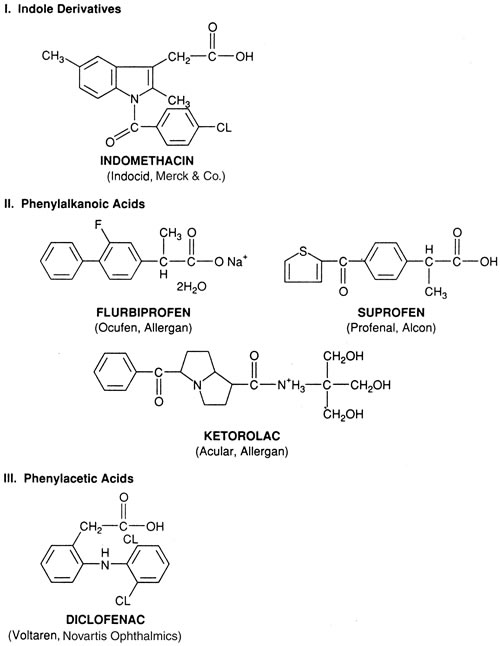
1. Goodman LS, Gilman A. Preface. In Hardman JG, Limbird LE, Gilman AG, eds. Goodman & Gilman's The Pharmacological Basis of Therapeutics, 10th ed. New York: McGraw-Hill, 2001:xxvii

2. Spector R, Roberts R, Vesell ES: Clinical pharmacology: A new specialty. Am J Med 70:221, 1981

3. Soine TO, Willette RE: Analgesic agents. In Wilson CC, Cisvoed O, eds. Textbook of Organic Medicinal and Pharmaceutical Chemistry, 4th ed. Philadelphia: JB Lippincott, 1962:592

4. Abrishami MA, Thomas J: Aspirin intolerance: A review. Ann Allergy 39:28, 1977

5. Roberts LJ, Morrow JD: Analgesic-antipyretics and anti-inflammatory agents: Drugs employed in the treatment of rheumatoid arthritis and gout. In Hardman JG, Limbird LE, Gilman AG, eds. Goodman & Gilman's The Pharmacological Basis of Therapeutics, 10th ed. New York: McGraw-Hill, 2001:687–732

6. Simon LS, Mills JA: Nonsteroidal anti-inflammatory drugs. N Engl J Med 302:1237, 1980

7. Day RO, Graham GG, William KM, et al: Clinical pharmacology of non-steroidal anti-inflammatory drugs. Pharmacol Ther 33:383, 1987

8. Magons K, Ball JP, Escale R, et al: Evidence for the existence of high affinity binding sites for indomethacin. Mol Pharmacol 29:39, 1985

9. Mochizuki M, Sawa M, Masuda K: Topical indomethacin in intracapsular extraction of senile cataract. Jpn J Ophthalmol 21:215, 1977

10. Brocks DR, Jamali F: Clinical pharmacokinetics of ketorolac tromethamine. Clin Pharmacokinet 23:415, 1992

11. Flurbiprofen—An ophthalmic NSAID. Med Lett Drugs Ther 29:58, 1987

12. Podos SM, Becker B: Comparison of ocular prostaglandin synthesis inhibitors. Invest Ophthalmol 15:841, 1976

13. Rooks WH, Maloney PJ, Shott LD, et al: The analgesic and anti-inflammatory profile of ketorolac and its tromethamine salt. Drugs Exp Clin Res 11:479, 1985

14. Skoutakis VA, Carter GA, Mickle TR, et al: Review of diclofenac and evaluation of its place in therapy as a nonsteroidal anti-inflammatory agent. Drug Intell Clin Pharm 22:850, 1988

15. Todd PA, Heel RC: Suprofen—A review of its pharmacodynamic and pharmacokinetic properties and analgesic efficacy. Drug Eval 30:514, 1985

16. Todd PA, Sarkin EM: Diclofenac sodium: A reappraisal of its pharmacodynamics and pharmacokinetic properties and therapeutic efficacy. Drugs 35:244, 1988

17. Benson MD, Aldo-Benson M, Brandt KD: Synovial fluid concentrations of diclofenac in patients with rheumatoid arthritis and osteoarthritis. Semin Arthritis Rheum 15:65, 1985

18. Buckley MMT, Brogden RN: Ketorolac. A review of its pharmacodynamic and pharmacokinetic properties and therapeutic potential. Drugs 39:86, 1990

19. Jallad NS, Garg DC, Martinez JJ, et al: Pharmacokinetics of single-dose oral and intramuscular ketorolac tromethamine in the young and elderly. J Clin Pharmacol 30:76, 1990

20. Tang-Lui DD, Liu SS, Weinkam RJ, et al: Ocular and systemic bioavailability of ophthalmic flurbiprofen. J Pharmacokinet Biopharm 12:611, 1984

21. Weintraub HS: Human acute pharmacokinetics and comparative bioavailability of suprofen from various dosage forms. Clin Res 26:595, 1978

22. Brune K, Glatt M, Graf P: Mini review: Mechanisms of action of anti-inflammatory drugs. Gen Pharmacol 7:27, 1976

23. Kaarela K, Lehtinen K, Makisara P, et al: Pharmacokinetics and tolerance of slow-released indomethacin tablets in rheumatoid arthritis. Eur J Clin Pharmacol 23:349, 1982

24. Huckey HB, Kwan KC, Duggan DE: Pharmacokinetics and metabolism of NSAIDs. Prog Drug Metab 5:165, 1980

25. Meffin PJ: The effect of renal dysfunction on the disposition of NSAIDs forming acyl-glucuronides. Agents Actions 17(suppl):85, 1985

26. Conquet PH, Plazonnet B, LeDourarec JC: Arachidonic acid-induced elevation of intraocular pressure and anti-inflammatory agents. Invest Ophthalmol 14:772, 1975

27. Hanna C, Sharp D: Ocular absorption of indomethacin by the rabbit. Arch Ophthalmol 88:196, 1972

28. Sanders DR, Kraff MC, Goldstick B: Aqueous penetration of oral and topical indomethacin in humans. Arch Ophthalmol 101:1614, 1983

29. Ling TL, Combs DL: Ocular bioavailability and tissue distribution of ketorolac tromethamine in rabbits. J Pharm Sci 76:289, 1987

30. Van Haeringen NJ: A comparison of the effects of nonsteroidal compounds on the disruption of the blood-aqueous barrier. Exp Eye Res 35:271, 1982

31. Miyake K, Sugiyama S, Noriamatsu I, et al: Prevention of cystoid macular edema after lens extraction by topical indomethacin—Radio-immunoassay measurement of prostaglandins in the aqueous during and after lens extraction. Albrecht von Graefes Arch Klin Exp Ophthalmol 209:83, 1978

32. Flower RJ, Blackwell GJ: Anti-inflammatory steroids induce biosynthesis of a phospholipase A2 inhibitor which prevents PG production. Nature 278:456, 1979

33. Mroszczak EJ, Ling T, Yee J, et al: Ketorolac absorption and pharmacokinetics in humans. Clin Pharmacol Ther 37:215, 1985

34. Fraunfelder FT: Extraocular fluid dynamics: How best to apply topical ocular medication. Am Ophthalmol Soc Trans 74:457, 1976

35. Zimmerman TJ, Kooner KS, Kandarakis AS: Improving the therapeutic index of topically applied ocular drugs. Arch Ophthalmol 102:551, 1984

36. Zipser RD, Laffi G: Prostaglandins, thromboxanes and leukotrienes in clinical medicine. West J Med 143:485, 1985

37. Ambache N: Irin, a smooth-muscle contracting substance present in rabbit iris. J Physiol 129:65, 1955

38. Eakins KE: Prostaglandins and prostaglandin synthetase inhibitors: Actions in ocular disease. In Robinson HJ, Vane JR, eds. Prostaglandin Synthetase Inhibitors. New York: Raven Press, 1974:343

39. Neufeld AH, Sears ML: Prostaglandins and the eye. Prostaglandins 4:157, 1973

40. Waitzman MB: Possible new concepts relating prostaglandins to various ocular functions. Surv Ophthalmol 14:301, 1970

41. Eakins KE, Whitelocke RAF, Bennett A, et al: Prostaglandin-like activity in ocular inflammation. Br Med J 3:452, 1972

42. Masuda K, Izawa Y, Mishima S: Prostaglandins and uveitis. Jpn J Ophthalmol 17:166, 1973

43. Abelson MB, Butrus SI, Weston JH: Aspirin therapy in vernal conjunctivitis. Am J Ophthalmol 95:502, 1983

44. Vane JR: Prostaglandins as mediators of inflammation. Prostaglandin Thromboxane Res 2:791, 1976

45. Boctor AM, Eickholt M, Pugsley TA: Meclofenamate is an inhibitor of both 5-lipoxygenase and cyclooxygenase in vitro. Prostaglandins Leukot Med 23:229, 1986

46. Siegel MI, McConnell RT, Porter NA: Aspirin-like drugs inhibit arachidonic acid metabolism via lipoxygenase and cyclo-oxygenase in rat neutrophils from carrageenan pleural exudates. Biochem Biophys Res Commun 92:688, 1980

47. Bonomi L, Perfetti S, Bellucci R, et al: Prevention of surgically induced sinuses by diclofenac eye drops. Ann Ophthalmol 19:142, 1987

48. Bomalaski JS, Hirata F, Clark MA: Aspirin inhibits phospholipase C. Biochem Biophys Res Commun 139:115, 1986

49. Ku EC, Lee W, Kothan HV, et al: Effect of diclofenac on rachidonic acid cascade. Am J Med 80(Suppl 4B):18, 1986

50. Abelson MB, Madrwale NA, Weston NH: The role of PGD2 in allergic ocular disease. In O'Connor GR, Chandler JM, eds. Advances in Immunology and Immunopathology of the Eye. New York: Masson, 1985:163–166

51. Abramson SB, Weissman G: The mechanisms of action of nonsteroidal anti-inflammatory drugs. Arthritis Rheum 32:1, 1989

52. Lee RF: The influence of psychotropic drugs on prostaglandin biosynthesis. Prostaglandins 5:63, 1974

53. Abramson S, Korchaktt LR, Edelson H, et al: Modes of action of aspirin-like drugs. Proc Natl Acad Sci USA 82:7227, 1985

54. Forrest M, Brooks PM: Mechanism of action of non-steroidal anti-rheumatic drugs. Baillieres Clin Rheumatol 2:275, 1988

55. Biemond P, Swaak AG, Penders JA, et al: Superoxide production by PMNs in rheumatoid arthritis and osteoarthritis. Ann Rheum Dis 45:249, 1986

56. Siegel M, McConnell R, Cuatrecasas A: Aspirin-like drugs interfere with arachidonate metabolism by inhibition of peroxidase activity of lipoxygenase pathway. Proc Natl Acad Sci USA 76:3774, 1979

57. Kim C, Tirama LA, Mann JD, et al: Saturable accumulation of an anion herbicide by rabbit choroid plexus. J Pharmacol Exp Ther 225:699, 1983

58. Herman JH, Appel AM, Khosia RC, et al: Cytokine modulation of chondrocyte metabolism. Inflammation 8:5125, 1984

59. Simchowitz L, Mehta J, Spilberg I: Chemotactic factor–induced generation of superoxide radicals by human neutrophils, effect of anti-inflammatory drugs. Arthritis Rheum 22:755, 1979

60. Perez HD, Elfman F, Marder S: Meclofenamate inhibits chemotactic factor–induced human PMN function. Arthritis Rheum 30:1023, 1987

61. Menta JO, Williams MD: Some NSAIDs inhibit the generation of superoxide anions by activated polymorphs by blocking ligand-receptor interactions. J Rheumatol 12:751, 1985

62. Silverstein FE, Graham DY, Senior JR, et al: Misoprostol reduced serious complications in patients with rheumatoid arthritis receiving nonsteroidal anti-inflammatory drugs. Ann Intern Med 123:241, 1995

63. Garcia L: Pain-free at last. Dallas Morning News. Sept 6, 1999:1C

64. Needleman P, Isakson PC: COX-1 and COX-2. J Rheumatol 24:6, 1997

65. Warner TD, Giuliano F, Vojnovic I, et al: Nonsteroid drug selectivities for cyclo-oxygenase-1 rather than clclooxygenase-2 are associated with human gastrointestinal toxicity. Proc Natl Acad Sci USA 96:7563, 1999.

66. Peterson WL, Cryer B: COX-1-Sparing NSAIDs-Is the enthusiasm justified? JAMA 2821961, 1999

67. Bombardier C, Laine L, Reicin A, et al: For VIGOR Study Group. Comparison of upper gastrointestinal toxicity of rofecoxib and naproxen in patients with rheumatoid arthritis. N Engl J Med 343:1520, 2000

68. Silverstein FE, Faich G, Goldstein JF, et al: Gastrointestinal toxicity with celecoxib vs NSAIDs for osteoarthritis and rheumatoid arthritis. The CLASS Study: A randomized controlled trial. JAMA 284:1247, 2000

69. Martinez B, Mathews AW, Lublin JS, et al: Merck pulls Vioxx from market after link to heart problems. Wall Street Journal October 1,2004:A1

70. Mukherjee D, Nissen SE, Topol EJ: Risk of cardiovascular events associated with selective COX-2 inhibitors. JAMA 286:954, 2001

71. Kloner DW: Relationship between cyclo-oxygenase-2 inhibition and thrombogenesis. J Cardiovasc Pharmacol Ther 9:51, 2004

72. Crofford LJ: Specific cyclo-oxygenase-2 inhibitors: What have we learned since they came into widespread clinical use? Curr Opin Rheumatol 13:225, 2002

73. Chiolero A, Maillard MP, Burnier M: Cardiovascular hazard of selective COX-2 inhibitors: myth or reality? Expert Opin Drug Saf 1:45, 2002

74. Hankey GJ, Eikelboom JW: Cyclooxygenase-2 inhibitors: Are they really atherothrombotic, and if not, why not? Stroke 34:2736, 2003

75. Proceeding of the National Academy of Science 10:1073, 2002

76. Duffin RM, Camras CB, Gardner SK: Inhibitors of surgically induced miosis. Ophthalmology 89:966, 1982

77. Camras CB, Miranda OC: The putative role of prostaglandins in surgical miosis. In Bito LZ, Stjernschantz J, eds. The Ocular Effects of Prostaglandins and Other Eicosanoids. New York: Alan R. Liss, 1989

78. Waitzman MB, King CD: Prostaglandin influences on intraocular pressure and pupil size. Am J Physiol 212:329, 1967

79. Kramer SG, Oyakawa RT, Drake M: Enhancement of pupillary dilation during intraocular surgery by prostaglandin inhibition. Invest Ophthalmol Vis Sci 15:63, 1976

80. Bito LZ: Species differences in the response of the eye to irritation and trauma: A hypothesis of divergence in ocular defense mechanisms, and the choice of experimental animals for eye research. Exp Eye Res 39:807, 1984

81. Mishima S, Masuda K: Clinical complications of prostaglandins and synthesis inhibitors. In Leopold IH, Burns RP, eds. Symposium on Ocular Therapy, Vol. 10. New York: John Wiley & Sons, 1977:1–19

82. ICL in Treatment of Myopia (ITM) Study Group: Postoperative inflammation after implantation of the implantable contact lens. Ophthalmology 110:2335, 2003

83. Dick HB, Schwenn O, Krummenauer F, et al: Inflammation after sclerocorneal versus clear corneal tunnel phacoemulsification. Ophthalmology 107:241, 2000

84. Dowler JGF, Hykin PG, Hamilton AMP: Phacoemulsification versus extracapsular cataract extraction in patients with diabetes. Ophthalmology 107:457, 2000

85. Friedlaender MH: Allergy and Immunology of the Eye. New York: Harper & Row, 1979

86. O'Connor GR: Corticosteroids and immuno-suppressives reviewed. In Srinivasan BD, ed. Ocular Therapeutics. New York: Masson, 1980:69

87. Dunne JA, Jacobs N, Morrison A, et al: Efficacy in anterior uveitis of two unknown steroids and topical tolmetin. Br J Ophthalmol 69:12, 1985

88. Hogan MJ, Kimura SJ, Thygeson P: Signs and symptoms of uveitis. Am J Ophthalmol 47:155, 1959

89. Yourston D, Whicher J, Chambers R, et al: The acute phase response in acute anterior uveitis. Trans Ophthalmol Soc UK 104:166, 1985

90. Fearnley IR, Spalton DJ, Smith SE: Anterior segment fluorophotometry in acute anterior uveitis. Arch Ophthalmol 105:1550, 1987

91. Flach AJ, Graham J, Kruger L, et al: Quantitative assessment of postsurgical breakdown of the blood aqueous barrier following administration of ketorolac tromethamine solution: A double-masked, paired comparison with vehicle-placebo solution study. Arch Ophthalmol 106:344, 1988

92. Mishima S, Tanishima T, Masuda K: Pathophysiology and pharmacology of intraocular surgery. Aust N Z J Ophthalmol 13:147, 1985

93. Podos SM. Discussion of nonsteroidal anti-inflammatory agents after argon laser trabeculoplasty. Ophthalmology 91:97, 1984

94. Sanders DR, Kraff MC: Steroidal and nonsteroidal anti-inflammatory agents. Effects on postsurgical inflammation and blood-aqueous barrier breakdown. Arch Ophthalmol 102:1453, 1984

95. Weinreb RN, Rohm AL, Baerveldt G, et al: Flurbiprofen pretreatment in argon laser trabeculoplasty for primary open angle glaucoma. Arch Ophthalmol 102:1629, 1984

96. Flach AJ, Stegman RC, Graham J: Prophylaxis of aphakic cystoid macular edema without corticosteroids. Ophthalmology 97:1253, 1990

97. Kuchle M, Naumann GOH: Intraocular inflammation after cataract surgery. Ophthalmology 110:632, 2003

98. Stjernschantz J: Autacoids and neuropeptides. In Sears ML (ed): Pharmacology of the Eye. New York: Springer-Verlag, 1984:311

99. Ma uger TF, Craig EL: Havener's Ocular Pharmacology, 6th ed. St. Louis: CV Mosby, 1994:364

100. Stewart R, Horwitz B, Howes J, et al: Double-masked, placebo-controlled evaluation of loteprednol etabonate 0.5% for postoperative inflammation. J Cataract Refract Surg 24:1480, 1998

101. The Loteprednol Etabonate Postoperative Inflammation Study Group 2: A double-masked, placebo-controlled evaluation of 0.5% loteprednol etabonate in the treatment of postoperative inflammation. Ophthalmology 105:1780, 1998

102. Fan DSP, Yu CBO, Chiu TYH: Ocular-hypertensive and anti-inflammatory response to rimexolone therapy in children. Arch Ophthalmol 121:1716, 2003

103. Floman N, Zor U. Mechanism of steroid action in ocular inflammation: Inhibition of prostaglandin production. Invest Ophthalmol 16:69, 1977

104. Kazuo S, Tsurufuji S: Mechanism of anti-inflammatory action of glucocorticoids: Re-evaluation of vascular constriction hypothesis. Br J Pharmacol 73:605, 1981

105. Stark WJ, Worthen DM, Halladay JT: The FDA report on intraocular lenses. Ophthalmology 90:311, 1983

106. Irvine AR: Cystoid maculopathy. Surv Ophthalmol 21:1, 1976

107. Jampol LM: Aphakic cystoid macular edema. A hypothesis. Arch Ophthalmol 103:1134, 1985

108. Miller D, Gruenberg P, Miller P, et al: Topical flurbiprofen or prednisolone: Effect on corneal wound healing in rabbits. Arch Ophthalmol 99:681, 1981

109. Yannuzzi LA: Perspective on the treatment of aphakic cystoid macular edema. Surv Ophthalmol 28(May Suppl):540, 1984

110. Flach AJ: The incidence, pathogenesis and treatment of cystoid macular edema following cataract surgery. Trans Am Ophth Soc 95:557, 1998

111. Cunha-Vaz JG, Travassos A: Breakdown of the blood-retinal barriers and cystoid macular edema. Surv Ophthalmol 28(May Suppl):485, 1984

112. Gass JDM, Norton EWD: Cystoid macular edema and papilledema following cataract extraction. Arch Ophthalmol 76:646, 1966

113. Irvine SR: A newly defined vitreous syndrome following cataract surgery: Interpreted according to recent concepts of the structure of the vitreous. Am J Ophthalmol 36:699, 1953

114. Martin WF, Green WR, Martin LW: Retinal phlebitis in the Irvine-Gass syndrome. Am J Ophthalmol 83:377, 1977

115. Norton AL, Brown WJ, Carson M: Pathogenesis of aphakic macular edema. Am J Ophthalmol 80:86, 1975

116. Obstbaum SA, Galen MA: Cystoid macular edema and ocular inflammation. The corneo-retinal inflammatory syndrome. Trans Ophthalmol Soc UK 99:187, 1979

117. Stark WJ, Maumenee AE, Fagadau W: Cystoid macular edema in pseudophakia. Surv Ophthalmol (May suppl) 28:442, 1984

118. Wilkinson CP: A long-term follow-up study of cystoid macular edema in aphakic and pseudophakic eyes. Trans Am Ophthalmol Soc 79:810, 1981

119. Sears ML: Aphakic cystoid macular edema: The pharmacology of ocular trauma. Surv Ophthalmol 28(May Suppl):525, 1984

120. Bonnet M, Bievelez B: Iris fluorescein angiography and Irvine-Gass syndrome. Albrecht von Graefes Arch Klin Exp Ophthalmol 213:187, 1980

121. Kondo T, Havashi M, Miki M: The results of intraocular lens implantation. The relationship between anterior segment fluorescein angiograph and cystoid macular edema. Acta Soc Ophthalmol Jpn 85:451, 1981

122. Kraff MC, Sanders DR, Jampol LM: Effect of an ultraviolet filtering intraocular lens on cystoid macular edema. Ophthalmology 92:366, 1985

123. Yamaaki H, Hendriks F, Deutman AF: Iris angiography after cataract extraction and the effect of indomethacin eye drops. Ophthalmologica 188:82, 1984

124. Kremer M, Baikoff G, Charbonnel B: The release of prostaglandins in human aqueous after intraocular surgery: Effect of indomethacin. Prostaglandins 23:695, 1982

125. Miyake K: Prostaglandins as a causative factor of the cystoid macular edema after lens extraction. Acta Soc Ophthalmol Jpn 81:1449, 1977

126. Worst JCF: Biotoxizitat des Kammerwassers. Klin Monatsbl Augenheilkd 167:376, 1975

127. Samuelsson B, Goldone M, Cranstrom E, et al: Prostaglandins and thromboxanes. Annu Rev Biochem 47:997, 1978

128. Eakins KE: Prostaglandin and non-prostaglandin mediated breakdown of the blood-aqueous barrier. Exp Eye Res 80(suppl):483, 1977

129. Kottow M, Hendrickson P: Iris angiography in cystoid macular edema after cataract extraction. Arch Ophthalmol 93:487, 1975

130. Neufeld AH, Jampol LM, Sears ML: Aspirin prevents the disruption of the blood-aqueous barrier in the rabbit eye. Nature 238:158, 1972

131. Bito LZ, Salvador EV: Intraocular fluid dynamics: The site of mechanism of prostaglandin transfer across the blood intraocular barriers. Exp Eye Res 14:233, 1972

132. Peyman GA, Sptiznas M, Straatsma BR: Chorioretinal diffusion of peroxidase before and after photocoagulation. Invest Ophthalmol 10:489, 1971

133. Gass JDM, Norton EWD: Follow-up of aphakic cystoid macular edema. Trans Am Acad Ophthalmol 73:665, 1969

134. Kaliner M, Eggleston PA, Mathews KP: Rhinitis and asthma. JAMA 258:2851, 1987

135. Leonardi A, Busato F, Fregona I, et al: Anti-inflammatory and antiallergic effects of ketorolac in the conjunctival provocation model. Br J Ophthalmol 84:1228, 2000

136. Okazaki T, Reisman RE, Arbesman CE: Prostaglandin E in the secretions of allergic rhinitis. Prostaglandins 13:681, 1977

137. Dhir SP, Garg SK, Sharma YR, et al: Prostaglandins in human tears. Am J Ophthalmol 87:403, 1979

138. Bhattacherjee P: The role of arachidonate metabolites in ocular inflammation. Prog Clin Biol Res 312:211, 1989

139. Ballas Z, Blumenthal M, Tinkelman D, et al: Clinical evaluation of ketorolac tromethamine 0.5% ophthalmic solution for the treatment of seasonal allergic conjunctivitis. Surv Ophthalmol 38(suppl):141, 1993

140. Tinkelman D, Rupp G, Kaufman H, et al: Ketorolac tromethamine 0.5% ophthalmic solution in treatment of seasonal allergic conjunctivitis. Surv Ophthalmol 38(suppl):133, 1993

141. Bazan HEP: The synthesis and effects of eicosanoids in avascular ocular tissues. In Bito LZ, Stjernschantz J, eds. Ocular Effects of Prostaglandins and Other Eicosanoids. New York: Alan R. Liss, 1989:73–84

142. Drews R, Knights K: Postulated reactive metabolites of NSAIDs. Agents Actions 17(suppl):127, 1985

143. Ferreira SH, Moncada S, Vane JR: Prostaglandins and the mechanism of analgesia, produced by aspirin-like drugs. Br J Pharmacol 49:86, 1973

144. Capitola JR, Rosenthal ME, Dubinski B, et al: Peripheral analgesics. A review. J Clin Pharmacol 23:545, 1983

145. Frucht-Pery J, Levinger S, Zaukerman H: The effect of topical administration of indomethacin on symptoms in corneal scars and edema. Am J Ophthalmol 112:186, 1991

146. Guzek JP, Holm M, Cotter JB, et al: Risk factors for intraoperative complications in 1000 extracapsular cataract cases. Ophthalmology 94:461, 1987

147. Summary Basis of Approval for Ocufen (Allergan's flurbiprofen) subsequent to New Drug Application 19-404. Washington, DC, Department of Health & Human Services, Food and Drug Administration, 1987

148. Summary Basis of Approval for Profenal (Alcon's suprofen) subsequent to New Drug Application 19-387. Washington, DC, Department of Health & Human Services, Food and Drug Administration, 1989

149. Keates RH, McGowan KA: Clinical trial of flurbiprofen to maintain pupillary dilation during cataract surgery. Ann Ophthalmol 16:919, 1984

150. Stark WJ, Fagadau WR, Stewart RH: Reduction of pupillary constriction during cataract surgery using suprofen. Arch Ophthalmol 104:364, 1986

151. Flach AJ: Cyclo-oxygenase inhibitors in ophthalmology. Surv Ophthalmol 36:259, 1992

152. Solomon KD, Turkalj JW, Whiteside SB, et al: Topical 0.5% ketorolac vs 0.03% flurbiprofen for inhibition of miosis during cataract surgery. Arch Ophthalmol 115:1119, 1997

153. Keates RH, McGowan KA: The effect of topical indomethacin in maintaining mydriasis during cataract surgery. Ann Ophthalmol 16:1116, 1984

154. Keulen-DeVos HC, van Rij JCG, deLavalette J, et al: Effect of indomethacin in preventing surgically induced miosis. Br J Ophthalmol 67:94, 1983

155. Gimbel H, Van Westenbrugge J, Chetham JK, et al: Intraocular availability and pupillary effect of flurbiprofen and indomethacin during cataract surgery. J Cataract Refract Surg 22:474, 1996

156. Gimbel HV: The effect of treatment with topical nonsteroidal anti-inflammatory drugs with and without intraoperative epinephrine on the maintenance of pupillary mydriasis during cataract surgery. Ophthalmology 96:585, 1989

157. Psilas K, Kalogeropoulos C, Loucatzicos E, et al: The effect of indomethacin, diclofenac, and flurbiprofen on the maintenance of mydriasis during extracapsular cataract extraction. Doc Ophthalmol 81:293, 1992

158. Sachdev MS, Mehta MR, Dada VK, et al: Pupillary dilatation during cataract surgery-relative efficacy of indomethacin and flurbiprofen. Ophthalmic Surg 21:557, 1990

159. Bito LZ: Surgical miosis: Have we been misled by a bunch of rabbits? Ophthalmology 97:1, 1990

160. Katz IM: Indomethacin. Ophthalmology 88:455, 1987

161. Sawa M, Masuda K: Topical indomethacin in soft cataract aspiration. Jpn J Ophthalmol 20:514, 1976

162. Roberts CW: Pretreatment with topical diclofenac sodium to decrease postoperative inflammation. Ophthalmology 103:636, 1996

163. Eguchi K, Ohara K, Tobari I, et al: Effects of topical indomethacin on suppression of inflammation after ocular surgeries—Double masked study of anti-inflammatory effects on the intracapsular lens extraction. Acta Soc Ophthalmol Jpn 86:2198, 1982

164. Araie M, Sawa M, Takase M: Effect of topical indomethacin on the blood aqueous barrier after intracapsular extraction of senile cataract—A fluorophotometric study. Jpn J Ophthalmol 25:237, 1981

165. Sanders DR, Kraff MC, Lieberman HL, et al: Breakdown and reestablishment of the blood aqueous barrier following implant surgery. Arch Ophthalmol 100:588, 1982

166. Garner A, Klintworth GK, eds: Pathobiology of Ocular Disease. New York: Marcel Dekker, 1982:59–89

167. Swan KC, Hart WM: A comparative study of the effects of Mecholyl, Doryl, Eserine, pilocarpine, atropine, and epinephrine on the blood-aqueous barriers. Am J Ophthalmol 23:1311, 1940

168. Van Alphen G, Macri FJ: Entrance of fluorescein into the aqueous humor of cat eye. Arch Ophthalmol 75:247, 1966

169. Adler FH: The action of atropine in ocular inflammations. Arch Ophthalmol 55:484, 1926

170. Novack GD, Leopold IH: The blood-aqueous and blood-brain barriers to permeability. Am J Ophthalmol 705:412, 1988

171. Miyake K, Kensaku K, Asakura M, et al: Consensual reactions of human blood-aqueous barrier to implant operations. Arch Ophthalmol 102:558, 1984

172. Albert DM, Diaz-Rohena R: A historical review of sympathetic ophthalmia and its epidemiology. Surv Ophthalmol 34:1, 1989

173. Kwito GM, Shin DH, Ahn RH, et al: Bilateral effects of long-term monocular timolol therapy. Am J Ophthalmol 104:591, 1987

174. Freddo TF, Barsotti M, Bartels SP, et al: Combined aqueous fluorophotometric and tracer localization studies of owl monkeys with endotoxin induced uveitis. Invest Ophthalmol 30:375, 1989

175. Hara Y, Yuasa T: Disturbances of the blood-ocular barrier in uveitis. Invest Ophthalmol 30:450, 1989

176. Komuro Y, Matsumoto S, Shirato S, et al: A new apparatus for automatic counting of aqueous floaters. Acta Soc Ophthalmol Jpn 89:556, 1985

177. Sawa M, Tsurimka , Tsuru T, et al: New quantitative method to determine protein and cells in aqueous in vivo. Jpn J Ophthalmol 32:132, 1988

178. DeBeijer-Dominicus JA, Van Rij G, DeLavalette JCC: Indomethacin eye drops and the postoperative irritation after extracapsular cataract extraction combined with intraocular lens implantation. Doc Ophthalmol 56:167, 1983

179. Kraff MC, Martin RG, Neumann AC, et al: Efficacy of diclofenac sodium ophthalmic solution versus placebo in reducing inflammation following cataract extraction and posterior chamber lens implantation. J Cataract Refract Surg 20:138, 1994

180. Goa KL, Chrisp P: Ocular diclofenac. Drugs Aging 2:473, 1992

181. Vickers FF, McGuigan LJB, Ford C, et al: The effect of diclofenac sodium ophthalmic on the treatment of postoperative inflammation [Abstract]. Invest Ophthalmol Vis Sci 32(suppl):793, 1991

182. Ronen S, Rozenman Y, Zylbermann R, et al: Treatment of ocular inflammation with diclofenac: Double blind trial following cataract surgery. Ann Ophthalmol 17:577, 1985

183. Noonan WD, Samples JR: New drugs in ophthalmology: Diclofenac sodium. J Toxicol Cut Ocular Toxicol 12:265, 1993

184. Flach AJ, Lavelle CJ, Olander KW, et al: The effect of ketorolac 0.5% solution in reducing postsurgical inflammation following ECCE with IOL: Double-masked, parallel comparison with vehicle. Ophthalmology 5:1279, 1988

185. Solomon KD, Cheetham JK, DeGryse R, et al: Topical ketorolac tromethamine 0.5% ophthalmic solution in ocular inflammation after cataract surgery. Ophthalmology 108:331, 2001.

186. Heier J, Cheetham JK, DeGryse R, et al: Ketorolac tromethamine 0.5% ophthalmic solution in the treatment of moderate to severe ocular inflammation after cataract surgery: A randomized, vehicle-controlled clinical trial. Am J Ophthalmol 127:253, 1999

187. Flach AJ, Dolan BJ, Donahue ME, et al: Comparative effects of ketorolac 0.5% or diclofenac 0.1% ophthalmic solutions on inflammation after cataract surgery. Ophthalmology 105:1775, 1998

188. Sabiston MB, Tessler D, Sumersk H, et al: Reduction of inflammation following cataract surgery by flurbiprofen. Ophthalmic Surg 18:873, 1987

189. Diestelhorst M, Aspacher F, Konen W, et al: The effect of flurbiprofen 0.03% eye drops on the blood aqueous barrier in extracapsular cataract extraction with IOL implantation. Int Ophthalmol 15:69, 1991

190. Blaydes JE, Kelley EP, Walt JG, et al: Flurbiprofen 0.03% for the control of inflammation following cataract extraction by phacoemulsification. J Cataract Refract Surg 22:481, 1993

191. Rossetti L, Bujtar E, Castoldi D, et al: Effectiveness of diclofenac eyedrops in reducing inflammation and the incidence of cystoid macular edema after cataract surgery. J Cataract Refract Surg 22:794, 1996

192. Italian Diclofenac Study Group. Efficacy of diclofenac eyedrops in preventing postoperative inflammation and long-term cystoid macular edema. J Cataract Refract Surg 23:1183, 1997

193. Kraff MC, Sanders DR, McGuigan L, et al: Inhibition of blood-aqueous humor barrier breakdown with diclofenac: A fluorophotometric study. Arch Ophthalmol 108:380, 1990

194. Alpar JJ, McCuigan L, Alpar AJ, et al: Effect of diclofenac sodium ophthalmic 0.1% on inflammation following cataract extraction: A fluorophotometry study. Ophthalmology 96(suppl):109, 1989

195. Flach AJ, Jaffe NS, Akers WA, et al: The effect of ketorolac 0.5% solution in reducing postoperative inflammation: Double mask parallel comparison with dexamethasone phosphate 0.1% solution. Ann Ophthalmol 21:407, 1989

196. Flach AJ, Kraff MC, Sanders DR, et al: The quantitative effect of 0.5% ketorolac tromethamine solution and dexamethasone phosphate 0.1% solution on postsurgical blood aqueous barrier. Arch Ophthalmol 106:480, 1988

197. Othenin-Girard P, Tritten J, Pittet N, et al: Dexamethasone versus diclofenac sodium eyedrops to treat inflammation after cataract surgery. J Cataract Refract Surg 20:9, 1994

198. Roberts CW, Brennan K: A comparison of topical diclofenac with prednisolone for postoperative inflammation after cataract surgery. Arch Ophthalmol 113:725, 1995

199. El-Harazi SM, Ruiz RS, Feldman RM, et al: A randomized double-masked trial comparing ketorolac tromethamine 0.5%, diclofenac sodium 0.1%, and prednisolone acetate 1% in reducing post-phacoemulsification flare and cells. Ophthalmic Surg Lasers 29:539, 1998

200. Simone JN, Pendelton RA, Jenkins JE: Comparison of the efficacy and safety of ketorolac tromethamine 0.5% and prednisolone acetate 1% after cataract surgery. J Cataract Refract Surg 25:699, 1999

201. Ostrov CS, Sirkin SR, Deutsch WE, et al: Ketorolac, prednisolone and dexamethasone for postoperative inflammation. Clin Ther 19:259, 1997

202. Diestelhorst M, Aspacher F, Konen W, et al: Effect of dexamethasone 0.1% and prednisolone acetate 1% eye drops on the blood-aqueous barrier after cataract surgery: A controlled randomized fluorophotometric study. Graefe's Arch Clin Exp Ophthalmol 230:451, 1992

203. Diestelhorst M, Schmidl B, Konen W, et al: Efficacy and tolerance of diclofenac sodium 0.1%, flurbiprofen 0.03%, and indomethacin 1.)% in controlling postoperative inflammation. J Cataract Refract Surg 22:788, 1996

204. Samiy N and Foster CS: The role of nonsteroidal anti-inflammatory drugs in ocular inflammation. Int Ophthalmol Clin 36:195, 1996

205. Hotchkiss IL, Robin AL, Pollack IP, et al: Nonsteroidal anti-inflammatory agents after argon laser trabeculoplasty: Flurbiprofen and indomethacin. Ophthalmology 91:969, 1984

206. Levine SB, Leopold IH: Advances in ocular corticosteroid therapy. Med Clin North Am 57:1167, 1973

207. Burde RM, Waltman SR: Topical corticosteroids after cataract surgery. Ann Ophthalmol 4:290, 1972

208. Corby JM: Corticosteroid therapy for the reduction of postoperative inflammation after cataract extraction. Am J Ophthalmol 82:923, 1976

209. Diamond JG, Kaplan HJ: Lensectomy and vitrectomy for complicated cataract secondary to uveitis. Arch Ophthalmol 96:1798, 1978

210. Huang K: Indomethacin inhibition of prostaglandin mediated inflammation following intraocular surgery. Invest Ophthalmol 16:760, 1977

211. Kaplan HJ, Diamond JG, Brown SA: Vitrectomy in experimental uveitis. Arch Ophthalmol 97:336, 1979

212. Knopf MM: A double-blind study of fluorometholone. Am J Ophthalmol 69:739, 1970

213. Mustaka A, Kaufman HE, Johnston G, et al: Corticosteroid efficacy in postoperative uveitis. Ann Ophthalmol 6:719, 1993

214. Assil KK, Massry G, Lehmann R, et al: Control of ocular inflammation after cataract extraction with rimexolone 1% ophthalmic suspension. J Cataract Refract Surg 23:750, 1997

215. Hoskins HD, Hetherington J, Minckler DS, et al: Complications of laser trabeculoplasty. Ophthalmology 90:796, 1983

216. Hurvitz LM, Spaeth GL, Zakhour I, et al: Comparison of the effect of flurbiprofen, dexamethasone and placebo on cyclocryotherapy-induced inflammation. Ophthalmic Surg 15:394, 1984

217. Mermond A, Pittet N, Herbort CP: Inflammation patterns after laser trabeculoplasty measured with the laser flare meter. Arch Ophthalmol 110:368, 1992

218. Herbort CP, Mermond A, Schnyder C, et al: Anti-inflammatory effect of diclofenac drops after argon laser trabeculoplasty. Arch Ophthalmol 111:481, 1993

219. Goethals M, The Belgian Study Group on Glaucoma: Efficacy and safety of indomethacin 0.1% versus flurbiprofen 0.03% eyedrops in inflammation after argon laser trabeculoplasty. Documenta Ophthalmologica 85:287, 1994

220. Diestelhorst M, Thull D and Krieglstein GK: The effect of argon laser trabeculoplasty on the blood-aqueous barrier and intraocular pressure in human glaucomatous eyes treated with diclofenac 0.1%. Graefe's Arch Clin Exp Ophthalmol 233:559, 1995

221. Cox SN, Hay E, Bird AC: Treatment of chronic macular edema with acetazolamide. Arch Ophthalmol 106:1190, 1988

222. Jampol LM, Sanders DR, Kraff MC: Prophylaxis and therapy of aphakic cystoid macular edema. Surv Ophthalmol 28(suppl):535, 1984

223. Jacobson DR, Dellaporta A: Natural history of cystoid macular edema after cataract extraction. Am J Ophthalmol 77:445, 1974

224. Drolsum L, Haaskjold E: Causes of decreased visual acuity after cataract extraction. J Cataract Refract Surg 21:59, 1995

225. Henkind P, ed: The First International Cystoid Macular Edema Symposium. Surv Ophthalmol 28(suppl):431, 1984

226. Jampol LM: Pharmacologic therapy of aphakic and pseudophakic cystoid macular edema: 1985 update. Ophthalmology 92:807, 1985

227. Jampol LM: Pharmacologic therapy of aphakic cystoid macular edema: A review. Ophthalmology 89:891, 1982

228. Miyake K: Indomethacin in the treatment of postoperative cystoid macular edema. Surv Ophthalmol 78(suppl):554, 1984

229. Rossetti L, Chaudhuri J, Dickersin K: Medical prophylaxis and treatment of cystoid macular edema after cataract surgery. Ophthalmology 105:397, 1998

230. Koay P: The emerging roles of topical non-steroidal anti-inflammatory agents in ophthalmology. Br J Ophthalmol 80:480, 1996

231. Rocha G, Deschenes J: Pathophysiology and treatment of cystoid macular edema. Can J Ophthalmol 31:282, 1996

232. Fechner PU: Die prophylaxe des zystoiden Makulaodems mit indomethacin–Augentropfen. Klin Monatsbl Augenheilkd 180:169, 1980

233. Hollwich F, Jocobi K, Kuchle HJ, et al: Zur prophylaxe des zystoiden makulaodems mit Indomethacin-Augentropfen. Klin Monatsbl Augenheilkd 183:477, 1983

234. Klein RM, Katzin HM, Yannuzzi LA: The effect of indomethacin pretreatment on aphakic cystoid macular edema. Am J Ophthalmol 87:487, 1979

235. Kraff MC, Sanders DR, Jampol LM, et al: Prophylaxis of pseudophakic cystoid macular edema with topical indomethacin. Ophthalmology 89:885, 1982

236. Miyake K: Prevention of cystoid macular edema after lens extraction by topical indomethacin. II. A control study in bilateral extractions. Jpn J Ophthalmol 2:80, 1978

237. Miyake K: Prevention of cystoid macular edema after lens extraction by topical indomethacin. Albrecht von Graefes Arch Klin Ophthalmol 203:81, 1977

238. Miyake K, Miyake Y, Makubo K, et al: Incidence of cystoid macular edema after retinal detachment surgery and the use of topical indomethacin. Am J Ophthalmol 95:451, 1983

239. Miyake K, Sakamura S, Miura H: Long-term follow-up study on the prevention of aphakic cystoid macular edema by topical indomethacin. Br J Ophthalmol 64:324, 1980

240. Sholiton DB, Reinhart WJ, Frank KE: Indomethacin as a means of preventing cystoid macular edema following intracapsular cataract surgery. Am Intraocular Implant Soc J 5:137, 1979

241. Tanake N, Saga Y, Hara K: Effect of topical indomethacin in treatment for cystoid macular edema after lens extraction. Folia Ophthalmol Jpn 34:178, 1983

242. Tennant JL: Is cystoid macular edema reversible by oral indomethacin, yes or no? In Emery JM, Paton D, eds. Current Concepts in Cataract Surgery. Selected Proceedings of the Fourth Biannual Cataract Surgical Congress. St. Louis: CV Mosby, 1976:310–312

243. Yannuzzi LA, Landau AN, Turtz AL: Incidence of aphakic cystoid macular edema with the use of topical indomethacin. Ophthalmology 88:947, 1981

244. Yannuzzi LA, Wallyn RH: Cystoid macular edema. A controlled treatment study utilizing indomethacin. In Emery JM, Paton D, eds. Current Concepts in Cataract Surgery. Selected Proceedings of the Fourth Biannual Cataract Surgical Congress. St. Louis: CV Mosby, 1976:313–316

245. Mizuno K, Miyake K: Pre- and postoperative therapy in cataract extraction. Ophthalmic Surg 18:143, 1987

246. Solomon LD: Flurbiprofen-CME Study Group I. Efficacy of topical flurbiprofen and indomethacin in preventing pseudophakic cystoid macular edema. J Cataract Refract Surg 21:73, 1995

247. Ibanez HE, Lesher MP, Singerman LJ, et al: Prospective evaluation of the effect of pseudophakic cystoid macula edema on contrast sensitivity. Arch Ophthalmol 111:1635, 1993

248. Waitzman MB: Topical indomethacin in treatment and prevention of intraocular inflammation. Ann Ophthalmol 11:489, 1979

249. Heier JS, Topping TM, Baumann W, et al: Ketorolac versus prednisolone versus combination therapy in the treatment of acute pseudophakic cystoid macular edema. Ophthalmology 107:2034, 2000

250. Yannuzzi LA, Klein RM, Wallyn RH: Ineffectiveness of indomethacin in the treatment of chronic cystoid macular edema. Am J Ophthalmol 84:517, 1977

251. Burnett J, Tessler H, Isenberg S, et al: Double-masked trial of fenoprofen sodium: Treatment of chronic aphakic cystoid macular edema. Ophthalmic Surg 14:150, 1983

252. Flach AJ, Dolan BJ, Irvine AR: Effectiveness of ketorolac 0.5% solution for chronic aphakic and pseudophakic cystoid macular edema. Am J Ophthalmol 103:479, 1987

253. Flach AJ, Jampol LM, Yannuzzi LA: Improvement in visual acuity in chronic aphakic and pseudophakic cystoid macular edema after treatment with topical 0.5% ketorolac tromethamine. Am J Ophthalmol 112:514, 1991

254. Epstein DL: Cystoid macular edema occurring 13 years after cataract extraction. Am J Ophthalmol 83:501, 1977

255. Weisz JM, Bressler NM, Bressler SB, et al: Ketorolac treatment of pseudophakic cystoid macular edema identified more than 24 months after cataract extraction. Ophthalmology 106:1656, 1999

256. Severin SL: Late cystoid macular edema in pseudophakia. Am J Ophthalmol 90:223, 1980

257. Flach AJ: Discussion of Ketorolac versus prednisolone versus combination therapy in the treatment of acute pseudophakic cystoid macular edema. Ophthalmology 107:2039, 2000

258. Smith RT, Campbell CJ, Koester CJ: The barrier function in extracapsular cataract surgery. Ophthalmology 97:90, 1990

259. Sorr EM, Everett WH, Hurite FC: Incidence of fluorescein angiographic subclinical macular edema following phacoemulsification of senile cataracts. Ophthalmology 86:201, 1979

260. Wetzig PC, Thatcher DB, Christiansen JM: The intracapsular versus extracapsular cataract technique in relationship to retinal problems. Trans Am Ophthalmol Soc 77:339, 1979

261. Gilbard SM, Peyman GA, Goldberg MF: Evaluation for cystoid maculopathy after pars plicata lensectomy-vitrectomy for congenital cataracts. Ophthalmology 90:1201, 1983

262. Hoyt CS, Nickel B: Aphakic cystoid macular edema: Occurrence in infants and children after transpupillary lensectomy and anterior vitrectomy. Arch Ophthalmol 100:746, 1982

263. Poer DV, Helveston EM, Ellis FD: Aphakic cystoid macular edema in children. Arch Ophthalmol 99:249, 1981

264. Stern AL, Taylor DM, Dalburg LA, et al: Pseudophakic cystoid maculopathy—Study of 50 cases. Ophthalmology 88:942, 1981

265. Federman JL, Annesley WH Jr, Sarin LK: Vitrectomy and cystoid macular edema. Ophthalmology 87:622, 1980

266. Fung WE: Surgical therapy for chronic aphakic cystoid macular edema. Ophthalmology 89:898, 1982

267. Fung WE: Anterior vitrectomy for chronic aphakic cystoid macular edema. Ophthalmology 87:189, 1980

268. Katzen LY, Fleischman J, Trokel S: YAG laser treatment of cystoid macular edema. Am J Ophthalmol 95:589, 1983

269. Pfoff DS, Thom SR: Cystoid macular edema treated with hyperbaric oxygen. Ophthalmology 97(suppl):120, 1990

270. Bito LZ, Nichols R, Siminoff S: Ocular effects of substance P and other biologically active peptides as compared to prostaglandins E2, (PGE2). Proc Int Soc Eye Res 1:53, 1980

271. Pendergast SD, Marcherio RR, Williams GA, et al: Vitrectomy for chronic pseudophakic cystoid macular edema. Am J Ophthalmol 128:317, 1999

272. Wilkinson CP: Vitrectomy for chronic pseudophakic cystoid macular edema. Am J Ophthalmol 129:560, 2000.

273. Peyman GA, Canakis C, Livir-Rallatos C, et al: The effect of internal limiting membrane peeling on chronic recalcitrant pseudophakic cystoid macular edema: A report of two cases. Am J Ophthalmol 133:571, 2002

274. Kobayashi H, Kobayashi K, Okinami S: Randomized clinical trial of topical betaxolol for persistent macular edema after vitrectomy and epiretinal membrane removal. Am J Ophthalmol 136:244, 2003

275. Tach AB, Dugel PU, Flindall RJ, et al: A comparison of retrobulbar versus sub-tenon's corticosteroid therapy for cystoid macular edema refractory to topical medications. Ophthalmology 104:2003, 1997

276. Abe T, Hayasaka S, Nagaki Y, et al: Pseudophakic cystoid macular edema treated with high-dose intravenous methylprednisolone. J Cataract Refract Surg 25:1286, 1999

277. Benhamou N, Massin P, Haouchine B, et al: Intravitreal triamcinolone for refractory pseudophakic macular edema. Am J Ophthalmol 135:246, 2003

278. Jonas JB, Kreissig I, Degenring RF: Intravitreal triamcinolone acetonide for pseudophakic cystoid macular edema. Am J Ophthalmol 136:384, 2003

279. Jonas JB, Kreissig I, Degenring RF: Secondary chronic open-angle glaucoma after intravitreal triamcinolone acetonide. Arch Ophthalmol 121:729, 2003

280. Friedlaender MH: Conjunctivitis of allergic origin: Clinical presentation and differential diagnosis. Surv Ophthalmol 38(suppl):105, 1993

281. Jackson WB: Differentiating conjunctivitis of diverse origins. Surv Ophthalmol 38(suppl):91, 1993

282. Leopold IH: Advances in ocular therapy noncorticosteroid anti-inflammatory agents. Am J Ophthalmol 78:759, 1974

283. Greaves MW, McDonald W: Itch: Role of prostaglandins. Br Med J 22:608, 1973

284. Pakes GE, Brogden RN, Heel RD, et al: Flunisolide: A review. Drugs 19:397, 1980

285. Meyer E, Kraus E, Zonis S: Efficacy of antiprostaglandin therapy in vernal conjunctivitis. Br J Ophthalmol 71:497, 1987

286. Laibovitz RA, Koester J, Schaich L, et al: Safety and efficacy of diclofenac sodium 0.1% ophthtalmic solution in acute seasonal allergic conjunctivitis. J Ocular Pharm Ther 11:361, 1995

287. Lewis RA, Roberts LJ, Holgate ST, et al: Immunologic generation of mast cell prostaglandins. J Allergy Clin Immunol 65:235, 1980

288. Gupta S, Khurang AK, Ahluwalia BK, et al: Topical indomethacin for vernal conjunctivitis. Acta Ophthalmol (Copenh) 69:95, 1991

289. Brooks CD, Nelson AL, Metzler C: Effect of flurbiprofen on induced allergic rhinitis. J Allergy Clin Immunol 73:584, 1984

290. Brooks CD, Karl KJ: Hay fever treatment in the combined antihistamine and cyclooxygenase-inhibiting drugs. J Allergy Clin Immunol 81:1110, 1988

291. Brooks CD, Nelson AL, Metzler C: Treatment of ragweed hay fever with flurbiprofen. Ann Allergy 55:557, 1985

292. Abelson MB, Schaefer K: Conjunctivitis of allergic origin: Immunologic mechanisms and current approaches to therapy. Surv Ophthalmol 38(suppl):115, 1993

293. Bishop K, Abelson MB, Cheetham J, et al: Evaluation of flurbiprofen in the treatment of antigen-induced allergic conjunctivitis. Invest Ophthalmol Vis Sci 31:487, 1990

294. Allansmith MR, Ross RN: Ocular allergy and mast cell stabilizers. Surv Ophthalmol 30:229, 1986

295. Wood TS, Stewart RH, Bowman RW, et al: Suprofen treatment of GPC. Ophthalmology 95:822, 1988

296. Seitz B, Sorken K, LaBree LD, et al: Corneal sensitivity and burning sensation-Comparing topical ketorolac and diclofenac. Arch Ophthalmol 114:921, 1996

297. Zaidman GW: Diclofenac and its effect on corneal sensation. Arch Ophthalmol 113:262, 1995.

298. Szerenyi K, Sorken K, Garbus JJ, et al: Decrease in normal human corneal sensitivity with topical diclofenac sodium. Am J Ophthalmol 118:312, 1994

299. Phillips AF, Szerenyi K, Campos M, et al: Arachidonic acid metabolites after excimer laser corneal surgery. Arch Ophthalmol 111:1273, 1993

300. Eiferman RA, Hoffman RS, Sher NA: Topical diclofenac reduces pain following photorefractive keratectomy. Arch Ophthalmol 111:1022, 1993

301. Gwon A, Vaughan ER, Cheetham JK, et al: Ocufen (flurbiprofen) in the treatment of ocular pain after radial keratotomy. CLAO J 20:2, 1994

302. Sher NA, Frantz JM, Talley A, et al: Topical diclofenac in the treatment of ocular pain after excimer photorefractive keratectomy. Refract Corneal Surg 9:425, 1993

303. Cherry PMH, Tutton MK, Adhikary H, et al: The treatment of pain following photorefractive keratectomy. J Cataract Refract Surg 10:S222, 1994

304. Yee RW, The Ketorolac Radial Keratotomy Study Group: Analgesic efficacy and safety of nonpreserved ketorolac tromethamine ophthalmic solution following radial keratotomy. Am J Ophthalmol 125:472, 1998

305. Stein R, Stein H, Cheskes A, et al: Photorefractive keratectomy and postoperative pain. Am J Ophthalmol 117:403, 1994

306. Arshinoff S, D'Addario D, Sadler C, et al: Use of topical nonsteroidal anti-inflammatory drugs in excimer laser photorefractive keratectomy. J Cataract Refract Surg 20:216, 1994

307. Maguen E, Salz JJ, Nesburn AB, et al: Results of excimer laser photorefractive keratectomy for the correction of myopia. Ophthalmology 101:1548, 1994

308. Epstein RL, Laurence EP: Effect of topical diclofenac solution on discomfort after radial keratotomy. J Cataract Refract Surg 20:378, 1994

309. Sher NA, Frantz JM, Talley A, et al: Topical diclofenac in the treatment of ocular pain after excimer photorefractive keratectomy. Refract Corneal Surg 9:425, 1993

310. Epstein RL, Laurence EP: Relative effectiveness of topical ketorolac and topical diclofenac on discomfort after radial keratotomy. J Cataract Refract Surg 21:156, 1995

311. Kaiser PK, Pineda R, Corneal Abrasion Patching Study Group: A study of topical nonsteroidal anti-inflammatory drops and no pressure patching in the treatment of corneal abrasions. Ophthalmology 104:1353, 1997

312. Donnenfeld ED, Selkin BA, Perry HD, et al: Controlled evaluation of a bandage contact lens and a topical nonsteroidal anti-inflammatory drug in treating traumatic corneal abrasions. Ophthalmology 102:9790, 1995

313. Salz JJ, Reader AL, Schwartz LJ, et al: Treatment of corneal abrasions with soft contact lenses and topical diclofenac. J Refract Corneal Surg 10:640, 1994

314. Verma S, Corbett MC, Marshall J: A prospective, randomized double-masked trial to evaluate the role of topical anesthetics in controlling pain after photorefractive keratectomy. Ophthalmology 102:1918, 1995

315. Rosenwasser GOD, Holland S, Pflugfelder SC, et al: Topical anesthetic abuse. Ophthalmology 97:967, 1990

316. Dornic DI, Thomas JM, Lass JH: Topical diclofenac sodium in the management of anesthetic abuse keratopathy. Am J Ophthalmol 125:719, 1998

317. Fry LL: Efficacy of diclofenac sodium solution in reducing discomfort after cataract surgery. J Cataract Refract Surg 21:187, 1995

318. Apt L, Voo I, Isenberg SJ: A randomized clinical trial of the nonsteroidal eyedrop diclofenac after strabismus surgery. Ophthalmology 105:1448, 1998

319. Morton WS, Benham SW, Lawson RA, et al: Diclofenac vs oxybuprocaine eye drops for analgesia in pediatric strabismus surgery. Paediatr Anaesth 7:221, 1997

320. Apple DJ, Solomon KD, Tetz MR, et al: Posterior capsule opacification. Surv Ophthalmol 37:73, 1992

321. Nishi O, Nishi K, Fujiwara T, et al: Effects of diclofenac sodium and indomethacin on proliferation and collagen synthesis of lens epithelial cells in vitro. J Cataract Refract Surg 21:461, 1995

322. Flach AJ: Incidence of postoperative posterior capsular opacification following treatment with diclofenac 0.1% and ketorolac 0.5% ophthalmic solutions: 3-year randomized, double-masked, prospective clinical investigation. Trans Am Ophth Soc 98:101, 2000

323. Snir M, Axer-Siegel R, Friling R: Efficacy of diclofenac versus dexamethasone for treatment after strabismus surgery. Ophthalmology 107:1884, 2000

324. Apt L, Voo I, Isenberg SJ: A randomized clinical trial of the nonsteroidal eyedrop diclofenac after strabismus surgery. Ophthalmology 105:1448, 1998

325. Wright M, Butt Z, McIlwaine G, et al: Comparison of the efficacy of diclofenac and betamethasone following strabismus surgery. Br J Ophthalmol 81:299, 1997

326. Morton NS, Benham SW, Lawson RA, et al: Diclofenac vs oxybuprocaine eye drops for analgesia in paediatric strabismus surgery. Paediatr Anesth 7:221, 1997

327. Gordon YJ, Araullo-Cruz T, Romanowski EG: The effects of topical nonsteroidal anti-inflammatory drugs on adenoviral replication. Arch Ophthalmol 116:900, 1998

328. Shiuey Y, Ambati BK, Adamis AP, et al: A randomized, double-masked trial of topical ketorolac versus artificial tears for treatment of viral conjunctivitis. Ophthalmology 107:1512, 2000

329. Gartry DS, Kerr Muir MG, Marshall J: Excimer laser photorefractive keratectomy: 18-month follow-up. Ophthalmology 99:1209, 1992

330. Tuft Sj, Gartry DS, Rawe IM, et al: Photorefractive keratectomy: implications of corneal wound healing. Br J Ophthalmol 77:243, 1993

331. Gartry DS, Kerr Muir MG, Marshall J: The effect of topical corticosteroids on refraction and corneal haze following excimer laser treatment of myopia: an update. Eye 7:584, 1993

332. Sher NA, Krueger RR, Teal P, et al: Role of topical steroids, corticosteroids and nonsteroidal anti-inflammatory drugs in the etiology of stromal infiltrates after excimer photorefractive keratectomy. J Refract Corneal Surg 10:587, 1994

333. Nassaralla BA, Szerenyi K, Wang XW, et al: Effect of diclofenac on corneal haze after photorefractive keratectomy in rabbits. Ophthalmology 102:469, 1995

334. O'Brart DPS, Lohmann CP, Klonos G, et al: The effects of topical corticosteroids and plasmin inhibitors on refractive outcome, haze, and visual performance after photorefractive keratectomy. Ophthalmology 101:1565, 1994

335. Talamo JH, Gollamudi S, Green WR, et al: Modulation of corneal wound healing after excimer laser keratomileusis using topical mitomycin C and steroids. Arch Ophthalmol 109:1141, 1991

336. Morlet N, Gillies MC, Crouch R, et al: Effect of topical interferon alpha 2 beta on corneal haze after excimer laser photorefractive keratectomy in rabbits. Refract Corneal Surg 9:443, 1993

337. Schaumberg DA, Sullivan DA, Dana MR: Epidemiology of dry eye syndrome. Adv Exp Med Biol 506:989, 2002

338. Avunduk AM, Avunduk MC, Varnell ED, et al: The comparison of efficacies of topical corticosteroids and nonsteroidal anti-inflammatory drops on dry eye patients. Am J Ophthalmol 136:593, 2003

339. Chan CKM, Lam DSC: The comparison of efficacies of corticosteroids and nonsteroidal anti-inflammatory drops on dry eye patients. Am J Ophthalmol 137:1157, 2004

340. Sall K, Stevenson OD, Mundorf , et al: Two multicenter, randomized studies of the efficacy and safety of cyclosporine ophthalmic emulsion in moderate to severe dry eye disease. Ophthalmology 107:631, 2000

341. Jaros PA, DeLuise VP: Pingueculae and pterygia. Surv Ophthalmol 33:41, 1988

342. FruchtPery J, Siganos CS, Solomon A, et al: Topical indomethacin solution versus dexamethasone solution for treatment of inflamed pterygium and pinquecula: A prospective randomized clinical study. Am J Ophthalmol 127:148, 1999

343. Manthey KF: Nonsteroidal anti-inflammatory drugs in the treatment of intermediate uveitis. Dev Ophthalmol 23:158, 1992

344. Maza MS, Jabbur NS, Foster CS: An analysis of therapeutic decision for scleritis. Ophthalmology 100:1372, 1993

345. Perianin A, Gougerot-Pocidalo ME, Giroud JP, et al: Diclofenac: A negative chemokinetic factor for neutrophil locomotion. Biochem Pharmacol 34:3433, 1985

346. Van Husen H: Lokale Behandlung mit Diclofenac Augentropfen bei Erkrankungen der vorderen Augenabschmitte. Klin Monatsbl Augenheilkd 188:615, 1986

347. Harth M: Rare miscellaneous adverse drug reactions and interactions of nonsteroidal anti-inflammatory drugs. In Borda T, Koff RS, eds. NSAIDs—A Profile of Adverse Effects. Philadelphia: Hanley and Belfur, 1992:219–231

348. Brooks PM: Side effects of non-steroidal anti-inflammatory drugs. Med J Aust 148:248, 1988

349. Calen A: In common clinical usage nonsteroidal anti-inflammatory drugs infrequently produce adverse effects on the kidney. Am J Kidney Dis 11:485, 1983

350. Heinrich WI: Nephrotoxicity of nonsteroidal anti-inflammatory agents. Am J Kidney Dis 11:485, 1983

351. Koch-Weser J: Non-steroidal anti-inflammatory drugs. N Engl J Med 302:1179, 1980

352. Masuda K: Anti-inflammatory agents: Nonsteroidal anti-inflammatory drugs. In Sears ML, ed. Pharmacology of the Eye. New York: Springer-Verlag, 1984:539–551

353. O'Brien WM: Pharmacology of nonsteroidal anti-inflammatory drugs. Am J Med 10:32, 1983

354. Stevenson DD: Diagnosis, prevention and treatment of adverse reactions to nonsteroidal anti-inflammatory drugs. J Allergy Clin Immunol 74:617, 1984

355. Wolfe F: Gastro protective therapy and risk of gastrointestinal ulcers: Risk reduction by COX-2. J. Rheumatol 29:3, 2002

356. Russell RI: Protective effects of prostaglandin on the gastric mucosa. Am J Med 81(Suppl 2A):2, 1986

357. Simmons TC, Weinstein WM, Shapiro N, et al: A therapeutic trial of 15-methyl prostaglandin E2 in rheumatoid arthritis patients with gastrointestinal lesions. Prostaglandins 21:165, 1981

358. Kivilaakso E, Silen W: Pathogenesis of experimental gastric-mucosal injury. N Engl J Med 301:364, 1979

359. Madhok R, MacKenzie JA, Lee FD: Small bowel ulceration in patients who received NSAIDs for rheumatoid arthritis. Q J Med 58:53, 1986

360. Somerville K, Faulkner G, Langman M: Non-steroidal anti-inflammatory drugs and bleeding ulcer. Lancet 1:462, 1986

361. Collier DSJ, Pain JA: NSAIDs and peptic ulcer perforation. Gut 26:359, 1985

362. Mitchell WS, Sturrock RD: Ulcers and anti-inflammatory agents. Br Med J 284:731, 1982

363. Carthbert MF: Adverse reactions to NSAIDs. Curr Med Res Opin 2:600, 1974

364. Rhymer AR, Gengos DC: Indomethacin. In Husklsson EC, ed. Antirheumatic Drugs. Eastbourne: Praeger, 1983:265–278

365. Collum LMT, Bowen DI: Ocular effects of ibuprofen. Br J Ophthalmol 55:472, 1971

366. Burns CA: Indomethacin reduced retinal sensitivity and corneal deposits. Am J Ophthalmol 66:825, 1968

367. O'Brien WM, Bigby GF: Rare adverse reactions to NSAIDs. J Rheumatol 12:13, 347, 562, 785, 1985

368. Ciabottoni G, Cinott GA, Prerucci A, et al: Effects of NSAIDs in patients with chronic glomerular disease. N Engl J Med 310:279, 1984

369. McGiff JC, Growshaw K, Itskovitz HD: Prostaglandins and renal function. Fed Proc 33:39, 1974

370. Clive DM, Stoff JS: Renal syndromes associated with NSAIDs. N Engl J Med 310:563, 1984

371. Koopmans PP, Thren TH, Thomas CMG, et al: The effects of NSAIDs on the anti-hypertensive and diuretic action of thiazides in patients with hypertension. Br J Clin Pharmacol 21:417, 1986

372. Prescott LF: Overdose with NSAIDs. Soc Med Int Congress Symp Series 52:21, 1982

373. Warren GV, Korbet SM, Schwartz MM, et al: Minimal change glomerulopathy associated with nonsteroidal anti-inflammatory drugs. Am J Kidney Dis 13:127, 1989

374. Porile JL, Bakris GL, Garella S: Acute interstitial nephritis with glomerulopathy due to nonsteroidal anti-inflammatory agents. J Clin Pharmacol 30:468, 1990

375. Radford MG, Holley KE, Grande JP: Reversible membranous nephropathy associated with the use of nonsteroidal anti-inflammatory drugs. JAMA 276:466, 1996

376. Mielke CH, Kaneshuo MM, Maher IA, et al: The standardized normal vey bleeding time and its prolongation by aspirin. Blood 34:204, 1969

377. Strom BL, Berlin JA, Kinman JL, et al: Parenteral ketorolac and risk of gastrointertinal and operative site bleeding: A postmarketing surveillance study. JAMA 275:376, 1996

378. Gallager JE, Blauth J, Fornadley JA: Perioperative ketorolac tromethamine and postoperative hemorrhage in cases of tonsillectomy and adenoidectomy. Laryngoscope 105:606, 1995

379. Strom BL, Berlin JA, Kinman JL: Risk of operative site bleeding with Parenteral ketorolac. JAMA 276:372, 1996

380. Lesko SM, Mitchell AA: An assessment of the safety of pediatric ibuprofen: a practitioner-based randomized clinical trial. JAMA 273:929, 1995

381. Lesko SM, Mitchell AA: Pediatric ibuprofen and leucopenia. JAMA 275:986, 1996

382. Inman WHW: Study of fatal bone marrow depression. Br Med J 1:1500, 1977

383. Szczeklik A, Gryglewski RJ, Czerniawska-Mysik G: Clinical patterns of hypersensitivity to nonsteroidal anti-inflammatory drugs and their pathogenesis. J Allergy Clin Immunol 60:276, 1977

384. Benson GD: Hepatotoxicity following therapeutic use of antipruritic analgesics. Am J Med 75:85, 1983

385. Juan J: Prostaglandins as modulators of pain. Gen Pharmacol 9:403, 1978

386. Bigby M, Stern R: Cutaneous reactions to NSAIDs. J Am Acad Dermatol 12:866, 1985

387. Sachs S. Phototoxicity to NSAIDs. N Engl J Med 309:187, 1983

388. Mahoney JM, Waterbury LD: RS 37619: A non-irritating ophthalmic anti-inflammatory agent. Invest Ophthalmol 24:151, 1983

389. Ocufen [product information package insert]. Alcon USA, Ft. Worth, Texas, 1990

390. Profenal [product information package insert]. Alcon USA, Ft. Worth, Texas, 1990

391. Araie M, Sawa M, Takase M: Topical flurbiprofen and diclofenac suppress blood-aqueous barrier breakdown in cataract surgery: A fluorophotometric study. Jpn J Ophthalmol 27:535, 1983

392. Gannon R: Focus on ketorolac. Hospital Formulary 24:695, 1989

393. Waterbury LD, Flach AJ: Efficacy of low concentrations of ketorolac tromethamine in animal models of ocular inflammation. J Ocular Pharm Ther 20:345, 2004

394. NSAID Adverse Reaction Report [press release). American Society of Cataract and Refractive Surgery, Aug 3, 1999

395. NSAID Update [press release]. American Society of Cataract and Refractive Surgery, American Society of Ophthalmic Administrators, Aug 11, 1999

396. Rosenthal KJ: ASCRS inquiry of NSAID problem leads to drug recall. Ophthalmol Times Nov 15. 1999

397. Gayton JL: Primary management problems symposium: Complications of NSAIDs. Presented at the 104th Annual Meeting of the American Academy of Ophthalmology, Oct 24, 2000

398. Flach AJ: Topically applied nonsteroidal anti-inflammatory drugs and corneal problems: An interim review and comment. Ophthalmology 107:1224, 2000

399. Congdon NG, Schein OD, von Kulajta P, et al: Corneal complications associated with topical ophthalmic use of nonsteroidal anti-inflammatory drugs. J Cataract Refract Surg 27:622, 2001

400. Flach AJ: Corneal melts associated with topically applied nonsteroidal anti-inflammatory drugs. (Discussion of comments) Tr Am Ophth Soc 99:210, 2001

401. Flach AJ: Corneal melts associated with topically applied nonsteroidal anti-inflammatory drugs. Tr Am Ophth Soc 99:205, 2001

402. Thygeson P: Controversies in Ophthalmology. Philadelphia, Pa: WB Saunders 450, 1977

403. O'Brien TP, Li QJ, Sauerburger F, et al: The role of matrix metalloproteinases in ulcerative keratolysis associated with perioperative diclofenac use. Ophthalmology 108:656, 2001

404. Lam S, Beck RW, Hall D, et al: Atonic pupil after cataract surgery. Ophthalmology 96:589, 1989

405. Percival SPB: Results after intracapsular extraction: The atonic pupil. Ophthalmic Surg 8:138, 1977

406. Sitenga GL, Ing EB, Van Dellen RG, et al: Asthma caused by topical application of ketorolac. Ophthalmology 103:890, 1996

407. Davies T, Lederer DA, Spencer AA, et al: The effect of flurbiprofen on platelet function and blood coagulation. Thromb Res 5:667, 1974

408. Brown SI, Weller CA, Vidrich AM: Effect of corticosteroids on corneal collagenase of rabbits. Am J Ophthalmol 70:744, 1970

409. Francois J: Cortisone et tension oculaire. Am Ocul 187:805, 1954

410. Nesburn AB: Immunological aspects of ocular herpes simplex disease. In Suran A, Gery I, Nussenblatt RB, eds. Immunology of the Eye: Workshop III. Washington, DC: Information Retrieval, 1979:21

411. Rooklin AR, Lampert SL, Jaeger EA, et al: Posterior subcapsular cataracts in steroid-requiring asthmatic children. J Allergy Clin Immunol 63:383, 1979

412. Schwartz B: Physiological effects of corticosteroids on the eye. In Schwartz B, ed. Corticosteroids and the Eye. Boston: Little, Brown & Co, 1980:753

413. Schwartz JT, Rueling FH, Feinleib M, et al: Twin study on ocular pressure following topically applied dexamethasone. Arch Ophthalmol 76:281, 1973

414. Skalka HW, Prichard JT: Effect of corticosteroids on cataract formation. Arch Ophthalmol 98:1773, 1980

415. Spaeth CL, Rodrigues MM, Weinreb S: Steroid induced glaucoma. Trans Am Ophthalmol Soc 75:353, 1977

416. Stewart RH, Kimbrough RL: Intraocular pressure response to topically administered fluorometholone. Arch Ophthalmol 97:2139, 1979

417. Urban RC, Cotlier E: Corticosteroid-induced cataracts. Surv Ophthalmol 31:102, 1986

418. Yablonski ME, Burde RM, Kolker AE, et al: Cataracts induced by topical dexamethasone in diabetics. Arch Ophthalmol 96:474, 1978

419. Raizman M: Corticosteroid therapy of eye disease-fifty years later. Arch Ophthalmol 114:1000, 1996.

420. Fan DSP, Ng JSK, Lam DSC: A prospective study on ocular hypertensive and anti-inflammatory response to different dosages of fluorometholone in children. Ophthalmology 108:12973, 2001

421. Leibowitz HM, Bartlett JD, Rich R: Intraocular pressure-raising potential of 1.0a% rimexolone in patients responding to corticosteroids. Arch Ophthalmol 114:933, 1996

422. Tan DTH, Chee SP, Lim L, et al: Randomized clinical trial of a new dexamethasone delivery system for treatment of post-cataract surgery inflammation. Ophthalmology 106:223, 1999

423. Chang DF, Garcia IH, Hunkeler JD, et al: Phase II results of an intraocular steroid delivery system for cataract surgery. Ophthalmology 106:1172, 1999

424. Gieser DK, Hoda PPE, Goldberg I, et al: Flurbiprofen and intraocular pressure. Ann Ophthalmol 13:831, 1981

425. Stjernschantz J, Sherk T, Sears M: Ocular responses to leukotriene C4 and D4 in the cat. Prostaglandins 27:5, 1984

426. Hersh PS, Rice BA, Baer JC, et al: Topical nonsteroidal agents and corneal wound healing. Arch Ophthalmol 108:577, 1990

427. Shimazaki j, Fujishima H, Yagi Y, et al: Effects of diclofenac eye drops on corneal epithelial structure and function after small-incision cataract surgery. Ophthalmology 103:50, 1996

428. Fraser-Smith EB, Matthews TR: Effect of ketorolac on herpes simplex virus type one ocular infection in rabbits. J Ocul Pharmacol 4:321, 1988

429. Fraser-Smith EB, Matthews TR: Effect of ketorolac on Candida albicans ocular infection in rabbits. Arch Ophthalmol 105:264, 1987

430. Hendricks RL, Barfknecht CF, Schoenwald RD, et al: The effect of flurbiprofen on herpes simplex virus type 1 stromal keratitis in mice. Invest Ophthalmol 31:1503, 1990

431. Klein BE, Klein R, Lee KE, et al: Drug use and five-year incidence of age-related cataracts. Ophthalmology 108:1670, 2001

432. Age-Related Eye Disease Study Research Group: Risk factors associated with age-related nuclear and cortical cataract. Ophthalmology 108:1400, 2001

433. Napoli SA, Helm C, Insler MS, et al: External ocular inflammatory effects of lipoxygenase enzyme products. Ann Ophthalmol 22:30, 1990

434. Palmer RMJ, Stepney RJ, Higg GA, et al: Chemokinetic activity of arachidonic acid lipoxygenase products on leukocytes of different species. Prostaglandins 20:411, 1980

435. Samuelsson B, Borgeat P, Hammarstrom SJ, et al: Leukotrienes: A new group of biologically active compounds. In Samuelsson B, Ramwell PW, Proletti R, eds. Leukotrienes and Other Lipoxygenase Products. New York: Raven Press, 1980:1–18

436. Rao NA, Patchett R, Fernandez MA, et al: Treatment of experimental granulomatous uveitis by lipoxygenase and cyclo-oxygenase inhibitors. Arch Ophthalmol 105:413, 1987

437. Moreira H, McDonnell PJ, Fasano AP, et al: Treatment of experimental Pseudomonas keratitis with cyclo-oxygenase and lipoxygenase inhibitors. Ophthalmology 98:1693, 1991

Back to Top