Chapter 20
Intraocular Tumors in Adults
RALPH C. EAGLE, JR
Main Menu   Table Of Contents

Search

MELANOCYTIC TUMORS OF THE UVEAL TRACT
THE DIFFERENTIAL DIAGNOSIS OF POSTERIOR UVEAL MELANOMA
REFERENCES

The ophthalmologist's primary duty is to preserve or improve vision. Occasionally, however, he or she may elicit symptoms or make observations that can save a patient's life. A variety of malignant tumors arise primarily within the eye or can involve its tissues secondarily. Ocular tumors generally are rare, but some have significant mortality. The retinal tumor retinoblastoma predominates in early childhood and is discussed in another chapter. In adults most intraocular tumors affect the uveal tract. Primary nevi and malignant melanomas arise from uveal melanocytes. The uvea, particularly the posterior choroid, is also involved secondarily by metastasis from distant primary tumors, mainly carcinomas, and also is affected by leukemia and lymphoma. Other primary tumors that affect the uveal stroma are hemangiomas and rare entities including leiomyomas, peripheral nerve sheath tumors, and hemangiopericytomas. Other rare intraocular tumors in adults arise from the neuroepithelial layers of the eye. The retina in adults may be affected by vascular tumors, most often capillary hemangiomas or hemangioblastomas, which often are associated with the tumor diathesis von Hippel-Lindau (VHL) disease. Acquired neoplasms of the iris and retinal pigment epithelium and the pigmented and nonpigmented epithelia of the ciliary body occur but are quite rare compared with melanoma. These are discussed in greater detail later.
Back to Top
MELANOCYTIC TUMORS OF THE UVEAL TRACT
In adults, most intraocular tumors arise from, or involve the uveal tract, the eye's middle coat comprising the stroma of the iris, ciliary body, and choroid. The uveal tract is highly vascular and pigmented. The pigment is contained within the cytoplasm of dendritic uveal melanocytes, which are derived embryologically from the neural crest. A similar number of uveal melanocytes are present in lightly and heavily pigmented eyes. Increasing intensity of uveal pigmentation (and eye color) are caused by a corresponding increase in the size and number of melanin pigment granules or melanosomes in the cytoplasm of the melanocytes.1 The pigment may protect against the development of uveal tumors, because uveal malignant melanoma occurs most often in patients with blue eyes and is rare in heavily pigmented individuals. The choroidal vessels occasionally undergo hamartomatous proliferation forming hemangiomas, and the rich vascular supply of the posterior choroid explains that region's predilection for blood-borne metastases.

NEVI

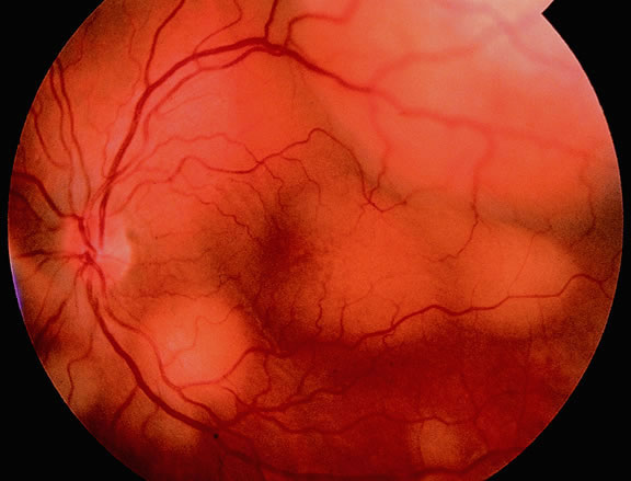
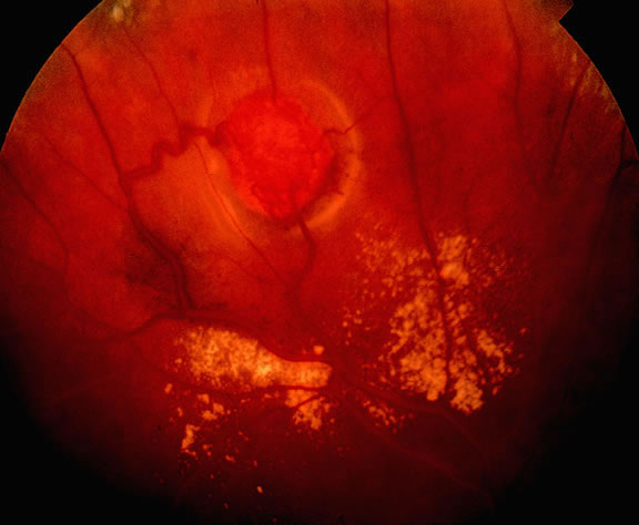
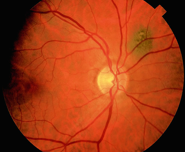
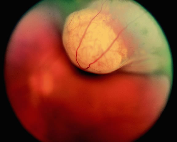
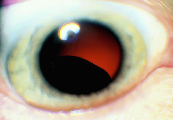
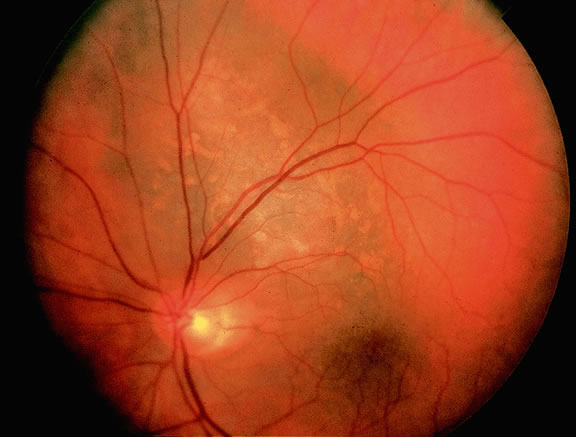
Uveal nevi are benign neoplasms derived from uveal melanocytes; 90% are located in the choroid posterior to the equator.2 Like nevi of the skin, uveal nevi are quite common; it has been estimated that they occur in more than 6% to 10% of the population.3,4 Nevi are often detected during routine fundus examinations (Fig. 1). Most choroidal nevi are flat or minimally elevated lesions that usually measure between 1.5 to 5 mm in diameter; most are less than 2 mm in thickness. Nevi may be pigmented or amelanotic, and they often have an irregular or jagged border. Drusen are quite common on the surface of nevi. Most nevi are stationary lesions that do not change on serial observation. Nevi have been observed to give rise to malignant melanomas, but this is a rare phenomenon. If one assumes the prevalence of uveal nevi to be 5% per year and the incidence of melanoma to be 7 per million per year, it can be estimated that the rate of transformation of nevi to melanoma is only 1 per 10,000 to 15,000 per year.5 Suspicious clinical features that suggest that a nevus actually may be a small melanoma that is likely to grow include the presence of orange pigment and subretinal fluid.6,7 It may be impossible to clinically differentiate large choroidal nevi from small malignant melanomas. The observation of growth is the only clinical criterion that is helpful.8

Fig. 1. Choroidal nevus. Small nonsuspicious pigmented nevus is flat and lacks drusen, orange pigment, and subretinal fluid.

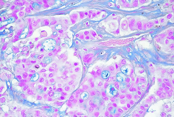
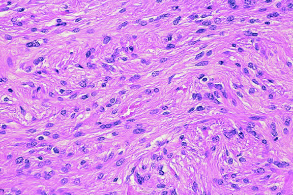
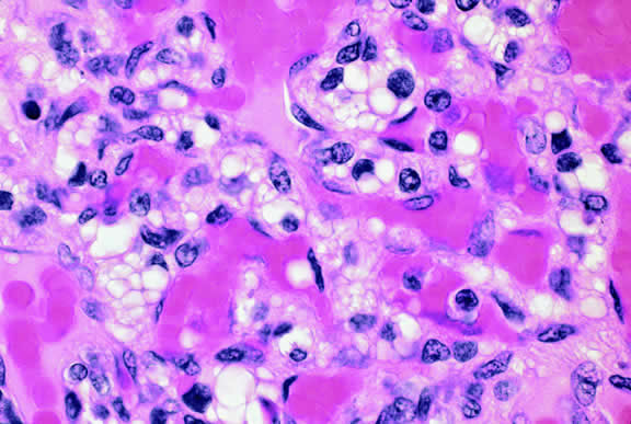
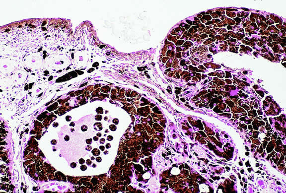
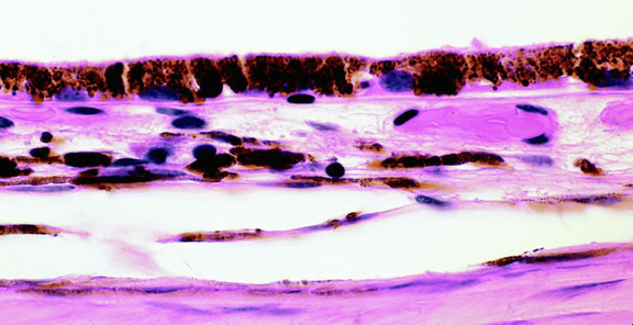
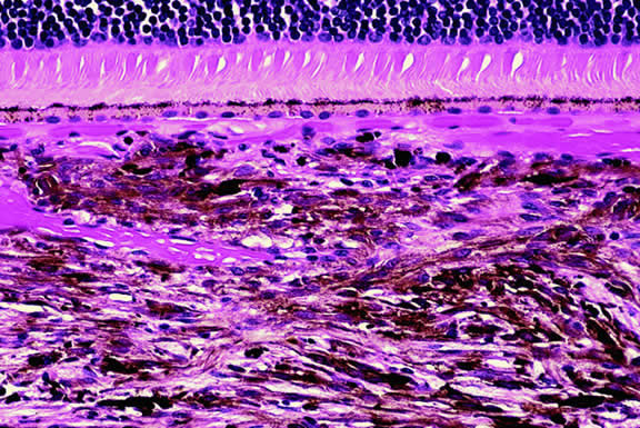
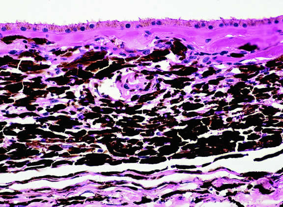
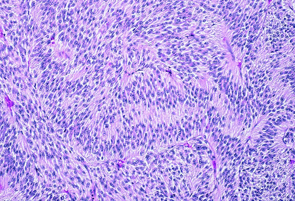
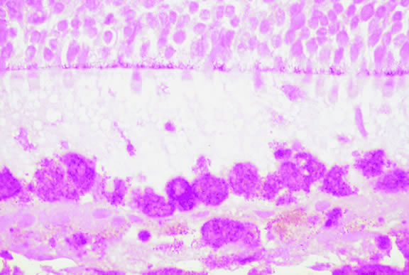
Nevi constitute the benign end of the biologic spectrum of melanocytic neoplasms. Most nevi are bland spindle cells tumors. Histopathologically, the choroidal stroma typically is replaced by a compact infiltrate of slender pigmented or nonpigmented spindle cells (Fig. 2). The nevus cells have bland oval or cigar-shaped nuclei that have finely dispersed chromatin and lack nucleoli or nuclear folds. Intranuclear cytoplasmic inclusions are common in some cases. Mitotic activity is absent. In some cases, the nevus cells are plump and dendritic in shape.9 Foamy balloon cells that appear to be undergoing lipoidal degeneration are found in 4% of nevi.4 Maximally pigmented, plump, polyhedral nevus cells comprise the magnocellular variant of nevus called melanocytoma (see later).

Fig. 2. Choroidal nevus. Choroidal stroma contains an infiltrate of pigmented spindle cells with bland nuclei. The retinal pigment epithelium is intact and the overlying retina remains attached. (Hematoxylin-eosin, × 100.)

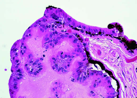
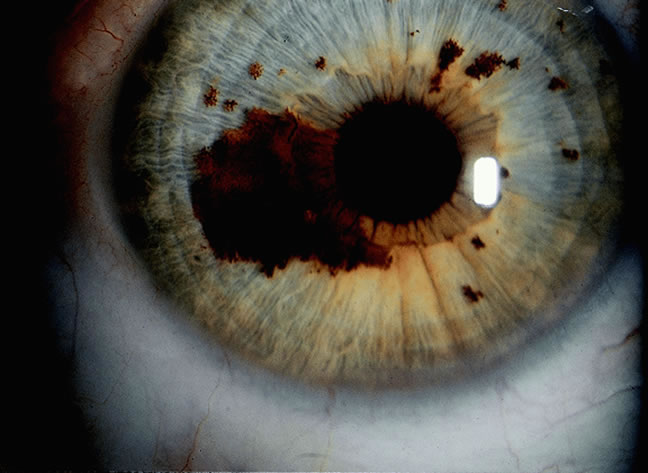
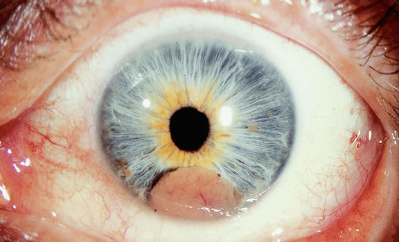
Most pigmented tumors of the iris are low-grade spindle cell nevi that remain stationary and do not enlarge on follow-up (Fig. 3). The iris freckle is a small, stationary form of iris nevus that is found in half of the adult population.1

Fig. 3. Iris nevus. Pigmented nevus does not distort pupil. Several iris freckles are present.

MELANOCYTOMA

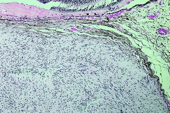
A melanocytoma is a characteristic type of uveal nevus that is composed of plump, polyhedral nevus cells filled with copious quantities of maximally pigmented cytoplasm.10,11 Melanocytomas also are called magnocellular nevi.12 In contrast to most nevi, melanocytoma may be relatively large and may be difficult to distinguish clinically from melanoma.

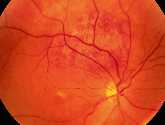
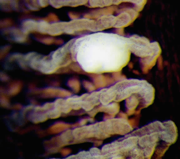
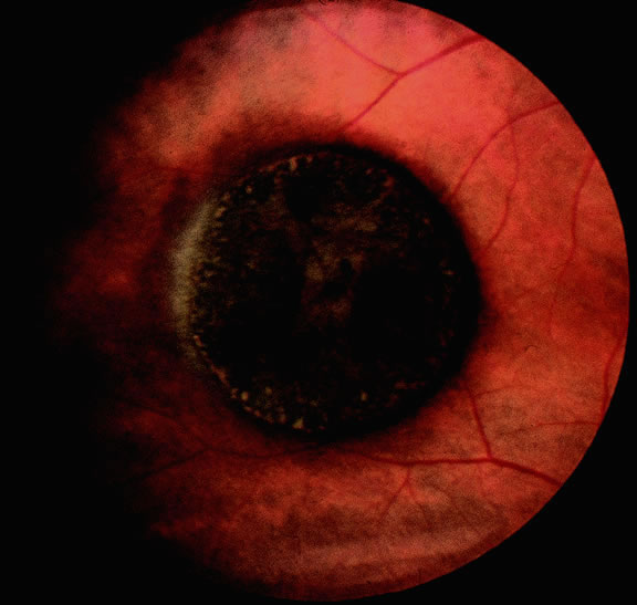
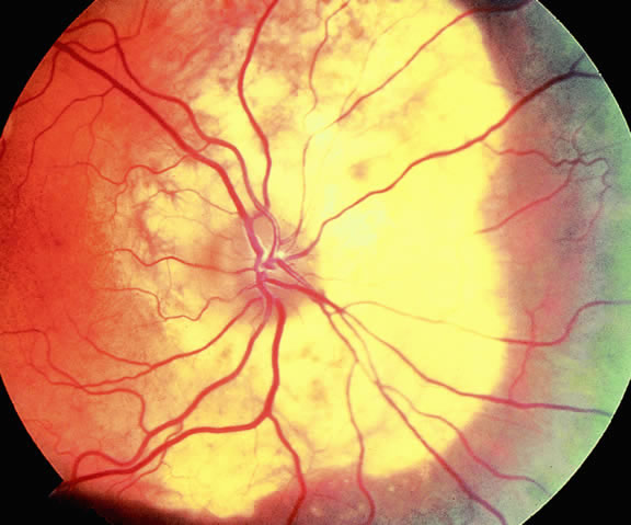
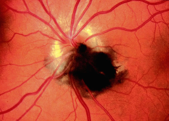
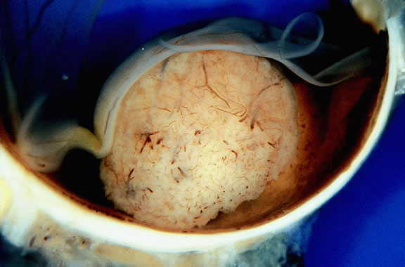
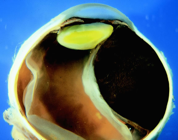
Although best know as a pigmented lesion of the optic disc (Fig. 4), melanocytoma can arise from the stroma of the iris, ciliary body, or choroid13, 14, 15 (Fig. 5). Clinically, melanocytomas are intensely pigmented and often occur in young patients. Unlike uveal melanoma, they do not have a predilection for Caucasians; 37% to 50% of optic disc melanocytomas have been reported in African Americans.10,16 Melanocytomas of the iris are uniformly dark brown in color and often have an irregular friable surface.

Fig. 4. Melanocytoma, optic nerve. The intensely pigmented magnocellular nevus partially obscures the optic nerve. Melanocytomas classically affect the optic nerve, but they may involve any part of the uvea.

Fig. 5. Ciliary body melanocytoma. Ciliary epithelium covers intensely pigmented tumor. Melanin pigment obscures nuclei. (Hematoxylin-eosin, × 50.)

Most melanocytomas of the optic disc have a feathery margin caused by infiltration of the nerve fiber layer. In approximately one half of cases, a juxtapapillary component in the choroid identical to a choroidal nevus is present.16 Visual acuity is often good, but 30% of patients have an afferent pupillary defect.17 Visual field examination may show enlargement of the blind spot or nerve fiber bundle defects.17 About 15% of optic disc melanocytomas enlarge on follow-up.16 Rare cases of malignant transformation have been reported.18, 19, 20 Central retinal vein occlusion has been reported in a patient with a necrotic optic disc melanocytoma and does not always imply malignant transformation.21 Primary malignant melanomas of the optic nerve head have been reported.22 Retinal pigment epithelial (RPE) adenoma may mimic optic disc melanocytoma.23

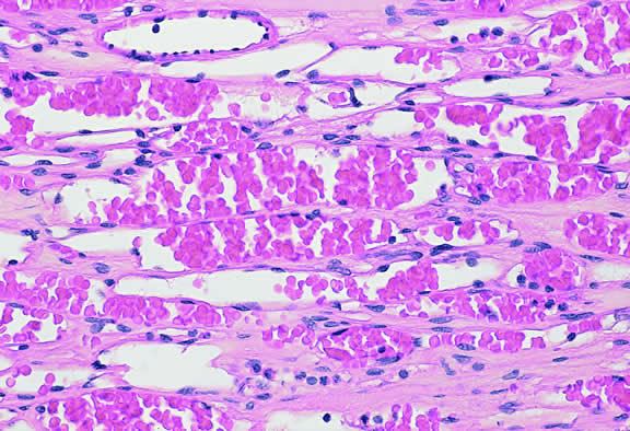
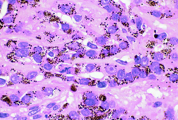
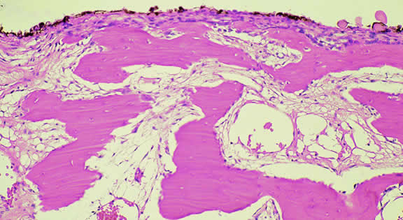
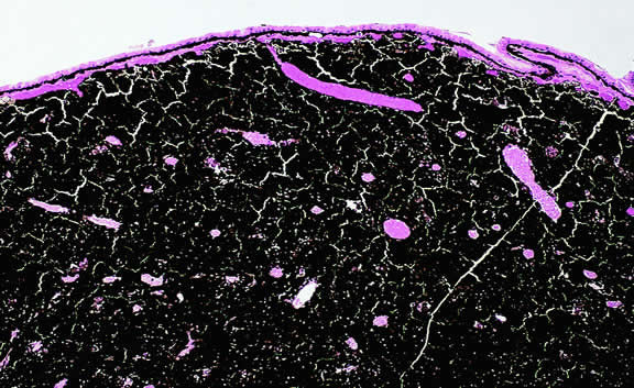
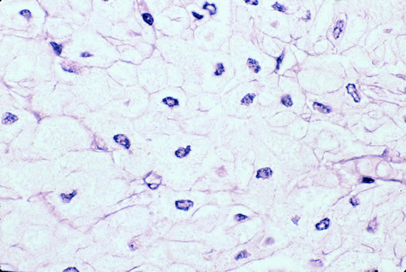
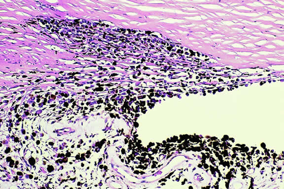
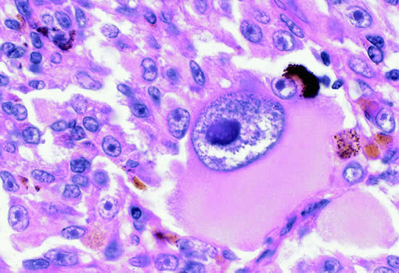
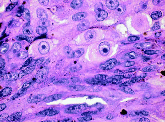
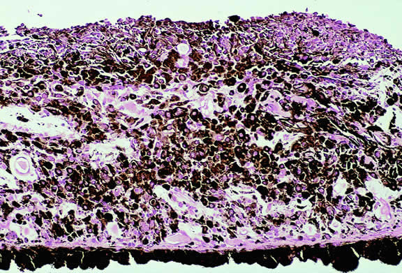
Histopathologically, melanocytomas are composed of cells that have abundant quantities of maximally pigmented cytoplasm that often obscures nuclear details, making microscopic assessment impossible11 (see Fig. 5). Bleached sections, which are a requisite for examination, show that the cells have bland nuclei and a low nuclear-to-cytoplasmic ratio (Fig. 6). Nucleoli usually are inconspicuous, but there are exceptions to the rule. Electron microscopic studies also have revealed a second population of spindle cells.15,24 Melanocytoma cells resemble the benign melanocytes found in the uvea of patients with ocular melanocytosis.

Fig. 6. Melanocytoma, depigmented section. Bleaching of melanin pigment discloses cells with bland nuclei and low nuclear-to-cytoplasmic ratio consistent with benign magnocellular nevus. (Bleach, × 250.)

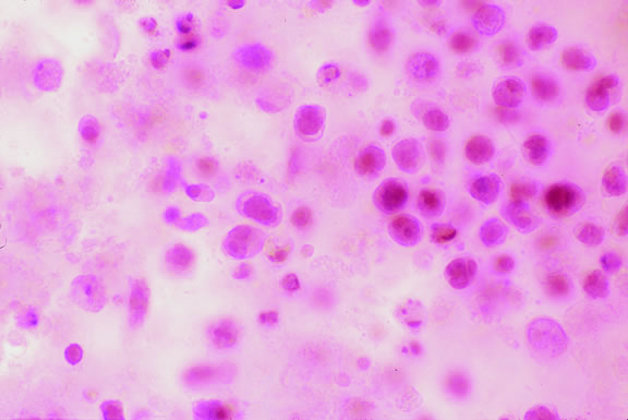
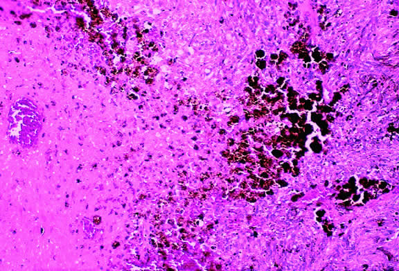
Melanocytomas are predisposed to undergo spontaneous necrosis.14 One is much more likely to find extensive tumor necrosis in a melanocytoma than a melanoma of comparable size, and totally necrotic melanocytomas occasionally are encountered. Numerous plump melanophages typically are found when necrosis is present. It may be difficult to differentiate the melanophages from magnocellular nevus cells in cytologic preparations.

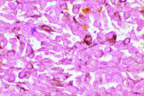
A rare type of secondary glaucoma called melanocytomalytic glaucoma may develop in patients who have partially necrotic melanocytomas of the iris and ciliary body.25, 26, 27 In such cases, the open angle is obstructed by macrophages that have ingested melanin pigment released by the necrotic tumor (Fig. 7).

Fig. 7. Melanocytomalytic glaucoma. Macrophages that have phagocytosed melanin pigment dispersed by necrotic melanocytoma fill peripheral anterior chamber and infiltrate trabecular meshwork.

UVEAL MELANOMA

On a worldwide basis, uveal melanoma is the most common primary intraocular tumor in adults. In the United States and Europe, uveal melanoma is also the most common primary intraocular tumor. In most of the rest of the world, the most common intraocular tumor is the pediatric retinal neoplasm retinoblastoma.28

Uveal melanoma is rare, even among persons of European ancestry. Less than 2000 cases occur yearly in the United States where the incidence is estimated to be 7 cases per million per year, less than 10% the incidence of lung cancer.29

Although pediatric and even rare congenital cases30,31 have been reported, uveal melanoma generally occurs in older persons. The mean age of patients eligible for treatment in the Collaborative Ocular Melanoma Study (COMS) I-125 brachytherapy study was 59 years.32 Less than 0.8% of cases occur in patients younger than age 20 years.33 In general, patients who are older tend to have larger tumors and are more likely to die from disseminated melanoma after enucleation. Recent data from COMS indicates that uveal melanoma affects both sexes equally.32

Predisposing Factors

Race is an important predisposing factor. Uveal malignant melanoma has a definite predilection for Europeans who have blue or lightly pigmented eyes.34, 35, 36 In a recent study in Florida, the risk of uveal melanoma in non-Hispanic white men was 72 times that in black men.29 Uveal melanoma also is less common in Latin and Native Americans29,37 and Asians. The incidence of uveal melanoma in the United States is 25 times greater than that in Taiwan, that is, 7 per million versus 0.28 per million.38 The prognosis of melanoma in black and white patients is similar. Tumors in black patients typically show more necrosis and heavy pigmentation.39

Caucasian patients who have congenital ocular or oculodermal melanocytosis (nevus of Ota) are especially at risk to develop uveal malignant melanoma.40 It has been estimated that about one in 400 white patients with oculodermal melanocytosis will develop uveal melanoma in his or her lifetime.41 This risk is about 25 times greater than the risk in unaffected patients. Melanomas have developed in young patients with Ota's nevus.33,42–44 Ocular and oculodermal melanocytosis are relatively common in Asians45 but do not appear to predispose them to melanoma. Several African Americans with nevus of Ota who developed uveal melanoma have been reported.46,47

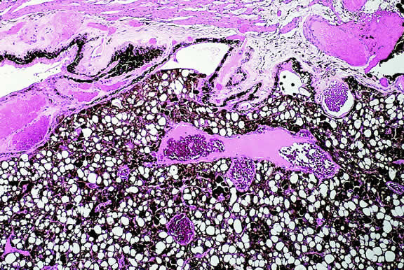
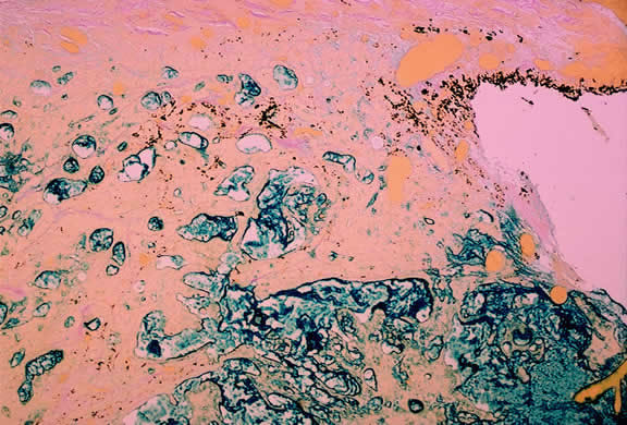
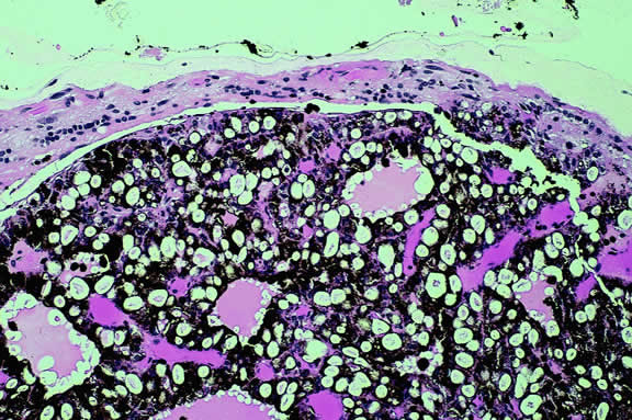
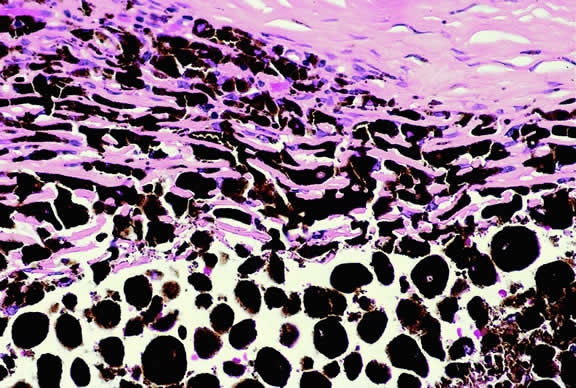
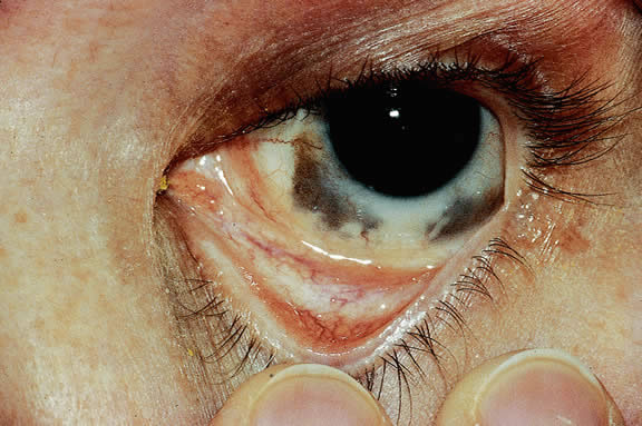
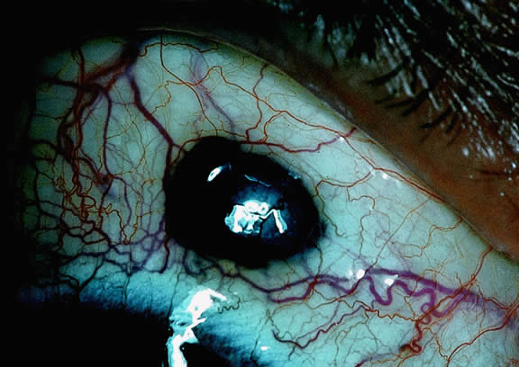
The substrate for the development of melanoma in white patients with ocular melanocytosis is a diffuse nevus that affects all, or part, of the uveal tract. Clinically, the nevus typically is evident as hyperchromic heterochromia iridum, patchy slate gray epibulbar pigmentation, and a darker aspect of the fundus compared with the fellow eye (Fig. 8). A sector of uvea is affected in some patients, however, and the nevus may spare the iris. The uvea in congenital melanocytosis is thickened by an increased number of heavily pigmented nevus cells similar to those found in melanocytomas (Fig. 9). RPE abnormalities such as drusen often develop on the surface of the thickened choroid.48 The sclera also contains patchy foci of pigmentation. The slate gray conjunctival pigment reflects the presence of dendritic melanocytes on the epibulbar tissues deep to the conjunctiva. Nevus of Ota does not predispose to conjunctiva melanoma. However, patients rarely may develop melanomas of the orbit49–51 and even leptomeninges.1,52,53 White patients who have congenital ocular or oculodermal melanocytosis should be followed periodically because of the increased risk of uveal melanoma.54

Fig. 8. Nevus of Ota. Caucasian patient who developed choroidal melanoma has dark brown iris and slate-gray pigmentation of sclera and periocular skin.

Fig. 9. Congenital ocular melanocytosis, choroid. Choroidal stroma is thickened by infiltrate of heavily pigmented benign melanocytes. Patient had iris heterochromia and skin pigmentation consistent with nevus of Ota. (Hematoxylin-eosin, × 100.)

Uveal melanoma occasionally arises from localized uveal nevi, but the estimated incidence of malignant transformation is quite low. Nevus-like cells observed histopathologically at the base of many uveal melanomas probably are a compression artifact and do no represent a pre-existing nevus.55 Cases of uveal melanoma have been reported in patients with von Recklinghausen's neurofibromatosis type I42,56–59 and the dysplastic nevus or familial atypical mole-melanoma syndrome.59–63 The tumor's propensity for light-eyed individuals and the exposed inferior part of the iris suggests that exposure to ultraviolet light could be a predisposing factor.35,36 “Conjugal melanoma,” that is, the occurrence of tumors in both husband and wife, may be coincidental but could be indicative of the role of shared environmental factors.64 An increased incidence of uveal melanoma has been reported in a group of chemical workers.65

Uveal melanoma can arise in patients who have a rare paraneoplastic syndrome called benign diffuse uveal melanocytic proliferation (BDUMP) syndrome.66–69 Unknown factors produced by a nonocular cancer stimulate diffuse proliferation of benign appearing nevoid or spindle cells throughout the uveal tract of both eyes. Often occult, the primary tumor is typically a poorly differentiated ovarian or uterine carcinoma in women or lung carcinoma in men. Patients typically present with rapid visual loss and usually have cataracts. Fundus examination shows multiple pigmented and nonpigmented placoid nodules and reddish patches in the choroid, which show striking early hyperfluorescence during intravenous fluorescein angiography (IVFA), as well as serous retinal detachment.67 One patient presented with nonocular pigmentation typical of the Peutz-Jeghers syndrome.68

Clinical Features

CLINICAL PRESENTATION.

The clinical signs and symptoms of uveal malignant melanoma depend largely on the location of the tumor and the extent of the disease when the patient initially seeks medical attention. More than a century ago, Fuchs70 described four characteristic stages in the natural history of uveal melanoma. These include a relatively asymptomatic stage; a symptomatic stage of glaucoma and/or inflammation; a stage of extraocular growth; and finally, distant metastasis. Today, most patients are encountered in the early stages of the disease. Although metastatic disease ensues in entirely too many patients regardless of therapy, metastatic disease is almost never evident when the patient initially presents or is treated.71

Totally asymptomatic tumors occasionally are detected on a routine ophthalmoscopic examination. However, most choroidal melanomas present with painless visual loss. In most cases, visual loss is caused by serous or solid detachment of the retina. In rare instances the pathway of light to the fovea is physically blocked by the mushrooming apex of an actively growing, anteriorly located tumor. Ciliary body melanomas often do not produce retinal detachment and, therefore, may remain relatively asymptomatic for some time. Ciliary body melanomas cause loss of vision by directly encroaching on the visual axis or by impinging on the lens causing visually significant distortion, subluxation, or secondary cataract formation. Peripheral melanomas also can cause decreased vision by inducing cystoid macular edema. Dilated episcleral and conjunctival vessels called sentinel vessels often mark the site of an underlying ciliary body tumor.

Vitreous hemorrhage is an extremely rare cause of visual loss in uveal melanoma because the retina serves as a barrier that prevents the blood from entering the vitreous. Retinal perforation is a requisite for vitreous hemorrhage. This occurs most often when highly elevated tumors are situated near the optic disc or ora serrata where the retina is attached. Perforation of the retina also can lead to extensive seeding of the vitreous and the inner surface of the retina.72 In rare instances, malignant epiretinal membranes of seeded tumor cells can produce a pseudoretinitis pigmentosa-like picture.73

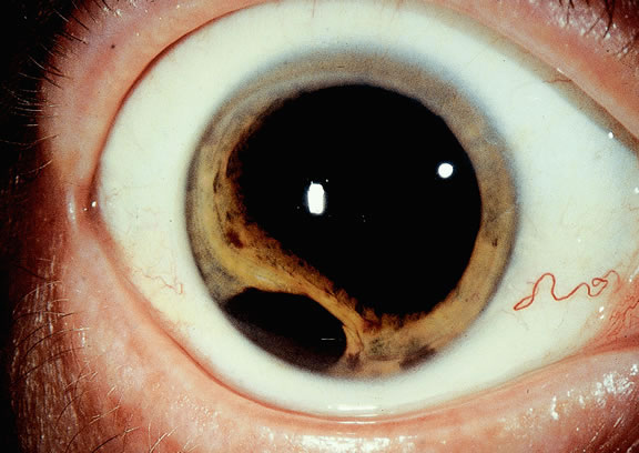
The ready visibility of many iris melanomas leads to their diagnosis at an earlier age and a smaller size (Fig. 10). Iris melanoma typically is discovered when a patient or his or her physician detects a focal pigmented blemish. Higher grade iris melanomas, or ciliary body tumors that invade the anterior chamber, can seed the iris and anterior chamber causing acquired hyperchromic heterochromia iridum (Fig. 11). Secondary glaucoma often accompanies the change in iris color. Not infrequently, the underlying diagnosis may be overlooked and the patient is treated for glaucoma, especially when a melanoma is nonpigmented or confined to the periphery of the iris or the trabecular meshwork.

Fig. 10. Iris melanoma. Amelanotic tumor fills inferior part of anterior chamber. Gonioscopy and transillumination disclosed no involvement of ciliary body.

Fig. 11. Patient with iris heterochromia caused by diffuse iris melanoma.

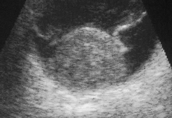
Most eyes with untreated uveal melanoma eventually develop glaucoma.74,75 Anterior tumors, that is, high-grade iris melanomas or ciliary body melanomas that invade the anterior chamber, typically cause secondary open-angle glaucoma by seeding or directly infiltrating the trabecular meshwork (Fig. 12). The latter often occurs in a circumferential or ring fashion. Posterior melanomas generally cause secondary angle closure glaucoma by stimulating iris neovascularization or by causing pupillary block. The pupil is blocked when large tumors or tumors that have caused highly elevated total retinal detachments displace the lens iris diaphragm forward (Fig. 13). The same mechanism also occurs in eyes with other neoplasms that typically are associated with high bullous retinal detachments such as uveal metastases, choroidal hemangiomas, and exophytic retinoblastomas. In such cases, severe pain often prompts a patient with a blind eye to seek medical care. More than a century ago, it initially was emphasized that a glaucomatous eye with a total retinal detachment harbors an intraocular tumor, typically a melanoma, until proven otherwise. Imaging studies such as ultrasonography are mandatory before therapy in such cases, especially if evisceration of a blind painful eye is contemplated instead of enucleation.

Fig. 12. Diffuse iris melanoma with secondary open-angle glaucoma. Tumor cells infiltrate trabecular meshwork and seed its inner surface. (Hematoxylin-eosin, × 50.)

Fig. 13. Large ciliochoroidal melanoma with total retinal detachment and secondary closed-angle glaucoma caused by anterior displacement of lens-iris diaphragm.

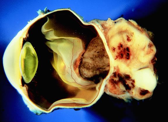
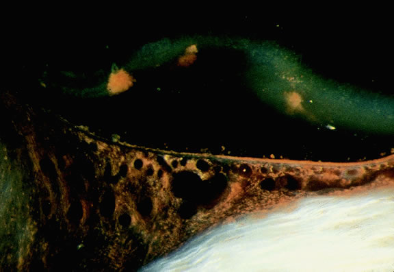
Rarely, patients with uveal melanoma may present with extraocular extension of their tumors.48,76,77 Epibulbar foci from ciliary body melanomas that have grown out of the eye through anterior emissarial canals or Axenfeld nerve loops can be confused clinically with conjunctival melanomas, but they are not attached to the conjunctival epithelium (Fig. 14). Although some posterior melanomas infiltrate directly through the sclera, transscleral extension typically occurs along the emissarial canals of vessels or nerves (Fig. 15). Rarely a posterior nodule of extrascleral extension develops that is much larger than the primary intraocular part of the tumor that spawned it.48,77,78 Such patients may present with ocular proptosis. In exceptional circumstances, patients with chronically neglected tumors, which have filled and destroyed the eye, will present with an orbital mass.

Fig. 14. Anterior epibulbar focus of transscleral extension from ciliary body melanoma. Conjunctiva covers tumor, which has grown through emissary canal.

Fig. 15. Massive posterior extrascleral extension, uveal melanoma. Pigmented choroidal melanoma has extended extrasclerally forming large nonpigmented orbital mass.

Necrotic melanomas may present with severe orbital and adnexal inflammation that can mimic infectious cellulitis or idiopathic orbital inflammation.48 Extensive necrosis typically is found in eyes with rapidly growing high-grade tumors. Marked elevation of intraocular pressure in eyes with secondary glaucoma may cause infarction of the tumor and other intraocular structures.

Unsuspected melanomas occasionally are discovered when blind painful eyes with opaque media are examined pathologically. Before the advent of ultrasonography as many as 10% of blind painful eyes were said to harbor previously undiagnosed tumors.79 The possibility of a tumor should always be entertained when cataract is unilateral or asymmetric. It is quite embarrassing to diagnose a melanoma during cataract surgery.

CLINICAL SIGNS.

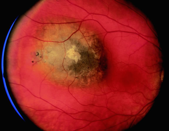
Ophthalmoscopic examination of a choroidal melanoma typically reveals a sessile or dome-shaped pigmented mass located deep to the retina (Figs. 16, 17, and 18). There often is an associated secondary nonrhegmatogenous serous detachment of the retina. Orange pigment may be located on the surface of smaller lesions.80,81 Tumors vary in pigment content and may be heavily pigmented or totally amelanotic. The differentiation of a totally amelanotic melanoma from uveal metastasis, hemangioma, lymphoma, or osteoma may be difficult. Compared with metastasis, melanoma usually is more highly elevated and drusen and RPE proliferation more evident on its surface. Ophthalmoscopy also typically shows well-defined vessels within melanoma.48 Ancillary studies such as fluorescein angiography and ultrasonography may be necessary to make the diagnosis.

Fig. 16. Small pigmented tumor thought to be choroidal nevus found on routine ophthalmoscopic examination. Nevus is slightly elevated and retinal pigment epithelial changes are present.

Fig. 17. Pigmented choroidal melanoma. Elevated tumor has broken through Bruch's membrane.

Fig. 18. Large ciliochoroidal melanoma. Wide-angle photo shows extent of lesion and associated inferior retinal detachment.

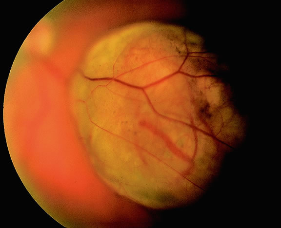
Many choroidal melanomas have a characteristic mushroom or collar button configuration that results when the tumor breaks through Bruch's membrane (Figs. 19, 20, 21, and 22). In such cases there is often secondary choroidal, subretinal, or vitreous hemorrhage. Dilated, intrinsic vessels often are evident in the mushrooming dome of nonpigmented melanomas. The vessels are obscured in more pigmented tumors.48

Fig. 19. Fundus photo of mushroom-shaped choroidal melanoma with nonpigmented dome. Configuration indicates that tumor has ruptured through Bruch's membrane.

Fig. 20. Gross photo of mushroom-shaped choroidal melanoma. Amelanotic dome of melanoma anterior to break in Bruch's membrane elevates and detaches retina. Mushrooming head of tumor has a rough papillary surface and contains many vessels.

Fig. 21. Gross photo, large mushroom-shaped ciliochoroidal melanoma. Pigmented tumor is highly elevated.

Fig. 22. Mushrooming head of choroidal melanoma contains dilated vessels which are located anterior to constricting gap in Bruch's membrane. (Hematoxylin-eosin, × 10.)

Although the diagnosis of melanoma often can be made by careful indirect ophthalmoscopy, ancillary studies such as intravenous fluorescein angiography, indocyanine green angiography, ultrasonography, computed tomography, magnetic resonance imaging, the radioactive phosphorus uptake test, and fine-needle aspiration biopsy (FNAB) occasionally are required to support or confirm the diagnosis.48,54

The characteristic fluorangiographic features of uveal melanoma include mottled hyperfluorescence in the vascular filling phases and diffuse late staining of the mass and surrounding subretinal fluid. A characteristic double circulation in which both the retinal vessels and the choroidal vessels in the tumor are readily evident may be shown by larger amelanotic tumors, particularly those that have perforated Bruch's membrane. The prominent blood vessels seen in amelanotic mushroom-shaped melanomas may impart a pseudoangiomatous appearance to the lesion.48

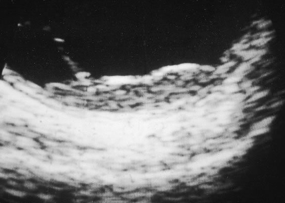
Ultrasonography can support the diagnosis of ciliary body or choroidal melanoma and is a particularly helpful way of disclosing tumors in eyes with opaque media.54 Ultrasonographic studies can alsodemonstrate areas of extrascleral extension. A scan ultrasonography shows a high internal spike and low internal reflectivity. B scan ultrasonography shows characteristic acoustic hollowness and choroidal excavation (Fig. 23). Hemangiomas and metastases usually appear acoustically solid because they contain multiple acoustic interfaces.

Fig. 23. B-scan ultrasound, choroidal melanoma.

Transvitreal FNAB occasionally is performed if the diagnosis remains uncertain after routine tests and the choice of therapy requires an accurate diagnosis.82,83 Potential candidates for FNAB include the patient who has a history of breast cancer and presents with a solitary amelanotic choroidal tumor that could be a primary amelanotic melanoma or the patient who is thought to have a choroidal metastasis but has no history of cancer.83

Gross Pathology

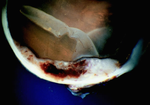
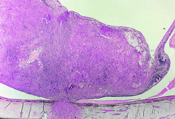
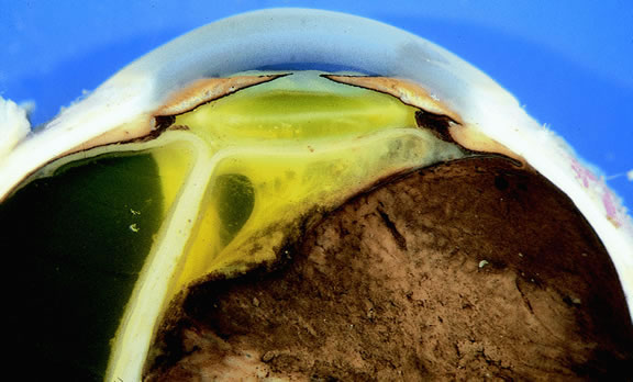
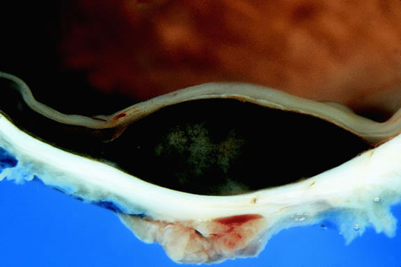
Choroidal melanomas initially arise in the stroma of the choroid. In early cases, the profile of the sectioned tumor is oval or almond-shaped, and its tissue usually appears relatively cohesive after fixation (Fig. 24). Although some tumors diffusely infiltrate the uvea, most uveal melanomas are relatively well-circumscribed tumors with distinct margins. In many cases the growing melanoma perforates Bruch's membrane and enters the subretinal space where its apex typically assumes a spherical shape that often is likened to a mushroom or collar button (see Figs. 19 to 22.). Dilated vessels often are found in the mushrooming head of the tumor because the ends of Bruch's membrane exert a compressive cinch-like effect on the waist of the tumor (see Fig. 22). Rupture of Bruch's membrane was present in 87.7% of 1527 large- or medium-sized melanomas examined in the COMS.84 Retinal invasion was present in nearly half (49.1%), and tumor cells were found in the vitreous body in one fourth.

Fig. 24. Cut surface of heavily pigmented choroidal melanoma is almond-shaped. Bruch's membrane is intact.

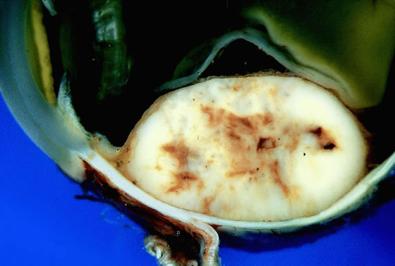
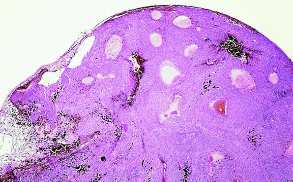
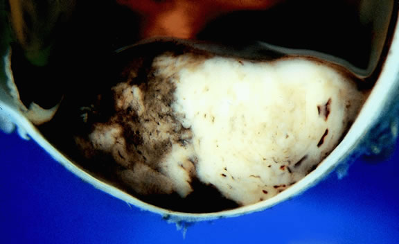
Uveal melanomas vary markedly in their pigment content. Some tumors are totally amelanotic; others are maximally pigmented. In some instances, distinct clones of tumor cells that vary in pigment content are evident on gross examination or even clinically (Fig. 25). A friable or granular appearance of the cut surface of a melanoma noted during gross examination may be indicative of necrosis or a tumor rich in poorly cohesive epithelioid cells. Melanomas that contain cystic cavities occasionally are encountered.85

Fig. 25. Large dome-shaped ciliochoroidal melanoma is partially amelanotic. Bruch's membrane is intact.

About 3% of melanomas have a diffuse growth pattern. These relatively flat tumors grow laterally without thickening the choroid.86,87 Most are about 2 mm in thickness. Diffuse melanomas often are of mixed cell type and, compared with localized lesions, are more apt to infiltrate the sclera and invade the optic nerve or orbit (Fig. 26). Delayed diagnosis or misdiagnosis is common.

Fig. 26. Diffuse choroidal melanoma thickens choroid. Massive invasion of optic nerve is seen at left. (Hematoxylin-eosin, × 50.)

Choroidal melanomas are classified as small, medium, or large based on the largest tumor diameter (LTD). Small choroidal melanomas are 10 mm in diameter or less and appear as focal discoid or oval areas of choroidal thickening. Medium-sized melanomas measure 11 to 15 mm, and large tumors are more than 15mm in largest basal tumor diameter. Larger tumors are more likely to have ruptured through Bruch's membrane.

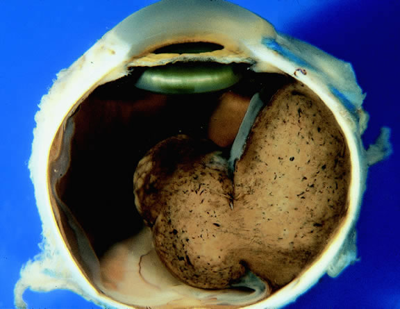
In the COMS, medium-sized tumors were located more posteriorly than large tumors.84 Presumably, more anteriorly situated ciliary body tumors are larger when they are first detected because they remain hidden behind the iris, cause late retinal detachment, and hence remain asymptomatic. Ciliary body melanomas (Figs. 27 to 29) are less common than choroidal melanomas and tend to be more spherical in shape. As noted previously, ciliary body melanomas can cause unilateral cataract, lens molding, or subluxation (see Fig. 28). In addition to causing secondary glaucoma by angle seeding or circumferential infiltration, ciliary body melanomas that invade the periphery of the anterior chamber often displace the iris root centrally causing a solid iridodialysis. Heterochromia iridum also is possible if there is extensive chamber seeding. Ciliary body tumors that perforate the iris initially may be misdiagnosed as a small iris tumor that in reality is the “tip of an iceberg” (see Fig. 29).

Fig. 27. Ciliary body melanoma. Pigmented tumor is seen behind lens.

Fig. 28. Large, heavily pigmented ciliochoroidal melanoma. Dome-shaped tumor is causing lens subluxation.

Fig. 29. Anterior chamber invasion by ciliary body melanoma. Part of the tumor is seen through pupil behind the iris.

Retinal detachment is the most common cause of visual loss in eyes with posterior melanoma. Initially solid, the detachment becomes exudative as serous fluid accumulates. Visual loss initially may be caused by induced hyperopia as a subfoveal tumor displaces the retina anteriorly. The exudative detachment initially is localized; fluid accumulates in the subretinal space tented up by the growing tumor. Failure to find a retinal hole in a patient with retinal detachment should always raise the possibility of a choroidal tumor or some other choroidal process known to produce an exudative detachment. Total retinal detachments are found in some eyes with choroidal melanomas, generally those with larger or neglected tumors (see Fig. 13). Melanoma-bearing eyes with total retinal detachments often have secondary closed-angle glaucoma caused by pupillary block or iris neovascularization.74,75 Clumps of orange pigment may be seen in the subretinal space overlying the tumor (see Fig. 40).

Fig. 40. Orange pigment. Clumps of orange pigment adhere to outer surface of detached retina overlying actively growing melanoma.

During gross examination, the external surface of an eye harboring a melanoma should be inspected carefully for signs of extraocular extension. The latter may be evident as an epibulbar nodule or a vortex vein that is dilated and heavily pigmented. Tumor infiltration of scleral emissarial canals may be evident on the cut surface of the globe at the base of the tumor.

Treatment effects are evident in eyes whose tumors have been treated previously with radioactive plaque brachytherapy or transpupillary thermotherapy (TTT).88,89 Prior plaque therapy often causes prominent areas of diffuse chorioretinal depigmentation and atrophy. TTT scars usually are white and sharply demarcated like a coloboma and often are rimmed by heavily pigmented foci of RPE hyperplasia.

Histopathology

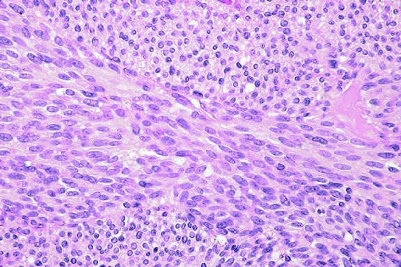
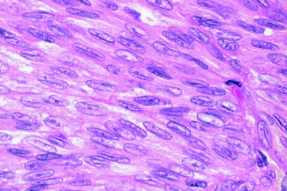
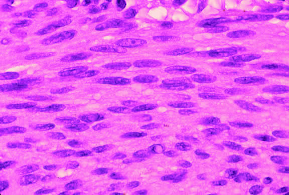
The cells comprising uveal melanoma constitute a biologic spectrum comprising bland spindle A melanoma cells at one end and wildly anaplastic epithelioid cells at the other. The term spindle cell is derived from the fusiform or spindled configuration of the cells' cytoplasmic outline. They are bipolar in shape, and many have long tapering processes that occasionally are highlighted when individual pigmented cells are seen in a largely amelanotic tumor. Spindle cells grow in a syncytial fashion forming interweaving fascicles of parallel oriented cells (Fig. 30). The cells can be pigmented or nonpigmented. Two types of spindle cells are recognized; spindle A and spindle B. These are distinguished by their nuclear characteristics. Spindle A nuclei are tapering ovals or cigar-shaped and have finely dispersed chromatin (Fig. 31). If a nucleolus is present, it usually is inconspicuous. Many spindle A cells have a longitudinally oriented chromatin stripe that actually is caused by a fold in the nuclear membrane. The nuclei of spindle B cells have distinct nucleoli and coarser chromatin and tend to be plumper and more oval in shape (Fig. 32).

Fig. 30. Amelanotic spindle cell melanoma. Tumor is composed of interweaving fascicles of spindle cells. Photomicrograph shows longitudinally and transversely sectioned fascicles. (Hematoxylin-eosin, × 100.)

Fig. 31. Low-grade spindle melanoma. Spindle A cells have bland, slender, cigar-shaped nuclei with finely dispersed chromatin and indistinct nucleoli. Longitudinal folds in the nuclear membrane are apparent microscopically as a chromatin stripe or line. Bland spindle B nuclei with distinct nucleoli also are present. (Hematoxylin-eosin, × 250.)

Fig. 32. Spindle B melanoma cells. Most of the cells in this field are spindle B melanoma cells. They have oval nuclei and an obvious nucleolus. Compared with spindle A cells, their chromatin is more coarsely clumped. The spindle cells form a syncytium and have indistinct cytoplasmic margins. (Hematoxylin-eosin, × 250.)

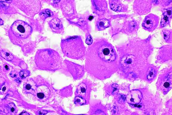
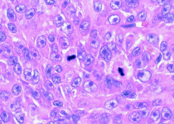
Epithelioid melanoma cells comprise the poorly differentiated end of the cytologic spectrum. Melanomas that contain epithelioid cells have a poorer prognosis. The term epithelioid meaning epithelial-like reflects the fanciful resemblance of the tumor cells to the cells of simple epithelia. Epithelioid cells have abundant cytoplasm and are often polygonal in shape (Fig. 33). They have distinct cytoplasmic margins, are poorly cohesive, and do not grow as a syncytium. The nuclei of epithelioid cells are usually round or oval and often appear vesicular because of margination or clumping of the chromatin along the inner side of the nuclear membrane. Epithelioid melanoma cells also have prominent nucleoli that are often large and reddish purple. Variants of epithelioid cells include relatively uniform small epithelioid cells (Fig. 34) and bizarre tumor giant cells that may appear wildly anaplastic (Fig. 35).

Fig. 33. Epithelioid melanoma cells. The cytoplasmic margins of these large, poorly cohesive epithelioid melanoma cells are easily discernible. Epithelioid cell nuclei are typically round and have peripheral margination of coarsely clumped chromatin. Epithelioid cells usually have prominent reddish purple nucleoli. They typically are polyhedral in shape and have copious amounts of cytoplasm. (Hematoxylin-eosin, × 250.)

Fig. 34. Small epithelioid cells. Cells are relatively small but are definitely epithelioid in character. They are polyhedral in shape and have distinct cytoplasmic outlines. The round nuclei contain prominent nucleoli. (Hematoxylin-eosin, × 250.)

Fig. 35. Tumor giant cell, uveal melanoma. Tumor giant cells are highly anaplastic epithelioid cells. They are relatively rare, and their prognostic significance is uncertain. (Hematoxylin-eosin, × 100.)

Melanoma cells should be classified by their nuclear characteristics. Spindle-shaped cells that have epithelioid nuclei occasionally are encountered; such cells are classified as epithelioid. In recent years, the term intermediate cell has been used increasingly. Intermediate cells are cells that have nuclear characteristics that are intermediate between spindle B and epithelioid. For example, one might apply the term intermediate cell to a spindle B cell that has a nucleus that is somewhat large and has a fairly prominent nucleolus.

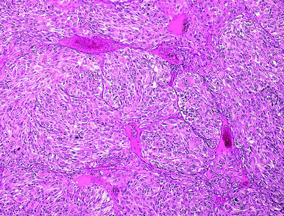
Occasionally, the spindle cells in a melanoma are arranged in a radial fashion around vessel or fibrovascular septa (vasocentric pattern), or the nuclei form rows that resemble the Verocay bodies or Antoni A pattern seen in schwannoma (Verocay pattern). Melanomas are called fascicular if these patterns dominate5 (Fig. 36). Fascicular melanoma was a category in Callender's initial classification90 that was dropped from McLean's 1983 modification.5,91

Fig. 36. Fascicular melanoma. This amelanotic melanoma has a striking fascicular appearance. The nuclei of its constituent spindle cells form rows that resemble the Antoni A pattern seen in schwannoma. The fascicular category of uveal melanoma has been removed from the modern revision of Callender's classification because cellular arrangement does not appear to affect prognosis. (Hematoxylin-eosin, × 50.)

Varying degrees of necrosis may be found (Figs. 37 and 38). Necrosis tends to be more prominent in rapidly growing high-grade tumors or tumors that have had prior brachytherapy. The necrosis may be patchy and focal or may involve extensive parts or even all of the tumor. Aggregates of melanophages typically are found in the necrotic areas. Total infarction of the tumor (and other intraocular structures) may occur in eyes with severe secondary closed-angle glaucoma. As mentioned previously, melanocytoma is prone to spontaneous necrosis. The latter diagnosis should always be considered when a totally necrotic, heavily pigmented tumor is found.

Fig. 37. Focus of necrosis in choroidal melanoma. Pigmented melanophages have accumulated at interface between viable tumor and necrotic focus (below). (Hematoxylin-eosin, × 100.)

Fig. 38. Necrotic uveal melanoma. The cells of this necrotic melanoma are eosinophilic because they have lost their basophilic nuclear DNA. The cell type of a necrotic melanoma often can be ascertained if careful microscopy with an oil-immersion lens is performed. Necrotic uveal melanomas tend to behave clinically like mixed cell type melanomas. (Hematoxylin-eosin, × 250.)

Choroidal melanomas produce abnormalities in the overlying retinal pigment epithelium including atrophy, hyperplasia, and the formation of drusen and drusenoid material.92 The overlying retina often shows photoreceptor loss and may develop cystoid edema. The latter tends to be more common in slower growing lesions, especially choroidal hemangiomas. After Bruch's membrane has ruptured, the vessels located in the mushrooming head of the tumor are often quite prominent, reflecting vascular stagnation caused by the compression at the waist of the tumor (see Fig. 22). Aggregates of macrophages that have ingested periodic acid-Schiff (PAS)-positive lipofuscin pigment and melanin from the damaged retinal pigment epithelium can be found in the subretinal fluid (Figs. 39 to 41). These are evident ophthalmoscopically as clumps of orange pigment that serve as a clinical marker for an actively growing neoplasm.80,81

Fig. 39. Juxtapapillary melanoma with orange pigment. Orange pigment is a clinical marker for an actively growing tumor.

Fig. 41. Orange pigment. Orange pigment is comprised of aggregates of macrophages that have phagocytized periodic acid-Schiff (PAS)-positive lipofuscin and melanin pigment released by retinal pigment epithelial cells that have been disrupted by the actively growing tumor. (Periodic acid-Schiff, × 100.)

Melanocytic tumors of the uvea are classified into four groups on the basis of cytology. Tumors composed entirely of spindle A cells or even blander nevus cells are classified as spindle cell nevi. Tumors composed of a mixture of malignant spindle A and spindle B cells are called spindle melanomas. Melanomas of mixed cell type contain a mixture of spindle and epithelioid melanoma cells (Fig. 42). Some laboratories specify the predominant cell type found in a mixed cell melanoma, for example, reporting mixed cell, predominantly spindle if only a few epithelioid cells are present. Epithelioid melanomas are composed predominantly of epithelioid cells. They are relatively rare and have the poorest prognosis. Most medium- and large-sized melanomas contain a mixture of spindle and epithelioid cells. In the COMS histopathology study, 86% of the posterior melanomas were classified as mixed cell type, 8% were of spindle cell type, and 5% were epithelioid.84 The association between cytology and mortality is known as the Callender classification.90 (See later section on prognostic factors.)

Fig. 42. Uveal melanoma, mixed cell type. Mixed cell melanomas are composed of a mixture and spindle and epithelioid cells. (Hematoxylin-eosin, × 250.)

Iris melanoma is relatively rare, constituting between 4% to 15% of uveal melanomas in various series.93 The studies with higher reported incidences may reflect inclusion of nevi. Iris melanomas differ in some respects from tumors of the posterior segment. Most are low-grade spindle cell tumors (Fig. 43). However, iris melanomas with epithelioid cells occasionally are encountered (Fig. 44). Many pigmented iris tumors actually are nevi,94 and relatively few of these enlarge when observed. Several studies have shown that the risk of enlargement in 5 years is only 5% to 6% after a subset of promptly treated lesions is excluded.95,96 One study showed that iris tumors are likely to be considered melanomas and be treated promptly if the basal diameter is greater than 3 mm, pigment dispersion or prominent tumor vascularity are present, the intraocular pressure is elevated, or there are tumor-related symptoms.95 The mean age of patients with iris melanomas is about 10 years younger (age 43 years) than the age of patients with posterior segment melanomas.97 The prognosis of iris melanoma is also relatively favorable compared with tumors of the posterior segment. Shields and coworkers97 studied 169 patients with histologically confirmed iris melanoma and found that distant metastases developed in 5% at 10-years follow-up. The relatively small size of most iris melanomas probably is a major factor in their good prognosis. The prognosis of iris melanomas actually may be similar to posterior segment tumors of similar size and cell type. Diffuse iris melanomas that cause heterochromia iridis and secondary glaucoma are a rare but clinically important group of iris tumors.98,99 Many diffuse iris melanomas are higher grade tumors that contain epithelioid cells, which are poorly cohesive and prone to aqueous dispersal. Glaucoma surgery should be avoided in such cases. It invariably fails and puts patients at greater risk for extraocular extension and metastasis.100

Fig. 43. Low-grade spindle cell iris melanoma. Cytology is very bland. Only a few small nucleoli are present. (Hematoxylin-eosin, × 250.)

Fig. 44. Iris melanoma, mixed cell type. Pigmented infiltrate replacing iris stroma contains many epithelioid cells. (Hematoxylin-eosin, × 250.)

Prognostic Factors

About one half of patients with choroidal and ciliochoroidal malignant melanomas die from their tumors.101,102 Because the eye and orbit lack lymphatics, uveal melanoma spreads via the bloodstream. Metastasis to the liver occur most often; more than 90% of cases with metastatic melanoma have liver metastases, and they are the first metastases detected in 80%. Other common sites of metastatic uveal melanoma include the lung (24%) and bone (16%).103 Multiple sites are found in 87%. Unfortunately, once distant metastases are manifest clinically, therapy generally is ineffective, and more than 50% of patients who have metastatic uveal melanoma die within 1 year. In recent years, there has been an effort to identify prognostic factors that could identify patients at high risk for metastatic disease who hopefully might benefit from prophylactic chemotherapy or immunotherapy.104

STANDARD HISTOPATHOLOGIC FACTORS.

The association between the cytologic characteristics of uveal melanoma (cell type) and mortality was initially reported in 1931 by Major George Russel Callender90 who examined a series of cases on file in the Registry of Ophthalmic Pathology at the Army Medical Museum in Washington, DC. Callender reported that melanomas were composed of two types of spindle cells that he designated spindle A and B and less differentiated epithelioid cells. He found that tumors that contained epithelioid cells had a poorer prognosis.

Ian McLean and his coworkers at the Armed Forces Institute of Pathology (AFIP) modified Callender's original classification in 1978. Spindle A and spindle B melanomas, which formed separate categories in Callender's original classification, were combined into the single category of spindle melanoma.105 Very low-grade spindle cell tumors classified as spindle cell nevi. McLean also introduced the concept of intermediate cells, that is, cells with nuclear characteristics that were intermediate between spindle B and epithelioid.

The presence or absence of epithelioid cells is an extremely important prognostic factor in uveal melanoma. In 1982, McLean and coworkers102 reviewed a series of 3432 cases of malignant melanoma of the choroid and ciliary body on file in the AFIP's Registry of Ophthalmic Pathology and found that 56% were mixed cell tumors composed of a mixture of spindle and epithelioid cells. The 15-year mortality of patients with melanomas of mixed cell type was three times that of patients whose tumors were composed solely of spindle cells. Tumor size, measured as LTD, was also highly correlated with mortality.

Cell type remains one of prognostic mainstays of surgical pathologists because assessment is relatively rapid and requires no special stains or equipment. However, application of the Callender classification unquestionably has limitations. First, determination of cell type is highly subjective and diagnostic accuracy of can vary with the expertise and experience of the pathologist. A masked study showed that even experienced ophthalmic pathologists disagree about their classification of individual tumor cells.106 Secondly, melanoma cells constitute a continuous biologic spectrum that includes extremely bland spindle A melanoma cells at one end and highly anaplastic epithelioid cells at the other. Despite this, only three categories—spindle, mixed, or epithelioid—are available for the classification of a given tumor, and tumors in a single category, for example, mixed cell type, can vary significantly in their apparent degree of differentiation.

MORPHOMETRY AND NUCLEOLAR PARAMETERS.

The limitations of Callender's classification prompted a search for more objective and reliable criteria for the histopathologic assessment of the malignant potential of uveal melanomas. Gamel and coworkers107–109 performed morphometric analysis on series of tumors with known survival and showed that certain nucleolar parameters, most notably the inverse of the standard deviation of the area of the nucleolus, were useful predictors of death from metastatic melanoma. Using the combination of nucleolar parameters and LTD, Gamel and colleagues108 were able to correctly predict the clinical course of 88% of cases. The technique subsequently was applied to 340 cases with known outcome from two independent laboratories, and successfully subdivided patients into groups that suffered a sixfold difference in mortality.109,110

Gamel's laboratory subsequently developed another simpler objective method of nucleolar assessment based on the measurement of the 10 largest nucleoli (MTLN).111 This was more reproducible than cell type and had substantial cost and labor advantages over the original computer-controlled system required to measure the standard deviation of nucleolar area (SDNA). The authors also used a digital filar micrometer to measure nucleoli. The latter method for measuring MTLN required less than 11 minutes per case, compared with an average of 28 minutes for the original method.112 The technique compared favorably with LTD, cell type, and SDNA in the prognostic assessment of a large series of patients with melanoma.113 Other researchers have had less success with MTLN, probably because of technical factors.114,115

Nucleolar parameters also have been assessed with the silver-stained nucleolar organizer region (AgNOR) technique that examines nucleolar organizing regions that have been stained with colloidal silver azotate.116–119 Spindle, mixed, and epithelioid melanomas were found to have progressively larger mean AgNOR values.117 and the COMS group showed that malignant lesions have higher mean AgNOR counts (4.347) than benign nevi119 (1.855). Automated image capture and analysis has been performed on melanomas stained with the AgNOR technique.116

The assessment of nucleolar parameters is one of the most accurate ways of predicting survival after enucleation using morphologic data contained within routine histologic slides. Unfortunately, the technique has not been widely adopted because it is labor-intensive and relatively time-consuming and requires special expertise and equipment.

Other attempts to make the assessment of cell type more objective and quantitative include counting the number of epithelioid cells and intermediate cells in 40 high-power fields (HPFs).110,120 The mitotic activity of uveal melanoma is routinely assessed by counting the number of mitotic figures in 40 HPFs.

MICROVASCULAR PATTERNS.

Certain microvascular patterns within uveal melanomas have been shown to be prognostic indicators for death from metastatic melanoma.121 Folberg and associates identified nine morphologic patterns of tumor vessels in eyes removed for ciliary body or choroidal melanoma and designated them: (1) normal, (2) silent, (3) straight, (4) parallel, (5) parallel with cross-linking, (6) arcs, (7) arcs with branching, (8) loops, and (9) networks. The presence of vascular loops and microvascular networks composed of back-to-back loops that encircle microdomains of tumor are strongly associated with death from metastatic melanoma122 (Fig. 45). Prognostic vascular patterns appear to be consistent throughout the depth of a tumor, and the cross-sectional area occupied by prognostic microvascular patterns also has prognostic value.123,124 Additional studies from Folberg's laboratory have compared the microcirculation architecture of nevi and melanomas125 and examined the relationship between microvascular architecture and the aggressive behavior of ciliary body melanomas.126 Attempts to detect prognostically significant microcirculatory patterns in vivo using noninvasive imaging techniques including ultrasonography127,128 and confocal angiography with fluorescein or indocyanine green have been made.129,130 A study of 496 posterior uveal melanomas at the AFIP confirmed that the presence of loops did indicate poor outcome but was not as good a prognostic indicator as the mean diameter of the largest nucleoli, cell type, or tumor size.131

Fig. 45. Networks of vascular loops, uveal melanoma. Fibrovascular septa divide parts of this predominantly epithelioid melanoma into roughly circular zones called vascular loops. Vascular networks are composed of adjacent vascular loops. Uveal melanomas that contain vascular loops and networks have a poorer prognosis. (Hematoxylin-eosin, × 50.)

Microvascular density is another prognostic factor based on tumor vessels.132 Less subjective than the assessment of microvascular patterns, this technique involves counting tumor vessels, which have been highlighted by immunostaining for endothelial markers like CD34, in the area of highest vessel density.

Blood-filled spaces within melanomas called venous lakes also may have an adverse effect on survival.133 Venous lakes are not lined by vascular endothelial cells. Folberg and colleagues134 at the University of Iowa Cancer Center recently have emphasized that highly invasive uveal melanoma cells are able to generate vascular channels that are not lined by endothelial cells. These vascular channels appear to be able to facilitate tumor perfusion independent of tumor angiogenesis.

MITOTIC ACTIVITY AND THE CELL CYCLE.

Ocular pathologists routinely assess the mitotic activity (Fig. 46) of uveal melanoma by counting the number of mitotic figures in 40 high power (“high dry”) microscopic fields. Forty fields are counted because most uveal melanomas contain relatively few mitoses, that is, only 5 or 10 per 40 HPFs. Not unexpectedly, patients whose tumors have more mitoses have a poorer prognosis.

Fig. 46. Mitotic figures, uveal melanoma. Microscopic field from mitotically active amelanotic melanoma contains two mitotic figures. Most uveal melanomas contain relatively few mitoses. (Hematoxylin-eosin, × 250.)

Other techniques have been used to more accurately assess the proliferative (cell cycling) capability of uveal melanomas. These include labeling cells with bromodeoxyuridine (BrdUrd-labeling index)85,135–137 and the use of immunohistochemistry with monoclonal antibodies such as Ki-67 and PC-10 that recognize antigens that are markers for cellular proliferation.114,138

The rate of cellular proliferation also can be assessed by using flow cytometry to measure the fraction of cells in the S phase of the cell cycle when a cell's deoxyribonucleic acid (DNA) content doubles before division. A significant correlation has been found between a high S-phase fraction and epithelioid cells and large tumors.139 Spindle cell tumors tend to have lower cell turnover rates.140

DNA CONTENT AND PLOIDY.

Flow cytometric analysis has been used to assess the effect of aneuploidy in melanoma cells on survival, with varying results.141–146 Most studies have been retrospective analyses of archival tissue. A prospective study that used fresh tissue showed that the estimation of DNA index or %SPF (S phase fraction) by flow cytometry was of little value in predicting survival from uveal melanomas after a minimum follow-up of 5 years.142 A study that used microspectrophotometry to measure DNA content from microscopic slides showed that the SDNA was a better predictor of metastasis than DNA content.147

ONCOGENES.

The hallmark of most, if not all, malignant neoplasms is an aberrant regulation of cellular proliferation caused by the overexpression of dominant oncogenes such as myc or the subversion of recessive tumor suppressor genes like the retinoblastoma gene and p53. A number of investigators have examined the expression of oncogenes and proliferation-associated proteins such as cyclin D1, p53, p16, c-myc, and bcl-2 in uveal melanoma.137,148–154 In one study, high expression of c-myc oncoprotein actually correlated with improved outcome in uveal melanoma. This contrasts with cutaneous melanoma, in which high levels of nuclear c-myc expression have been found to correlate with poor outcome in primary and metastatic disease.148,155 The expression of cyclin D1 in uveal melanoma appears to be associated with a more aggressive course and histologically unfavorable disease.149,150

CHROMOSOMAL AND CYTOGENETIC ABNORMALITIES.

The identification of nonrandom cytogenetic abnormalities in a specific type of neoplasm may serve to identify the location of genes that are involved in the molecular pathogenesis of that lesion. For example, the occurrence of retinoblastoma in patients with a deletion of the long arm of chromosome 13 (13q- syndrome) initially suggested that the retinoblastoma gene was located on the 13th chromosome.156 Although the pathogenesis of uveal melanoma remains uncertain, recent studies have suggested that cytogenetic analysis of uveal melanoma may provide prognostic information. Many uveal melanoma cells harbor recurrent nonrandom clonal abnormalities involving chromosomes 3, 6, and 8. These cytogenetic changes include monosomy 3, trisomy 8, and structural or numerical abnormalities of chromosome 6.59,157 Monosomy 3, which is present in approximately 60% of cases, is the most frequent karyotypic abnormality.158 Monosomy 3 has been shown to be a significant predictor of poor prognosis in uveal melanoma.159,160 Another study indicated that abnormalities of chromosomes 3 and 8 were associated with a poor prognosis, but only when these two chromosomal abnormalities were present together.158 Chromosomal abnormalities have been identified using fluorescence in situ hybridization (FISH) with an alpha-satellite probe for chromosome 3 on uveal melanoma imprints.161

OTHER MOLECULAR MARKERS.

Cell adhesion mechanisms and molecules theoretically are involved in the development and establishment of metastases. Adhesion molecules are thought to play an important role in the detachment of cells from the primary tumor, the interaction of tumor cells with the extracellular matrix, and the adhesion of circulating tumor cells to vascular endothelium. Anastassiou and colleagues162 showed that the loss of ICAM-1 expression by melanoma cells was associated with an increased risk of metastasis within the first 5 years after diagnosis, but NCAM and VCAM-1 expression was not. VLA-2, VLA-3, and alpha (v) integrin receptors also had no prognostic value.163

Proteases such as plasminogen activators (PAs) and metalloproteinases are involved in degradation of the extracellular matrix and other tissue barriers during tumor development. DeVries assessed the expression and distribution of various components of the PA system and the presence of PA enzyme in 45 freshly frozen primary uveal melanomas with known follow-up. Preliminary data suggest that the expression of urokinase PA (u-PA) correlates with the occurrence of metastasis. Metastatic melanoma also expresses tissue-type PA and plasminogen activator inhibitors.164

Vaisanen and coworkers165 showed that melanomas that expressed matrix metalloproteinase-2 (MMP-2) had a dismal prognosis: The 5-year overall survival rate for MMP-2-positive cases was 49% compared with 86% for MMP-2 negative cases. Patients with MMP-2-positive nonspindle cell uveal melanomas were at high risk of metastatic disease; only 38% survived for 5 years.

The expression of epidermal growth factor receptor (EGFR) has been correlated with the development of metastases in various malignancies. In one small study, EGFR expression by melanoma cells correlated significantly with death resulting from metastatic disease. Five of six patients whose tumors were immunoreactive for EGFR (83%) died because of metastases, compared with 2 (17%) of 12 patients whose tumors did not express EGFR expression.166

Primary melanomas that express cytokeratins CK 8 and 18 are more likely to metastasize.167 These cytokeratins, which are detected by monoclonal antibody CAM 5.2, occur more often in mixed cell melanomas. Cytokeratin expression may contribute to the epithelioid phenotype of melanoma cells because these intermediate filaments are normal cytoskeletal constituents of epithelial cells.

Hendrix and coworkers169 have shown that so-called interconverted melanoma cells that express CK 8 and 18 as well as vimentin, the intermediate filament typically found in melanoma cells, are six times more invasive in an in vitro system, compared with uveal melanoma cells that express solely vimentin.168 The interconverted cells have receptors for hepatocyte growth factor/scatter factor (HGF/SF) (also known as the c-met proto-oncogene). HGF/SF may play a role in local invasion and targeted dissemination to the liver.

IMMUNOLOGIC FACTORS.

In 1978, Zimmerman and colleagues170 hypothesized that enucleation of an eye containing a malignant melanoma might accelerate the dissemination of tumor cells. Although Zimmerman's hypothesis has never been proved or disproved, it is now believed that many uveal melanomas continuously shed tumor cells into the circulation. Host immunologic responses undoubtedly are a major factor in determining which patients develop overt metastatic disease and which do not.

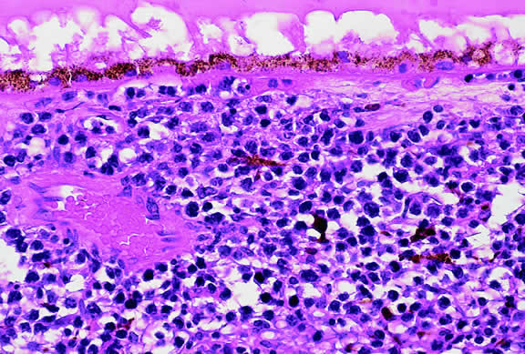
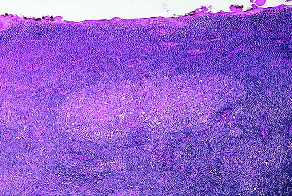
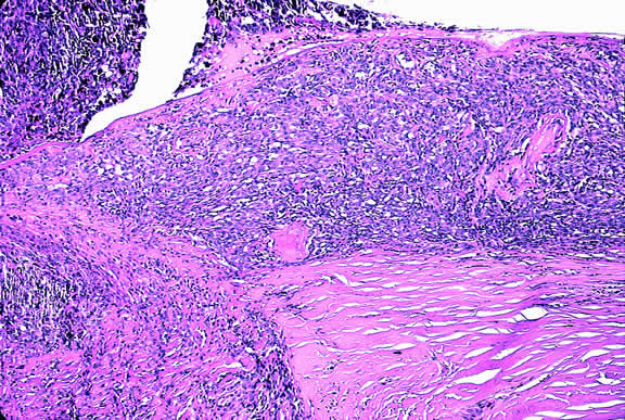
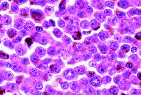
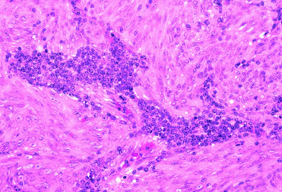
Lymphocytic infiltration of the tumor is one facet of the host response that can be assessed during the routine histopathologic examination (Fig. 47). In most parts of the body, a lymphocytic response to a malignant tumor generally is associated with an improved prognosis. In uveal melanoma, however, the presence of tumor infiltrating lymphocytes is associated with decreased survival.171 De la Cruz and coworkers used light microscopy to examine 1078 cases of uveal melanoma with known survival and found that 12.4% harbored 100 or more lymphocytes per 20 high-power (× 400) microscopic fields. The survival rate at 15 years was 36.7% for patients in the high lymphocytic group and 69.6% for patients in the low lymphocytic group. This seemingly counterintuitive observation is explained by the fact that extraocular dissemination of tumor cells is a requisite for stimulation of a T lymphocyte-mediated immune response. Ocular antigens are processed primarily in the spleen because the eye lacks lymphatics. Using immunohistochemistry, Whelchel and colleagues172 confirmed that lymphocytic infiltration is associated with death from metastasis and showed that T lymphocytes were the predominant cell type in about three fourths of the tumors.

Fig. 47. Uveal melanoma with prominent focus of lymphocytic infiltration. The presence of tumor infiltrating lymphocytes in uveal melanoma is associated with decreased survival. (Hematoxylin-eosin, × 100.)

Genetic determinants such as the genes for human leukocyte antigen (HLA) antigens also appear to be important in the development and clinical behavior of uveal melanoma. An association between the presence of HLA-B40 and death resulting from metastasis of uveal melanoma has been reported.173,174 High expression of class I HLA antigen HLA-B significantly correlates with the presence of epithelioid cells in the tumor, and high expression of HLA-A and -B are associated with decreased survival. Patients had improved survival when their tumors did not express HLA-A and -B. These data suggest that the low expression of HLA class I on micrometastases shed from uveal melanomas facilitates their removal from the systemic circulation and prevents the development of metastases. These findings support a protective role for natural killer cells in the development of metastatic disease.

Therapy

Most posterior melanomas are treated by enucleation or radiotherapy, typically plaque brachytherapy.48,54,175,176 Irradiation therapy with beams of protons or helium ions is performed at a few centers.177–179 Larger tumors or tumors that have caused secondary glaucoma generally are enucleated. Medium-sized tumors can be treated with brachytherapy. The COMS has shown that the mortality rates after I-125 plaque therapy and enucleation are similar.180 Another arm of the COMS study showed that pre-enucleation external beam radiotherapy of large choroidal melanomas does not improve survival.181 Although plaque brachytherapy conserves eyes, radiation retinopathy or papillopathy occurs commonly leading to loss of useful vision.89,182 Almost half of treated eyes have 20/200 vision 3 years after therapy.182 Some smaller tumors can be locally resected by partial lamellar sclerouvectomy. Today, this technique generally is performed on iridociliary tumors. Small relatively thin tumors of the posterior choroid can be treated with transpupillary thermotherapy (TTT), a infrared diode laser therapy that kills tumors cells by slowly heating them.89 A sandwich technique combining TTT and plaque brachytherapy has been used prevent tumor recurrence from cells sheltered in scleral canals at the base of a tumor.

If a pigmented lesion of the iris is thought to be a melanoma based on documented growth or other clinical factors, it can be locally excised by iridectomy, or by iridocyclectomy if there is focal angle and ciliary body involvement. Enucleation is often necessary when an iris melanomas has caused glaucoma or is unresectable.98 Enucleation also may be done after histopathologic examination has shown that a previously resected tumor is a high-grade lesion.98 Plaque brachytherapy occasionally is used to treat unresectable iris melanomas.54

Back to Top
THE DIFFERENTIAL DIAGNOSIS OF POSTERIOR UVEAL MELANOMA
A wide variety of neoplastic and non-neoplastic conditions must be differentiated from posterior uveal melanoma.48,54 Other intraocular neoplasms include benign melanocytic nevi, choroidal hemangiomas, metastases from distant nonocular primary neoplasms, and a variety of relatively rare intraocular tumors. The latter include peripheral nerve sheath tumors, leiomyomas, hemangiopericytomas, osteomas, adenomas and adenocarcinomas of the retinal and ciliary epithelia, fibrous histiocytoma, juvenile xanthogranuloma and the Letterer-Siwe variant of Langerhans' cell histiocytosis. Most of these tumors are discussed below.

Non-neoplastic lesions that can be confused with melanoma include choroidal hemorrhage, age-related macular degeneration, peripheral disciform degeneration, massive gliosis, and idiopathic sclerochoroidal calcification. It is uncertain whether vasoproliferative tumors of the fundus are truly neoplastic.183

UVEAL METASTASES

Although ocular oncologists see many more patients with uveal melanoma, metastatic nonocular cancer probably is the most common malignant intraocular neoplasm. A prospective postmortem study of 716 eyes obtained from patients who had malignant neoplasms at the time of death showed that the overall incidence of ocular metastases among all fatal cases of cancer was 9.3%.184 The total incidence of ocular metastases in patients dying of all types of carcinoma was 4.0%. The authors estimated that the incidence of ocular metastases is more than 10 times greater that the incidence of uveal melanoma. However, it must be emphasized that most of these secondary tumors occur in terminally ill patients, and few are detected clinically or referred to ophthalmologists.

Most of the solid tumors that metastasize to the uvea are carcinomas; sarcomas rarely metastasize to the eye. Although any part of the eye may be involved by metastatic tumor, the uveal tract, especially the posterior part of the choroid, is affected most often (Fig. 48). A retrospective review of 520 eyes with uveal metastases in 420 patients evaluated by the Oncology Service at the Wills Eye Hospital during a 20-year period showed that 88% of 950 metastatic foci involved the choroid.185 The iris was involved in 9% and the ciliary body in 2%. Most of the choroidal metastases involved the macula (12%) or the region between the macula and the equator (80%). The posterior choroid is affected most often because its blood supply is greater. Metastases to the retina, vitreous, and optic disc are relatively uncommon.186–190

Fig. 48. Metastatic breast carcinoma. The posterior pole contains multiple nodules of amelanotic tumor. The patient was known to have metastatic mammary carcinoma. (Photo courtesy of Dr. Carol L. Shields, Wills Eye Hospital)

Choroidal metastases typically appear as yellow or creamy yellow, nummular, sessile dome- or plateau-shaped masses (see Fig. 48). A secondary serous retinal detachment usually is present. The metastases may be solitary or multiple and occasionally may involve both eyes. In a study by Shields and associates,185 29% of the eyes contained 2 or more metastases, and 17% contained three or more foci of metastatic tumor. Visual loss usually is caused by the exudative retinal detachment, which often has shifting subretinal fluid. Metastases usually are creamy yellow in color but can appear partially pigmented if necrosis has produced dispersion of uveal pigment. Metastases from skin melanomas are often grayish or brown. Metastases from carcinoid tumors, thyroid carcinoma, or renal cell carcinomas may be orange in color.

Iris metastases appear as one or more yellow, pink, or white nodules in the iris stroma. The nodules are often friable and seed the aqueous humor with tumor cells simulating inflammation.191 A pseudohypopyon of tumor cells may develop, and secondary glaucoma is common. The nodules often contain blood vessels and hemorrhage may develop. Ciliary body metastases often involve the inferior quadrant and are difficult to visualize. They can produce inflammatory signs and simulate iridocyclitis.48 Ocular metastases from cutaneous melanoma often cause vitreous seeding by pigmented tumor cells.189,192,193 Retinal metastases are quite rare and can masquerade as an inflammatory process.48,186,187 Shields and colleagues190 have reported 30 patients who had metastatic tumors in the optic disc.

Breast and lung carcinomas are the primary tumors that metastasize to the uvea most often and are responsible for more than two thirds of cases.185,194 Nearly half (47%) of the 420 patients reported by Shields and associates had breast carcinoma, and about one fifth (21%) had lung cancer. Other primary tumors included gastrointestinal tract (4%), kidney (2%), skin (2%), prostate (2%), and other cancers (4%).185 The primary tumor may be occult when the patient presents to the ophthalmologist. In Shields' series, one third of the patients had no history of a primary cancer at the time of ocular diagnosis. Subsequent evaluation showed that the occult primary tumor was lung carcinoma in 35% and breast carcinoma in 7%. However, no primary tumor was discovered in 51%. Women with metastases from breast carcinoma usually have a prior history of mastectomy; only 5% in the Shields' series presented with an occult mammary tumor. In contrast, metastasis often was the presenting manifestation of an occult lung cancer; only 45% of the cases with metastatic lung cancer were know to have cancer when the ocular diagnosis was made. Metastatic lung cancer is more common in men.

The prognosis of patients with uveal metastasis generally is quite poor; the mean survival has been reported to be approximately 9 to 10 months.195,196 Treatment is recommended if the metastasis appears active and is threatening vision or the globe.185 Radiation therapy is recommended if the metastasis is symptomatic or continues to grow despite systemic chemotherapy. Plaque brachytherapy is advantageous compared with external beam radiotherapy because it can be delivered over a relatively short period of the patient's limited remaining life span.

The diagnosis of intraocular metastasis usually is made by slit lamp biomicroscopy and ophthalmoscopy in the patient who has been carefully questioned about a past medical history of cancer. Ancillary techniques such as intravenous fluorescein angiography and ultrasonography often can assist in making the diagnosis. Metastases generally begin to show hyperfluorescence in the late venous phase fluorescein angiography, somewhat later than most melanomas or hemangiomas.48,54 Metastases have many acoustical interfaces because they are composed of nests, cords, and island of tumor cells surrounded by stroma. Hence, they show high internal reflectivity on A scan ultrasonography and appear acoustically solid in B scan, characteristics they share with hemangiomas (Fig. 49). In cases in which the results from other studies are equivocal, cytopathologic examination of material obtained by fine-needle aspiration biopsy may establish the diagnosis.82,83

Fig. 49. B-scan ultrasound, choroidal metastasis. Placoid tumor has high internal reflectivity.

Today, relatively few eyes with metastatic carcinoma are accessioned by ophthalmic pathology laboratories because diagnosis is made clinically and most eyes are treated with radiotherapy and/or chemotherapy. Relief of pain is a major indication for enucleation of eyes with advanced disease.

Macroscopically, the appearance of eyes with uveal metastases is somewhat variable. In most instances the uveal tract is diffusely thickened by an infiltrate of white, pink, or yellow tissue (Fig. 50). Metastases occasionally have a multinodular growth pattern, and a few larger lesions are oval in configuration (Fig. 51). Bruch's membrane almost always remains intact, however. Although there are exceedingly rare exceptions to the rule, one generally can conclude that a mushroom-shaped tumor of the choroid is a malignant melanoma.

Fig. 50. Uveal metastasis. Uveal stroma in enucleated eye is diffusely thickened by focally hemorrhagic infiltrate of metastatic tumor.

Fig. 51. Large oval uveal metastasis. Nonpigmented tumor contains foci of hemorrhage. Few eyes with metastases currently are enucleated; most contain large tumors that have caused blindness and pain.

Microscopically, the uveal stroma is infiltrated by nest, cords, islands, and sheets of tumor cells whose general appearance and arrangement is dependent on the identity of the primary neoplasm. Most of the breast and lung tumors that metastasize to the eye are mucous-secreting adenocarcinomas. In such cases special stains such as Alcian blue, PAS, or mucicarmine are used to demonstrate the presence of intracytoplasmic mucin (Fig. 52). In most instances the pathologist can readily distinguish between a primary uveal melanoma and metastatic carcinoma in routine sections. Immunohistochemistry may be helpful in exceptional cases. Carcinomas are distinguished by positive immunoreactivity for epithelial markers such as cytokeratins and epithelial membrane antigen. Most melanomas stain with S-100 protein and vimentin and a variety of other markers including so-called melanoma specific antigen HMB-45, melan A, and microphthalmia factor. Occasionally, immunohistochemistry can confirm or provide clues to the identity of the primary tumor by demonstrating the presence of tumor specific antigens such as prostate specific antigen (PSA) in prostatic carcinoma or thyroglobulin in thyroid. Distinguishing between a primary uveal melanoma and choroidal metastasis from skin melanoma or another primary nonocular pigmented neoplasm may be challenging.197,198

Fig. 52. Mucin-secreting adenocarcinoma metastatic to the uvea. Alcian blue stain highlights mucin vacuoles in nests of tumor cells. (Alcian blue, × 100.)

LYMPHOPROLIFERATIVE TUMORS

Leukemia

Secondary ocular involvement is relatively common in patients with advanced leukemia; Leonardy and coworkers199 found ocular involvement in 31% of 135 eyes obtained postmortem from leukemic patients. Ocular tissues may involved primarily by infiltrates of leukemic cells or secondarily by manifestations of the disease including hemorrhage, ischemia, or secondary infection related to primary immunosuppression or the effect of immunosuppressive drugs.200,201 Although leukemic cells can infiltrate almost any ocular tissue, choroidal infiltration occurs most often. Kinkaid and Green201 found some degree of choroidal infiltration in 65% of 367 pairs of postmortem eyes from leukemic patients examined histopathologically. Significant choroidal involvement often is accompanied by a shallow serous retinal detachment at the posterior pole. Secondary retinal changes include retinal hemorrhages, white centered hemorrhages, cotton-wool spots, nonperfusion, vitreoretinal neovascularization, and central retinal vein occlusion. Vitreous and choroidal hemorrhage also may occur. Infiltration of the optic nerve may produce an optic neuropathy.202–204 Iris infiltration may manifest clinically as iris heterochromia, a pseudohypopyon of leukemic cells, or hyphema.

Lymphoma

Although non-Hodgkin's lymphoma often affects the orbit and conjunctiva, intraocular involvement by lymphoma is relatively rare and occurs in two basic ways. First, patients who have widely disseminated systemic lymphoma may develop secondary ocular involvement whose typical manifestation is uveal infiltration. Second, in the variant of central nervous system (CNS) lymphoma that has been called primary lymphoma of the CNS and retina, the vitreous characteristically is involved and the uvea is spared. The latter entity also has been called vitreous large cell lymphoma or ocular “reticulum cell sarcoma.”205–208 In addition to the vitreous, the lymphoma cells also infiltrate the retina, and typically collect between Bruch's membrane and the retinal pigment epithelium, forming solid yellowish RPE detachments, which are highly suggestive of the diagnosis. This rare variant of primary CNS lymphoma should be suspected in older patients who have chronic vitritis that is unresponsive to therapy. Cytologic examination of diagnostic vitrectomy specimens reveals a highly cellular and extensively necrotic infiltrate that contains atypical lymphocytes with prominent nucleoli and protrusions of the nuclear membrane (Fig. 53). The significance of elevated vitreous interleukin 10 levels as a diagnostic marker for lymphoma is controversial.209,210

Fig. 53. Primary central nervous system lymphoma, vitreous. Diagnostic vitrectomy specimen contains large atypical lymphocytes, necrotic lymphoid cells, and nuclear debris. Lymphoma cells in inset have nuclear membrane protrusions and prominent nucleoli. Main figure, Millipore filter. (Hematoxylin-eosin, × 250.)

Many patients who undergo diagnostic vitrectomy to exclude lymphoma actually are found to have a form of granulomatous vitreitis termed idiopathic senile vitritis.211 Cytologically, the latter lacks necrosis and contains a mixture of well-differentiated lymphocytes and epithelioid histiocytes with a spindled or dendritiform configuration. Careful cytologic screening and follow-up are warranted in such cases, however, because vitreous lymphoma occasionally presents with chronic inflammation. Imaging studies and spinal fluid examination should be performed to exclude CNS involvement if vitreous lymphoma is diagnosed.206 CNS lymphoma may produce dementia and other neurologic signs. The prognosis is poor with a mean survival of 22 months. Intraocular Whipple's disease may mimic vitreous lymphoma.212,213

The vitreous usually is spared when disseminated, non-CNS, non-Hodgkin's visceral lymphomas involve the eye secondarily (Fig. 54). Such lymphomas usually involve the uvea, but vitreous infiltration does occur sporadically. Occasional patients with intraocular lymphoma may present with iris heterochromia or a pseudo-hypopyon of lymphoma cells.

Fig. 54. Secondary choroidal involvement by disseminated large cell non-Hodgkin's lymphoma. Eye was obtained postmortem. (Hematoxylin-eosin, × 100.)

Recent studies have shown that the rare disorder previously called reactive lymphoid hyperplasia of the uvea probably is a form of lymphoma214,215 (Fig. 55). Affected patients may have massive infiltration of the uveal tract by low-grade non-Hodgkin's B cell lymphoma that may have germinal centers. Contiguous extraocular foci of tumor also may be present.216 The latter provide an opportunity to establish the diagnosis without an intraocular biopsy.

Fig. 55. Reactive lymphoid hyperplasia of the uvea. An infiltrate of well-differentiated lymphocytes thickens the choroid. The paler focus is a large germinal center. Additional foci of lymphoid cells were present on the epibulbar surface of the eye. Reactive lymphoid hyperplasia of the uvea is now thought to be a low-grade lymphoid neoplasm. (Hematoxylin-eosin, × 50.)

Lymphoid tumors of the iris and choroid have been reported in immunosuppressed patients with posttransplantation lymphoproliferative disorder.217–219 The lymphoid tumors are caused by Ebstein Barr virus, and some improve if immunosuppression is reduced.219

Juvenile xanthogranuloma generally affects the iris stroma in infants but can infiltrate the posterior uvea.220 It is an important cause of spontaneous hyphema. The infiltrate contains Touton giant cells rimmed with lipid. Several cases of posterior uveal involvement by Letterer-Siwe disease have been reported.221,222

LEIOMYOMA

Leiomyoma is a relatively rare nonpigmented tumor of the uveal tract that usually involves the ciliary body and typically affects young women.223,224 During transillumination, leiomyomas often transmit light vividly. The latter attribute, occurring in a young woman, should suggest that a nonpigmented ciliary body tumor may be a leiomyoma and not an amelanotic melanoma. Although leiomyoma is benign, local resection is recommended to prevent complications from the growing tumor.223

Histopathologically, leiomyoma is relatively paucicellular compared with amelanotic melanoma, and its cells have fibrillar eosinophilic cytoplasm and bland nuclei with finely dispersed chromatin (Fig. 56). Positive immunoreactivity for smooth muscle actin (Fig. 57) and other muscle markers serves to differentiate leiomyoma from uveal melanoma and rare neurogenic tumors such as schwannoma, which also are paucicellular. The latter distinction is important because some leiomyomas have a distinctly neural appearance on routine light microscopy. The term mesectodermal leiomyoma has been applied to such tumors, emphasizing the derivation of intraocular smooth muscle from neural crest.225

Fig. 56. Leiomyoma, ciliary body. This benign spindle cell tumor of smooth muscle derivation is relatively paucicellular compared with an amelanotic spindle cell melanoma. The spindle cells have bland nuclei and finely fibrillar cytoplasm. (Hematoxylin-eosin, × 100.)

Fig. 57. Leiomyoma, ciliary body. Positive immunoreactivity for smooth muscle actin confirms that tumor is composed of smooth muscle cells. (Immunoperoxidase, × 250.)

VASCULAR TUMORS

Choroidal Hemangioma

Choroidal hemangiomas occur sporadically as circumscribed tumors or as diffuse lesions in patients with Sturge-Weber syndrome. Sporadic circumscribed or solitary hemangiomas of the choroid appear clinically as subtle red-orange tumefactions in the posterior choroid48,226,227 (Fig. 58). In contrast, the hemangiomas in patients with Sturge-Weber Syndrome typically are diffuse lesions that obscure normal choroidal landmarks and impart a “tomato-ketchup” appearance to the fundus on ophthalmoscopy. Both types of hemangiomas often have an associated serous detachment of the neurosensory retina that involves the fovea.227 Hemangiomas can produce visual loss if they are located beneath the fovea and induce hyperopia or if they produce a retinal detachment.

Fig. 58. Solitary choroidal hemangioma. Localized tumor is orange-red in color.

Choroidal hemangiomas are benign vascular hamartomas composed of relatively large, thin-walled vascular channels lined by endothelial cells (Fig. 59). Based on the size of the vascular channels, choroidal hemangiomas have been classified as cavernous, capillary, or mixed. In contrast to cavernous hemangiomas in the orbit, choroidal hemangiomas are relatively devoid of stroma. They lack the thick fibrous septa found in the orbital lesions and the individual vascular channels almost appear to abut each other. Solitary tumors have clearly demarcated pushing margins that cause compression of adjacent melanocytes and choroidal lamellae.228 Patients with Sturge-Weber syndrome have a diffuse angiomatosis that involves more than half of the choroid and shows intermixture of engorged preexisting vessels with the vascular tumor.

Fig. 59. Choroidal hemangioma. Tumor is composed of relatively large, thin-walled vessels with little intervening stroma. (Hematoxylin-eosin, × 100.)

Choroidal hemangiomas often produce secondary changes in adjacent ocular structures in addition to retinal detachment. In addition to photoreceptor degeneration, the overlying retina often shows marked cystoid degeneration that may progress to retinoschisis in chronic cases. Varying degrees of RPE hyperplasia and metaplasia may develop; in rare instance circumscribed hemangiomas may be capped by dense plaques of fibrous metaplasia or even bone.229,230

Although choroidal hemangiomas are benign neoplasms, they can prove “fatal” to the eye that harbors one. Vision is lost to chronic retinal detachment, and the eye, as well, can be lost to secondary closed-angle glaucoma caused by iris neovascularization or pupillary block resulting when a high bullous retinal detachment displaces the lens-iris diaphragm anteriorly. About three quarters of eyes with the diffuse hemangiomas of Sturge-Weber syndrome have glaucoma. Mechanisms include iris neovascularization, maldevelopment of the angle, and elevation of episcleral venous pressure related to associated epibulbar vascular hamartomatous changes.228,231

Circumscribed choroidal hemangiomas are often misdiagnosed clinically as melanoma or metastasis.227 Intravenous fluorescein angiography is often helpful in establishing the correct diagnosis. IVFA typically shows lacy hyperfluorescence of vessels in the tumor in the prearterial phase and diffuse late staining of the mass.48 Cystoid edema within the overlying retina may be prominent. Indocyanine green angiography shows early filling and a characteristic “washout” of hyperfluorescence in the later frames.232

A scan ultrasonography shows high internal reflectivity within the tumor, and B scan shows a placoid or round choroidal mass that is acoustically solid. A highly reflective cap may be present on the surface of hemangiomas with fibrous or osseous metaplasia.229 Magnetic resonance imaging shows a choroidal hemangioma to be hyperintense to the vitreous on T1-weighted images and isointense to vitreous on T2-weighted images.227

Treatment of the benign vascular hamartoma is designed to preserve the eye by controlling exudative retinal detachment. Treatment modalities include delimiting laser photocoagulation, low-dose plaque radiotherapy, lens-sparing external beam radiotherapy, TTT, and photodynamic therapy using verteporfin.54,230,233 Visual prognosis is guarded, however. More than 60% of patients have poor visual acuity 10 years after treatment despite successful control of associated subretinal fluid.227

Hemangiopericytoma

Rare cases of hemangiopericytoma of the ciliary body and choroid have been reported.48,234,235 Clinically it is nearly impossible to differentiate these exceedingly rare nonpigmented vascular tumors from choroidal hemangiomas. Hemangiopericytomas typically are well-circumscribed neoplasms that contain pericytes, capillaries, and larger branching vascular sinusoidal vessels with a “stag horn” configuration. The pericytes typically are surrounded by capsules of reticulin and are immunoreactive for vascular marker CD34.

Retinal Capillary Hemangioma

Vascular tumors of the retina called retinal capillary hemangiomas or hemangioblastomas occur sporadically or in association with von Hippel-Lindau (VHL) disease, a hereditary cancer syndrome caused by germline mutations in a tumor suppressor gene located on chromosome 3p25.236–239 Ocular lesions classically appear as reddish-pink, globular cherry angiomas in the peripheral retina that have dilated and tortuous feeding and draining vessels48,54 (Fig. 60). The hemangiomas may arise from the outer retinal layers in an exophytic fashion or from the surface or parenchyma of the optic disc. The tumors can produce exudative retinal detachment and can present with Coats'-like exudative maculopathy.

Fig. 60. Retinal hemangioblastoma, von Hippel-Lindau disease. Classic endophytic cherry angioma has prominent retinal vessels. Retinal exudates are present nearby. (Photo courtesy of Dr. Arun Singh)

Histopathologically, the retinal angiomas appear identical to lesions of VHL disease that are called hemangioblastomas when they occur elsewhere in the CNS, particularly the cerebellum, spinal cord, and brainstem. In addition to capillary vessels, the tumors have a second population of stromal cells with foamy cytoplasm (Fig. 61). Atypical appearing glial cells also are common. Molecular genetic studies have demonstrated that loss of heterozygosity is confined to the stromal cells, suggesting that the latter actually are the neoplastic component in the tumors.240 Proliferation of capillaries is thought to be a secondary response to vascular endothelial growth factor (VEGF) produced by the stromal cells.

Fig. 61. Retinal hemangioblastoma, von Hippel-Lindau disease. Retinal tumor is comprised of capillaries and foamy lipidized stromal cells. Histology of retinal tumor is identical to central nervous system hemangioblastoma. (Hematoxylin-eosin, × 250.)

The retinal hemangioblastomas are bilateral in 50% of cases. Patients who are diagnosed with retinal capillary hemangiomas and have VHL are, on average, almost 18 years younger than those without VHL disease.241 Approximately half of the patients presenting with a solitary retinal capillary hemangioma are expected to have underlying VHL disease; the risk progressively diminishes with increasing age at diagnosis.242

Patients with VHL disease are at risk for developing hemangioblastomas of the CNS, classically cystic tumors of the cerebellum. The tumor suppressor gene mutation also predisposes them to pheochromocytoma and retinal cell carcinoma. Clear cell renal cell carcinoma occurs in up to 70% of patients with VHL and is a frequent cause of death.239

Vasoproliferative Tumor of the Ocular Fundus

Vasoproliferative tumor of the ocular fundus (VPTOF) is the term applied by Shields and associates183 to a vascularized fundus lesion that they sought to differentiate from retinal capillary hemangioma and melanoma. Generally these tumors appear ophthalmoscopically as an elevated reddish or yellow-pink mass that has a retinal feeding artery and draining vein. The vessels typically are not as dilated and tortuous as those associated with the retinal angiomas of VHL disease. Exudation is also characteristic but is located within the retina posterior to and continuous with the mass and does not have an affinity for the macula as does the Coats'-like exudative response seen in VHL disease. The authors initially suggested that VPTOF were acquired retinal hemangiomas, but they now believe that the lesion may be a reactive phenomenon and not a primary vascular neoplasm.

Primary and secondary variants of VPTOF are recognized.183 The primary or idiopathic type generally is a unilateral, solitary lesion found in the inferotemporal fundus between the equator and the ora serrata. Affected patients often are hypertensive. The secondary type occurs in eyes that have predisposing conditions such as intermediate uveitis, retinitis pigmentosa, ocular toxocariasis, Coats' disease, chronic retinal detachment, and other conditions associated with chronic inflammation or ocular trauma. Scant histopathologic data suggest that the tumor is a reactive process rather than a true neoplasm.243,244 The tumefactions appear to be composed primarily of benign glial cell proliferation with secondary vasoproliferation. RPE hyperplasia is an additional component in some cases. Although some vasoproliferative tumors remain asymptomatic and some regress, others produce exudation and vitreous hemorrhage warranting cryotherapy, laser photocoagulation, or plaque brachytherapy.48,245 Local resection has been performed in several cases in which melanoma could not be excluded on clinical grounds.244

NEUROGENIC TUMORS

A diffuse infiltrate of hamartomatous tissue may diffusely thicken the choroid in patients with von Recklinghausen's neurofibromatosis (NF-1). The infiltrate often contains increased numbers of melanocytes, which predispose to the development of uveal melanoma, and characteristic ovoid bodies composed of concentric lamellae of Schwann cells.93 Increased numbers of ganglion cells occur in some cases. A ganglioneuroma of the choroid has been reported in a young woman with NF-1.246

Benign peripheral nerve sheath tumors such as neurofibromas or schwannomas or neurilemomas are extremely rare uveal tumors that may be impossible to distinguish clinically from amelanotic malignant melanoma.247–250 Histopathologically, schwannomas generally are paucicellular compared with melanoma and may show palisading of nuclei (Fig. 62). Immunohistochemical data must be interpreted with care because these neural tumors, like melanomas, are immunoreactive for S-100 protein. They do not stain positively for other melanoma markers such as HMB-45 and melan A, however. A melanotic schwannoma of the choroid has been reported.251

Fig. 62. Benign peripheral nerve sheath tumor, choroid. The cytology of this bland paucicellular choroidal tumor is consistent with a schwannoma. Schwannomas are extremely rare intraocular tumors. Immunohistochemical stains or electron microscopy are necessary to confirm the diagnosis. (Hematoxylin-eosin, × 50.)

NEUROECTODERMAL TUMORS IN ADULTS

Most retinal tumors are retinoblastomas that occur in children before 3 years of age. However, older children may present with the relatively rare diffuse infiltrating form of retinoblastoma. Diffuse infiltrating retinoblastoma is universally unilateral and sporadic and the mean age of affected children is 6 years. Many cases are initially thought to have inflammation.252 An intraocular tumor should also be considered in a child with unilateral neovascular glaucoma.

Medulloepithelioma generally is a pediatric neoplasm that presents at about age 4 years. Rare cases have been reported in adults, however.253 Most cases are ciliary body tumors.254,255 Benign and malignant and teratoid and nonteratoid variants occur. Teratoid medulloepitheliomas contain foci of heteroplastic tissue including cartilage, brain, and striated muscle. Exceptionally rare intraocular rhabdomyosarcoma reported in young adults may represent one-sided differentiation of malignant teratoid medulloepithelioma.256

Astrocytic hamartomas of the retina are a characteristic finding in patients with the tuberous sclerosis complex (TSC).257–259 In infancy, they may be very difficult to distinguish clinically from small retinoblastomas. Calcospherites often accumulate within maturing tumors giving rise to the appearance of mulberry nodules.48,54 Some tumors in patients with TSC appear to be giant cell astrocytomas, which are similar histologically to the characteristic subependymal lesions that occur in the walls of the cerebral ventricles.258 They may be extensively necrotic and produce total retinal detachment.260 Acquired retinal astrocytomas also occur sporadically in patients who do not have TSC259 (Fig. 63).

Fig. 63. Retinal astrocytoma. Spindle cell tumor replaces posterior retina. The patient did not have tuberous sclerosis complex. (Hematoxylin-eosin, × 5.)

Primary acquired neoplasms occasionally arise from the retinal pigment epithelium and iris pigment epithelium and the pigmented and nonpigmented epithelia of the ciliary body. All of these layers are derived embryologically from the neuroectodermal optic cup. These neuroectodermal tumors include adenomas and adenocarcinomas.

Adenomas of the iris pigment epithelium are sharply demarcated black tumors that push aside, rather than infiltrate, the iris stroma (Fig. 64). The cytoplasm of these exceedingly rare tumors contains large round pigment granules.261

Fig. 64. Adenoma of iris pigment epithelium. Circumscribed lobule of iris pigment epithelial cells protrudes through anterior border layer of iris (top right) pushing normal stroma aside. (Hematoxylin-eosin, × 50.)

Ciliary epithelial tumors (Figs. 65 to 69) can be predominantly pigmented or nonpigmented. Small pseudoadenomatous proliferations of the nonpigmented ciliary epithelium called Fuchs' or coronal adenomas are a common incidental finding in older adult eyes262,263 (see Figs. 65 and 66). The cytoplasm of many pigmented ciliary epithelial tumors contains multiple small cystoid spaces264 (see Fig. 67). Nonpigmented tumors are white or yellowish white in color and often produce focal cataract or lens dislocation. Many contain pools of hyaluronic acid, which can be extensive in some cases265 (see Figs. 68 and 69). In contrast to melanoma, which arises from the uveal stroma, ciliary epithelial tumors arise from the epithelium lining the inner surface of the ciliary body. Local resection usually is curative. Many ciliary epithelial tumors are benign, but malignant variants have been reported.265–271 Invasion, mitotic activity, and nuclear pleomorphism are signs of malignancy.

Fig. 65. Coronal (Fuchs') adenoma. Adenoma is evident as white nodule on ciliary process in high magnification macrophoto.

Fig. 66. Coronal (Fuchs') adenoma. Hyperplastic nonpigmented ciliary epithelium surrounds acellular stroma of extracellular matrix material. (Hematoxylin-eosin, × 100.)

Fig. 67. Cystic adenoma of pigmented ciliary epithelium. The darkly pigmented tumor contains many small cystoid spaces. The tumor rests on the inner surface of the pars plicata. (Hematoxylin-eosin, × 50.)

Fig. 68. Adenoma of nonpigmented ciliary epithelium. Nonpigmented ciliary epithelial tumor rests on inner surface of ciliary body (above). Clear spaces contain hyaluronic acid. (Hematoxylin-eosin, × 50.)

Fig. 69. Adenoma of nonpigmented ciliary epithelium. Colloidal iron stain confirms that clear spaces contain mucopolysaccharide, which was hyaluronidase-sensitive. (Hale's colloidal iron stain, × 50.)

RETINAL PIGMENT EPITHELIAL TUMORS

Primary neoplasms of the retinal pigment epithelium are exceedingly rare. This fact is somewhat surprising because the retinal pigment epithelium readily undergoes reactive hyperplasia and metaplasia forming extensive amounts of fibrous tissue and bone. Although amelanotic tumors have been reported, RPE tumors are typically jet black in color and have abruptly elevated margins. They often perforate the overlying retina and often are associated with retinal exudation. RPE tumors initially are situated on the inner surface of the choroid. Malignant variants can invade the choroidal stroma. It generally is thought that RPE carcinomas do not metastasize.

Histopathologically, tumors arising from the anterior part of the retinal pigment epithelium have a vacuolated pattern similar to adenomas of the pigmented ciliary epithelium (Fig. 70). The cells of tumors arising from the posterior part of the retinal pigment epithelium are often arranged in strands and may form tubules or pseudoglands (Fig. 71). The cells typically rest on the surface of prominent PAS-positive connective-tissue septa.272 Epipapillary RPE adenoma may simulate optic disc melanocytoma.23

Fig. 70. Adenoma, peripheral retinal pigment epithelium. Cystic spaces in tumor arising from peripheral retinal pigment epithelium resemble those found in cystic adenoma of pigmented ciliary epithelium. Tumor is infiltrating overlying retina. (Hematoxylin-eosin, × 50.)

Fig. 71. Retinal pigment epithelial adenocarcinoma. Low-grade carcinoma is composed of cords of pigmented epithelial cells. A few cells have prominent nucleoli. A mitotic figure is present. Tumor arose from congenital hypertrophy of retinal pigment epithelium (CHRPE) (see Fig. 74). (Hematoxylin-eosin, × 250.)

Congenital hypertrophy of the retinal pigment epithelium (CHRPE) appears ophthalmoscopically as a flat, round or oval pigmented spot (Fig. 72), Many lesions are surrounded by a depigmented halo and most develop depigmented lacunae with time. Before the advent of the modern binocular indirect ophthalmoscope, CHRPE occasionally were confused with choroidal melanomas. Light microscopy of CHRPE discloses a focus of tall hypertrophic RPE cells packed with melanin pigment including large round macromelanosomes273 (Fig. 73). Within lacunae, the atrophic outer retina adheres to the denuded inner surface of Bruch's membrane. Although CHRPE originally was considered to be stationary, some lesions have been documented to grow by serial photography.54 Recently, Shields has observed that CHRPE rarely may give rise to solid tumors274,275 (Fig. 74). Histopathology (see Fig. 71) showed that one of these lesions was a low-grade adenocarcinoma of the retinal pigment epithelium.276

Fig. 72. Congenital hypertrophy of the retinal pigment epithelium (CHRPE). Round, flat and intensely pigmented lesion lacks lacunae.

Fig. 73. Congenital hypertrophy of the retinal pigment epithelium (CHRPE). Lesion is composed of tall hypertrophic retinal pigment epithelial (RPE) cells that are intensely pigmented. Large round macromelanosomes are present. (Hematoxylin-eosin, × 250.)

Fig. 74. Retinal pigment epithelial (RPE) adenocarcinoma arising from congenital hypertrophy of retinal pigment epithelium (CHRPE). Nodule of amelanotic RPE tumor partially obscures CHRPE. (Photo courtesy of Dr. Jerry A. Shields)

A variant of CHRPE is congenital grouped pigmentation (“bear tracks”).277 Recently, multiple bilateral pigmented RPE lesions that bear a superficial resemblance to CHRPE were reported as an ocular marker for the heritable cancer diathesis Gardner's syndrome (familial adenomatous polyposis with extracolonic manifestations).278,279

CHOROIDAL OSTEOMA

Choroidal osteoma is a benign tumor composed of bone, which usually is found in young women.48,280 It may be unilateral or bilateral and may occur in siblings.281 Choroidal osteoma appears clinically as a yellow-orange placoid tumor with sharply defined scalloped margins (Fig. 75). It typically is located next to the optic disc or in the macula. Osteomas may enlarge,282 become decalcified,283,284 undergo spontaneous involution,285 or develop choroidal neovascularization.48 Osteomas may be confused ophthalmoscopically with metastases but are easily distinguished by ultrasonography, which shows a highly reflective plaque that persists at lower sensitivity, or by computed tomography that reveals a plaque with bone density. Serum calcium, phosphorus, and alkaline phosphatase levels usually are normal. The visual prognosis is unpredictable.286 Only a single case of choroidal osteoma has been examined histopathologically.287 A plaque of mature bone was found within the stroma of the choroid (Fig. 76). In contrast, bone is found on the inner surface of Bruch's membrane in osseous metaplasia of the retinal pigment epithelium, which is a relatively common occurrence in phthisical eyes with chronically detached retinas.

Fig. 75. Choroidal osteoma. Yellow-orange peripapillary tumor has characteristic scalloped margins.

Fig. 76. Choroidal osteoma. The tumor is composed of irregular spicules of bone surrounded by an areolar stroma containing large vascular channels. The bone is within the choroid, deep to Bruch's membrane, choriocapillaris and an intact layer of retinal pigment epithelium. The intrachoroidal location of the bone distinguishes choroidal osteoma from osseous metaplasia of the retinal pigment epithelium. (Hematoxylin-eosin, × 50.)

Idiopathic uveoscleral calcification occurs occasionally in the eyes of older patients and may be confused with choroidal metastasis or an amelanotic melanoma.288–290 It usually is found in the superotemporal quadrant in the vicinity of the insertion of the superior oblique tendon.

Back to Top
REFERENCES

1. Eagle RC Jr. Iris pigmentation and pigmented lesions: An ultrastructural study. Trans Am Ophthalmol Soc 1988;86:581

2. Naumann GOH, Hellner HA, Naumann LR. Pigmented nevi of the choroid: Clinical study of secondary changes in the overlying tissue. Trans Am Acad Ophthalmol Otolaryngol 1971;75:110

3. Sumich P, Mitchell P, Wang JJ. Choroidal nevi in a white population: The Blue Mountains Eye Study. Arch Ophthalmol 1998;116:645

4. Naumann G. [Pigmented nevi of the choroid and ciliary bodies. A clinical and histopathological study]. Adv Ophthalmol 1970;23:187

5. McLean IW. Uveal nevi and malignant melanomas. In Spencer WH (ed). Ophthalmic Pathology: An Atlas and Textbook. Philadelphia: WB Saunders:2121–2217

6. Augsburger JJ, Schroeder RP, Territo C et al. Clinical parameters predictive of enlargement of melanocytic choroidal lesions. Br J Ophthalmol 1989;73:911

7. Shields CL, Cater J, Shields JA et al. Combination of clinical factors predictive of growth of small choroidal melanocytic tumors. Arch Ophthalmol 2000;118:360

8. Gass JD. Problems in the differential diagnosis of choroidal nevi and malignant melanomas. The XXXIII Edward Jackson Memorial Lecture. Am J Ophthalmol 1977;83:299

9. Naumann G, Yanoff M, Zimmerman LE. Histogenesis of malignant melanomas of the uvea. I. Histopathologic characteristics of nevi of the choroid and ciliary body. Arch Ophthalmol 1966;76:784

10. Zimmerman LE, Garron LK. Melanocytoma of the optic disk. Int Ophthalmol Clin 1962;2:431

11. Zimmerman LE. Melanocytes, melanocytic nevi and melanocytomas. Invest Ophthalmol Vis Sci 1965;4:11

12. Cogan DG. Discussion. In Boniuk M (ed). Ocular and Adnexal Tumors: New and Controversial Aspects. St Louis: CV Mosby, 1964:385

13. Shields JA, Augsburger JJ, Bernardino V Jr et al. Melanocytoma of the ciliary body and iris. Am J Ophthalmol 1980;89:632

14. Frangieh GT, el Baba F, Traboulsi EI et al. Melanocytoma of the ciliary body: Presentation of four cases and review of nineteen reports. Surv Ophthalmol 1985;29:328

15. LoRusso FJ, Boniuk M, Font RL. Melanocytoma (magnocellular nevus) of the ciliary body: Report of 10 cases and review of the literature. Ophthalmology 2000;107:795

16. Joffe L, Shields JA, Osher RH et al. Clinical and follow-up studies of melanocytomas of the optic disc. Ophthalmology 1979;86:1067

17. Osher RH, Shields JA, Layman R. Pupillary and visual field evaluation in patients with melanocytoma of the optic disc. Arch Ophthalmol 1979;97:1096

18. Shields JA, Shields CL, Eagle RC Jr et al Malignant melanoma associated with melanocytoma of the optic disc. Ophthalmology 1990;97:225

19. Apple DJ, Craythorn JM, Reidy JJ et al. Malignant transformation of an optic nerve melanocytoma. Can J Ophthalmol 1984;19:320

20. Mansour AM, Zimmerman L, La Piana FG et al. Clinicopathological findings in a growing optic nerve melanocytoma. Br J Ophthalmol 1989;73:410

21. Shields JA, Shields CL, Eagle RC Jr et al. Central retinal vascular obstruction secondary to melanocytoma of the optic disc. Arch Ophthalmol 2001;119:129

22. De Potter P, Shields CL, Eagle RC Jr et al. Malignant melanoma of the optic nerve. Arch Ophthalmol 1996;114:608

23. Shields JA, Eagle RC Jr, Shields CL et al. Pigmented adenoma of the optic nerve head simulating a melanocytoma. Ophthalmology 1992;99:1705

24. Juarez CP, Tso MO. An ultrastructural study of melanocytomas (magnocellular nevi) of the optic disk and uvea. Am J Ophthalmol 1980;90:48

25. Fineman MS, Eagle RC Jr, Shields JA et al. Melanocytomalytic glaucoma in eyes with necrotic iris melanocytoma. Ophthalmology 1998;105:492

26. Teichmann KD, Karcioglu ZA. Melanocytoma of the iris with rapidly developing secondary glaucoma. Surv Ophthalmol 1995;40:136

27. Bhorade AM, Edward DP, Goldstein DA. Ciliary body melanocytoma with anterior segment pigment dispersion and elevated intraocular pressure. J Glaucoma 1999;8:129

28. Lee SB, Au Eong KG, Saw SM et al. Eye cancer incidence in Singapore. Br J Ophthalmol 2000;84:767

29. Margo CE, Mulla Z, Billiris K. Incidence of surgically treated uveal melanoma by race and ethnicity. Ophthalmology 1998;105:1087

30. Broadway D, Lang S, Harper J et al. Congenital malignant melanoma of the eye. Cancer 1991;67:2642

31. Shields CL, Shields JA, Milite J et al. Uveal melanoma in teenagers and children. A report of 40 cases. Ophthalmology 1991;98:1662

32. Diener-West M, Earle JD, Fine SL et al. The COMS randomized trial of iodine 125 brachytherapy for choroidal melanoma, II: Characteristics of patients enrolled and not enrolled. COMS Report No. 17. Arch Ophthalmol 2001;119:951

33. Singh AD, Shields CL, Shields JA et al. Uveal melanoma in young patients. Arch Ophthalmol 2000;118:918

34. McLean IW, Burnier MNJ, Jakobiec FA et al. Atlas of Tumor Pathology. Tumors of the Eye and Ocular Adnexa, Fascicle 12. Washington, DC: Armed Forces Institute of Pathology, 1994

35. Vajdic CM, Kricker A, Giblin M et al. Eye color and cutaneous nevi predict risk of ocular melanoma in Australia. Int J Cancer 2001;92:906

36. Holly EA, Aston DA, Char DH et al. Uveal melanoma in relation to ultraviolet light exposure and host factors. Cancer Res 1990;50:5773

37. Wells CG, Bradford RH, Fish GE et al. Choroidal melanomas in American Indians. COMS Group. Collaborative Ocular Melanoma Study. Arch Ophthalmol 1996;114:1017

38. Cheng C-Y, Hsu W-M. Malignant tumors of the eye in a Chinese population: A 17-year review. Scientific Poster 87, Annual Meeting, American Academy of Ophthalmology, 2000 Annual Meeting, Dallas, TX

39. Margo CE, McLean LW. Malignant melanoma of the choroid and ciliary body in black patients. Arch Ophthalmol 1984;102:77

40. Gonder JR, Shields JA, Albert DM et al. Uveal malignant melanoma associated with ocular and oculodermal melanocytosis. Ophthalmology 1982;89:953

41. Singh AD, De Potter P, Fijal BA et al. Lifetime prevalence of uveal melanoma in white patients with oculo(dermal) melanocytosis. Ophthalmology 1998;105:195

42. Honavar SG, Singh AD, Shields CL et al. Iris melanoma in a patient with neurofibromatosis. Surv Ophthalmol 2000;45:231

43. Ticho BH, Tso MO, Kishi S. Diffuse iris nevus in oculodermal melanocytosis: A light and electron microscopic study. J Pediatr Ophthalmol Strabismus 1989;26:244

44. Gunduz K, Shields JA, Shields CL et al. Choroidal melanoma in a 14-year-old patient with ocular melanocytosis. Arch Ophthalmol 1998;116:1112

45. Leung AK, Kao CP, Cho HY et al. Scleral melanocytosis and oculodermal melanocytosis (nevus of Ota) in Chinese children. J Pediatr 2000;137:581

46. Bordon AF, Wray ML, Belfort R et al. Choroidal malignant melanoma in association with oculodermal melanocytosis in a black patient. Br J Ophthalmol 1995;79:191

47. Nik NA, Glew WB, Zimmerman LE. Malignant melanoma of the choroid in the nevus of Ota of a black patient. Arch Ophthalmol 1982;100:1641

48. Shields JS, Shields CL. Atlas of Intraocular Tumors. Philadelphia: Lippincott Williams & Wilkins, 1999

49. Dutton JJ, Anderson RL, Schelper RL et al. Orbital malignant melanoma and oculodermal melanocytosis: Report of two cases and review of the literature. Ophthalmology 1984;91:497

50. Rice CD, Brown HH. Primary orbital melanoma associated with orbital melanocytosis. Arch Ophthalmol 1990;108:1130

51. Wilkes TD, Uthman EO, Thornton CN et al. Malignant melanoma of the orbit in a black patient with ocular melanocytosis. Arch Ophthalmol 1984;102:904

52. Balmaceda CM, Fetell MR, O'Brien JL et al. Nevus of Ota and leptomeningeal melanocytic lesions. Neurology 1993;43:381

53. Sang DN, Albert DM, Sober AJ et al. Nevus of Ota with contralateral cerebral melanoma. Arch Ophthalmol 1977;95:1820

54. Shields JA, Shields CL. Intraocular Tumors: A Text and Atlas. Philadelphia: WB Saunders, 1992

55. Albert DM, Lahav M, Packer S et al. Histogenesis of malignant melanomas of the uvea. Occurrence of nevus-like structures in experimental choroidal tumors. Arch Ophthalmol 1974;92:318

56. Antle CM, Damji KF, White VA et al. Uveal malignant melanoma and optic nerve glioma in von Recklinghausen's neurofibromatosis. Br J Ophthalmol 1990;74:502

57. Bacin F, Kemeny JL, D'Hermies F et al. [Malignant melanoma of the choroid associated with neurofibromatosis]. J Fr Ophthalmol 1993;16:184

58. Rehany U, Rumelt S. Iridocorneal melanoma associated with type 1 neurofibromatosis: A clinicopathologic study. Ophthalmology 1999;106:614

59. Singh AD, Wang MX, Donoso LA et al. Genetic aspects of uveal melanoma: A brief review. Semin Oncol 1996;23:768

60. Albert DM, Chang MA, Lamping K et al. The dysplastic nevus syndrome. A pedigree with primary malignant melanomas of the choroid and skin. Ophthalmology 1985;92:1728

61. Rodriguez-Sains RS. Ocular findings in patients with dysplastic nevus syndrome. Ophthalmology 1986;93:661

62. Singh AD, Shields JA, Eagle RC et al. Iris melanoma in a ten-year-old boy with familial atypical mole-melanoma (FAM-M) syndrome. Ophthalmic Genet 1994;15:145

63. Singh AD, Shields CL, Shields JA et al. Uveal melanoma and familial atypical mole and melanoma (FAM-M) syndrome. Ophthalmic Genet 1995;16:53

64. Shields JA, Augsburger JJ, Arbizo V et al. Malignant melanoma of the choroid in a husband and wife. Br J Ophthalmol 1984;68:623

65. Albert DM, Robinson NL, Fulton AB et al. Epidemiological investigation of increased incidence of choroidal melanoma in a single population of chemical workers. Int Ophthalmol Clin 1980;20:71

66. Barr CC, Zimmerman LE, Curtin VT et al. Bilateral diffuse melanocytic uveal tumors associated with systemic malignant neoplasms. A recently recognized syndrome. Arch Ophthalmol 1982;100:249

67. Gass JD, Gieser RG, Wilkinson CP et al. Bilateral diffuse uveal melanocytic proliferation in patients with occult carcinoma. Arch Ophthalmol 1990;108:527

68. Gass JD, Glatzer RJ. Acquired pigmentation simulating Peutz-Jeghers syndrome: Initial manifestation of diffuse uveal melanocytic proliferation. Br J Ophthalmol 1991;75:693

69. Chahud F, Young RH, Remulla JF et al. Bilateral diffuse uveal melanocytic proliferation associated with extraocular cancers: Review of a process particularly associated with gynecologic cancers. Am J Surg Pathol 2001;25:212

70. Fuchs E. Sarcom des Uvealtractus. Wien: Wilhelm Braumüller, 1882

71. Zimmerman LE, McLean LW. Metastatic disease from untreated uveal melanomas. Am J Ophthalmol 1979;88:524

72. Eagle RC Jr, Brucker AJ. Choroidal melanoma with retinal perforation and vitreous hemorrhage: A scanning electron microscopic study. Jpn J Ophthalmol 1983;27:512

73. Eagle RC Jr, Shields JA. Pseudoretinitis pigmentosa secondary to preretinal malignant melanoma cells. Retina 1982;2:51

74. Yanoff M. Glaucoma mechanisms in ocular malignant melanomas. Am J Ophthalmol 1970;70:898

75. Shields CL, Shields JA, Shields MB et al. Prevalence and mechanisms of secondary intraocular pressure elevation in eyes with intraocular tumors. Ophthalmology 1987;94:839

76. Shields JA, Shields CL. Massive orbital extension of posterior uveal melanomas. Ophthal Plast Reconstr Surg 1991;7:238

77. Shields CL, Santos MC, Shields JA et al. Extraocular extension of unrecognized choroidal melanoma simulating a primary optic nerve tumor: Report of two cases. Ophthalmology 1999;106:1349

78. Shields CL, Shields JA, Yarian DL et al. Intracranial extension of choroidal melanoma via the optic nerve. Br J Ophthalmol 1987;71:172

79. Zimmerman LE. Problems in the diagnosis of malignant melanomas of the choroid and ciliary body. The 1972 Arthur J Bedell Lecture. Am J Ophthalmol 1973;75:917

80. Font RL, Zimmerman LE, Armaly MF. The nature of the orange pigment over a choroidal melanoma. Histochemical and electron microscopical observations. Arch Ophthalmol 1974;91:359

81. Shields JA, Rodrigues MM, Sarin LK et al. Lipofuscin pigment over benign and malignant choroidal tumors. Trans Am Acad Ophthalmol Otolaryngol 1976;81:871

82. Augsburger JJ, Shields JA, Folberg R et al. Fine needle aspiration biopsy in the diagnosis of intraocular cancer. Cytologic-histologic correlations. Ophthalmology 1985;92:39

83. Shields JA, Shields CL, Ehya H et al. Fine-needle aspiration biopsy of suspected intraocular tumors. The 1992 Urwick Lecture. Ophthalmology 1993;100:1677

84. Histopathologic characteristics of uveal melanomas in eyes enucleated from the Collaborative Ocular Melanoma Study. COMS report no. 6. Am J Ophthalmol 1998;125:745

85. Lois N, Shields CL, Shields JA et al. Cavitary melanoma of the ciliary body. A study of eight cases. Ophthalmology 1998;105:1091

86. Font RL, Spaulding AG, Zimmerman LDM. Diffuse malignant melanoma of the uveal tract. A clinicopathologic report of 54 cases. Trans Am Acad Ophthalmol Otolaryngol 1968;72:877

87. Shields CL, Shields JA, De Potter P et al. Diffuse choroidal melanoma. Clinical features predictive of metastasis. Arch Ophthalmol 1996;114:956

88. Oosterhuis JA, Journee-de Korver HG, Kakebeeke-Kemme HM et al. Transpupillary thermotherapy in choroidal melanomas. Arch Ophthalmol 1995;113:315

89. Shields CL, Shields JA, Cater J et al. Transpupillary thermotherapy for choroidal melanoma: Tumor control and visual results in 100 consecutive cases. Ophthalmology 1998;105:581

90. Callender G. Malignant melanotic tumors of the eye: A study of histologic types in 111 cases. Trans Am Acad Ophthalmol Otolaryngol 1931;36:131

91. McLean IW, Foster WD, Zimmerman LE et al. Modifications of Callender's classification of uveal melanoma at the Armed Forces Institute of Pathology. Am J Ophthalmol 1983;96:502

92. Tso MO, Albert DM. Pathological condition of the retinal pigment epithelium. Neoplasms and nodular non-neoplastic lesions. Arch Ophthalmol 1972;88:27

93. Green WR. The uveal tract. In Spencer WH (ed). Ophthalmic Pathology: An Atlas and Textbook. vol 3. Philadelphia: WB Saunders, 1996:1466–1477

94. Jakobiec FA, Silbert G. Are most iris “melanomas” really nevi? A clinicopathologic study of 189 lesions. Arch Ophthalmol 1981;99:2117

95. Harbour JW, Augsburger JJ, Eagle RC Jr. Initial management and follow-up of melanocytic iris tumors. Ophthalmology 1995;102:1987

96. Territo C, Shields CL, Shields JA et al. Natural course of melanocytic tumors of the iris. Ophthalmology 1988;95:1251

97. Shields CL, Shields JA, Materin M et al. Iris melanoma: Risk factors for metastasis in 169 consecutive patients. Ophthalmology 2001;108:172

98. Shields CL, Materin MA, Shields JA et al. Factors associated with elevated intraocular pressure in eyes with iris melanoma. Br J Ophthalmol 2001;85:666

99. Brown D, Boniuk M, Font RL. Diffuse malignant melanoma of iris with metastases. Surv Ophthalmol 1990:34:357

100. Grossniklaus HE, Brown RH, Stulting RD et al. Iris melanoma seeding through a trabeculectomy site. Arch Ophthalmol 1990;108:1287

101. Jensen OA. Malignant melanomas of the human uvea: 25-year follow-up of cases in Denmark, 1943-1952. Acta Ophthalmol Copenh 1982;60:161

102. McLean IW, Foster WD, Zimmerman LE. Uveal melanoma: Location, size, cell type, and enucleation as risk factors in metastasis. Hum Pathol 1982;13:123

103. Assessment of metastatic disease status at death in 435 patients with large choroidal melanoma in the Collaborative Ocular Melanoma Study (COMS): COMS report no. 15. Arch Ophthalmol 2001;119:670

104. Mooy CM, De Jong T. Prognostic parameters in uveal melanoma: A review. Surv Ophthalmol 1996;41:215

105. McLean IW, Zimmerman LE, Evans RM. Reappraisal of Callender's spindle a type of malignant melanoma of choroid and ciliary body. Am J Ophthalmol 1978;86:557

106. Gamel JW, McLean IW. Quantitative analysis of the Callender classification of uveal melanoma cells. Arch Ophthalmol 1977;95:686

107. Gamel JW, McLean IW, Foster WD et al. Uveal melanomas: Correlation of cytologic features with prognosis. Cancer 1978;41:1897

108. Gamel JW, McLean IW, Greenberg RA et al. Computerized histologic assessment of malignant potential: A method for determining the prognosis of uveal melanomas. Hum Pathol 1982;13:893

109. Gamel JW, Greenberg RA, McLean IW e t al. A clinically useful method for combining gross and microscopic measurements to select high-risk patients after enucleation for ciliochoroidal melanoma. Cancer 1986;57:1341

110. Seddon JM, Polivogianis L, Hsieh CC et al. Death from uveal melanoma. Number of epithelioid cells and inverse SD of nucleolar area as prognostic factors. Arch Ophthalmol 1987;105:801

111. Huntington A, Haugan P, Gamel J et al. A simple cytologic method for predicting the malignant potential of intraocular melanoma. Pathol Res Pract 1989;185:631

112. McCurdy J, Gamel J, McLean I. A simple, efficient, and reproducible method for estimating the malignant potential of uveal melanoma from routine H & E slides. Pathol Res Pract, 1991;187:1025

113. Gamel JW, McCurdy JB, McLean IW. A comparison of prognostic covariates for uveal melanoma. Invest Ophthalmol Vis Sci 1992;33:1919

114. Seregard S, Spangberg B, Juul C et al. Prognostic accuracy of the mean of the largest nucleoli, vascular patterns, and PC-10 in posterior uveal melanoma. Ophthalmology 1998;105:485

115. Pe'er J, Rummelt V, Mawn L et al. Mean of the ten largest nucleoli, microcirculation architecture, and prognosis of ciliochoroidal melanomas. Ophthalmology 1994;101:1227

116. McLean IW, Sibug ME, Becker RL et al. Uveal melanoma: The importance of large nucleoli in predicting patient outcome—an automated image analysis study. Cancer 1997;79:982

117. Mera M. AgNOR values in Callender histopathological types of malignant uveal melanomas. Rom J Morphol Embryol 1995;41:125

118. Staibano S, Orabona P, Mezza E et al. Morphometric analysis of AgNORs in uveal malignant melanoma [see comments]. Anal Quant Cytol Histol 1998;20:483

119. Marcus DM, Minkovitz JB, Wardwell SD et al. The value of nucleolar organizer regions in uveal melanoma. The Collaborative Ocular Melanoma Study Group. Am J Ophthalmol 1990;110:527

120. Bechrakis N, Weng Sehu K, Lee W et al. Transformation of cell type in uveal melanomas. Arch Ophthalmol 2000;118:1406

121. Folberg, R, Pe'er J, Gruman LM et al. The morphologic characteristics of tumor blood vessels as a marker of tumor progression in primary human uveal melanoma: A matched case-control study. Hum Pathol 1992;23:1298

122. Folberg R, Rummelt V, Parys-Van Ginderdeuren R et al. The prognostic value of tumor blood vessel morphology in primary uveal melanoma. Ophthalmology 1993;100:1389

123. Mehaffey MG, Folberg R, Meyer M et al. Relative importance of quantifying area and vascular patterns in uveal melanomas. Am J Ophthalmol 1997;123:798

124. Mehaffey MG, Gardner LM, Folberg R. Distribution of prognostically important vascular patterns across multiple levels in ciliary body and choroidal melanomas. Am J Ophthalmol 1998;126:373

125. Rummelt V, Folberg R, Rummelt C et al. Microcirculation architecture of melanocytic nevi and malignant melanomas of the ciliary body and choroid. A comparative histopathologic and ultrastructural study. Ophthalmology 1994;101:718

126. Rummelt V, Folberg R, Woolson RF et al. Relation between the microcirculation architecture and the aggressive behavior of ciliary body melanomas. Ophthalmology 1995;102:844

127. Coleman DJ, Rondeau MJ, Silverman RH et al. Correlation of microcirculation architecture with ultrasound backscatter parameters of uveal melanoma. Eur J Ophthalmol 1995;5:96

128. Silverman RH, Folberg R, Boldt HC et al. Correlation of ultrasound parameter imaging with microcirculatory patterns in uveal melanomas. Ultrasound Med Biol 1997;23:573

129. Mueller AJ, Folberg R, Freeman WR et al. Evaluation of the human choroidal melanoma rabbit model for studying microcirculation patterns with confocal ICG and histology. Exp Eye Res 1999;68:671

130. Mueller AJ, Bartsch DU, Folberg R et al. Imaging the microvasculature of choroidal melanomas with confocal indocyanine green scanning laser ophthalmoscopy. Arch Ophthalmol 1998;116:31

131. McLean IW, Keefe KS, Burnier MN. Uveal melanoma. Comparison of the prognostic value of fibrovascular loops, mean of the ten largest nucleoli, cell type, and tumor size. Ophthalmology 1997;104:777

132. Makitie T, Summanen P, Tarkkanen A et al. Microvascular density in predicting survival of patients with choroidal and ciliary body melanoma. Invest Ophthalmol Vis Sci 1999;40:2471

133. Kantelip B, Albuisson E, Bacin F et al. [Intratumoral blood pools and prognosis of malignant uveal melanoma treated with enucleation]. Ophtalmologie 1989;3:49

134. Maniotis AJ, Folberg R, Hess A et al. Vascular channel formation by human melanoma cells in vivo and in vitro: Vasculogenic mimicry [see comments]. Am J Pathol 1999;155:739

135. Ghazvini S, Kroll S, Char DH et al. Comparative analysis of proliferating cell nuclear antigen, bromodeoxyuridine, and mitotic index in uveal melanoma. Invest Ophthalmol Vis Sci 1995;36:2762

136. Lattman J, Kroll S, Char DH et al. Cell cycling and prognosis in uveal melanoma. Clin Cancer Res 1995;1:41

137. Lawry J, Currie Z, Smith MO et al. The correlation between cell surface markers and clinical features in choroidal malignant melanomas. Eye 1999;13:301

138. Pe'er J, Gnessin H, Shargal Y et al. PC-10 immunostaining of proliferating cell nuclear antigen in posterior uveal melanoma. Enucleation versus enucleation postirradiation groups. Ophthalmology 1994;101:56

139. Karlsson M, Boeryd B, Carstensen J et al. Correlations of Ki-67 and PCNA to DNA ploidy, S-phase fraction and survival in uveal melanoma. Eur J Cancer 1996;32A:357

140. Rennie IG, Rees RC, Parsons MA et al. Estimation of DNA content in uveal melanomas by flow cytometry. Eye 1989;3:611

141. Hodge WG, Duclos AJ, Rocha G et al. DNA index and S phase fraction in uveal malignant melanomas [see comments]. Br J Ophthalmol 1995;79:521

142. Richardson RP, Lawry L, Rees RC et al. DNA index and % S phase fraction in posterior uveal melanoma: A 5 year prospective study of fresh tissue using flow cytometry. Eye 1997;11:629

143. Coleman K, Baak JP, van Diest J et al. DNA ploidy status in 84 ocular melanomas: A study of DNA quantitation in ocular melanomas by flow cytometry and automatic and interactive static image analysis. Hum Pathol 1995;26:99

144. Elavathil LJ, LeRiche J, Rootman J et al. Prognostic value of DNA ploidy as assessed with flow cytometry in uveal melanoma. Can J Ophthalmol 1995;30:360

145. Meecham WJ, Char DH. DNA content abnormalities and prognosis in uveal melanoma. Arch Ophthalmol 1986;104:1626

146. Mooy C, Vissers K, Luyten G et al. DNA flow cytometry in uveal melanoma: The effect of pre-enucleation irradiation. Br J Ophthalmol 1995:79:174

147. McLean IW, Gamel JW. Prediction of metastasis of uveal melanoma: Comparison of morphometric determination of nucleolar size and spectrophotometric determination of DNA. Invest Ophthalmol Vis Sci 1988;29:507

148. Chana JS, Wilson GD, Cree IA et al. c-myc, p53, and Bcl-2 expression and clinical outcome in uveal melanoma. Br J Ophthalmol 1999;83:110

149. Coupland SE, Bechrakis N, Schuler A et al. Expression patterns of cyclin D1 and related proteins regulating G1-S phase transition in uveal melanoma and retinoblastoma. Br J Ophthalmol 1998;82:961

150. Coupland SE, Anastassiou G, Stang A et al. The prognostic value of cyclin D1, p53, and MDM2 protein expression in uveal melanoma [see comments]. J Pathol 2000;191:120

151. Kishore K, Ghazvini S, Char DH et al. p53 gene and cell cycling in uveal melanoma. Am J Ophthalmol 1996;121:561

152. Merbs SL, Sidransky D. Analysis of p16 (CDKN2/MTS-1/INK4A) alterations in primary sporadic uveal melanoma. Invest Ophthalmol Vis Sci 1999;40:779

153. Royds JA, Sharrard RM, Parsons MA et al. C-myc oncogene expression in ocular melanomas. Graefes Arch Clin Exp Ophthalmol 1992;230:366

154. Mooy CM, Luyten GP, de Jong T et al. Immunohistochemical and prognostic analysis of apoptosis and proliferation in uveal melanoma. Am J Pathol 1995;147:1097

155. Cree IA. Cell cycle and melanoma—two different tumours from the same cell type [editorial; comment]. J Pathol 2000;191:112

156. Weichselbaum RR, Zakov ZN, Albert DM et al. New findings in the chromosome 13 long-arm deletion syndrome and retinoblastoma. Ophthalmology 1979;86:1191

157. Wiltshire RN, Elner VM, Dennis T et al. Cytogenetic analysis of posterior uveal melanoma. Cancer Genet Cytogenet 1993;66:47

158. White VA, Chambers JD, Courtright D et al. Correlation of cytogenetic abnormalities with the outcome of patients with uveal melanoma. Cancer 1998;83:354

159. Prescher G, Bornfeld N, Hirche H et al. Prognostic implications of monosomy 3 in uveal melanoma. Lancet 1996;347:1222

160. Sisley K, Rennie IG, Parsons MA et al. Abnormalities of chromosomes 3 and 8 in posterior uveal melanoma correlate with prognosis. Genes Chromosomes Cancer 1997;19:22

161. McNamara M, Felix C, Davison EV et al. Assessment of chromosome 3 copy number in ocular melanoma using fluorescence in situ hybridization. Cancer Genet Cytogenet 1997;98:4

162. Anastassiou G, Schilling H, Stang A et al. Expression of the cell adhesion molecules ICAM-1, VCAM-1 and NCAM in uveal melanoma: A clinicopathological study. Oncology 2000;58:83

163. Anastassiou G, Schilling H, Djakovic S et al. Expression of VLA-2, VLA-3, and alpha(v) integrin receptors in uveal melanoma: Association with microvascular architecture of the tumour and prognostic value. Br J Ophthalmol 2000;84:899

164. De Vries TJ, Mooy CM, Van Balken MR et al. Components of the plasminogen activation system in uveal melanoma—a clinico-pathological study. J Pathol 1995;175:59

165. Vaisanen A, Kallioinen M, von Dickhoff K et al. Matrix metalloproteinase-2 (MMP-2) immunoreactive protein—a new prognostic marker in uveal melanoma? J Pathol 1999;188:56

166. Monique H, Hurks H, Metzelaar-Blok JA et al. Expression of epidermal growth factor receptor: Risk factor in uveal melanoma. Invest Ophthalmol Vis Sci 2000;41:2023

167. Fuchs U, Kivela T, Summanen P et al. An immunohistochemical and prognostic analysis of cytokeratin expression in malignant uveal melanoma. Am J Pathol 1992;141:169-

168. Hendrix MJ, Seftor EA, Seftor RE et al. Biologic determinants of uveal melanoma metastatic phenotype: Role of intermediate filaments as predictive markers. Lab Invest 1998;78:153

169. Hendrix MJ, Seftor EA, Seftor RE et al. Regulation of uveal melanoma interconverted phenotype by hepatocyte growth factor/scatter factor (HGF/SF). Am J Pathol 1998;152:855

170. Zimmerman LE, McLean IW, Foster WD. Does enucleation of the eye containing a malignant melanoma prevent or accelerate the dissemination of tumour cells. Br J Ophthalmol 1978;62:420

171. de la Cruz O Jr, Specht CS, McLean IW. Lymphocytic infiltration in uveal malignant melanoma. Cancer 1990;65:112

172. Whelchel JC, Farah SE, McLean IW et al. Immunohistochemistry of infiltrating lymphocytes in uveal malignant melanoma. Invest Ophthalmol Vis Sci 1993;34:2603

173. Jager,MJ, Volker-Dieben HJ, de Wolff-Rouendaal D et al. Possible relation between HLA and ABO type and prognosis of uveal melanoma. Doc Ophthalmol 1992;82:43

174. Blom DJ, Luyten GP, Mooy C et al. Human leukocyte antigen class I expression. Marker of poor prognosis in uveal melanoma. Invest Ophthalmol Vis Sci 1997;38:1865

175. Shields JA, Shields CL. Current management of posterior uveal melanoma. Mayo Clin Proc 1993;68:1196

176. Shields JA, Shields CL, De Potter P et al. Diagnosis and treatment of uveal melanoma. Semin Oncol 1996;23:763

177. Gragoudas ES. 1996 Jules Gonin Lecture of the Retina Research Foundation. Long-term results after proton irradiation of uveal melanomas. Graefes Arch Clin Exp Ophthalmol 1997;235:265

178. Char DH, Kroll SM, Castro J. Ten-year follow-up of helium ion therapy for uveal melanoma. Am J Ophthalmol 1998;125:81

179. Char DH, Kroll SM, Castro J. Long-term follow-up after uveal melanoma charged particle therapy. Trans Am Ophthalmol Soc 1997;95:171

180. Diener-West M, Earle JD, Fine SL et al. The COMS randomized trial of iodine 125 brachytherapy for choroidal melanoma, III: Initial mortality findings. COMS Report No. 18. Arch Ophthalmol 2001;119:969

181. The Collaborative Ocular Melanoma Study (COMS) randomized trial of pre-enucleation radiation of large choroidal melanoma II: Initial mortality findings. COMS report no. 10. Am J Ophthalmol 1998;125:779

182. Melia BM, Abramson DH, Albert DM et al. Collaborative ocular melanoma study (COMS) randomized trial of I-125 brachytherapy for medium choroidal melanoma. I. Visual acuity after 3 years COMS report no. 16. Ophthalmology 2001:108:348

183. Shields CL, Shields JA, Barrett J et al. Vasoproliferative tumors of the ocular fundus. Classification and clinical manifestations in 103 patients. Arch Ophthalmol 1995;113:615

184. Nelson CC, Hertzberg BS, Klintworth GK. A histopathologic study of 716 unselected eyes in patients with cancer at the time of death. Am J Ophthalmol 1983;95:788

185. Shields CL, Shields JA, Gross NE et al. Survey of 520 eyes with uveal metastases. Ophthalmology 1997;104:1265

186. Leys AM, Van Eyck LM, Nuttin BJ et al. Metastatic carcinoma to the retina. Clinicopathologic findings in two cases. Arch Ophthalmol 1990;108:1448

187. Young SE, Cruciger M, Lukeman J. Metastatic carcinoma to the retina: Case report. Ophthalmology 1979;86:1350

188. Gunduz K, Shields JA, Shields CL et al. Lung carcinoma metastatic to the vitreous cavity. Retina 1998;18:285

189. Gunduz K, Shields JA, Shields CL et al. Cutaneous melanoma metastatic to the vitreous cavity. Ophthalmology 1998;105:600

190. Shields JA, Shields CL, Singh AD. Metastatic neoplasms in the optic disc: The 1999 Bjerrum Lecture: Part 2. Arch Ophthalmol 2000;118:217

191. Shields JA, Shields CL, Kiratli H et al. Metastatic tumors to the iris in 40 patients. Am J Ophthalmol 1995;119:422

192. Robertson DM, Wilkinson CP, Murray JL et al. Metastatic tumor to the retina and vitreous cavity from primary melanoma of the skin: Treatment with systemic and subconjunctival chemotherapy. Ophthalmology 1981;88:1296

193. Spraul CW, Martin DF, Hagler WS et al. Cytology of metastatic cutaneous melanoma to the vitreous and retina. Retina 1996;16:328

194. Ferry AP, Font RL. Carcinoma metastatic to the eye and orbit II. A clinicopathological study of 26 patients with carcinoma metastatic to the anterior segment of the eye. Arch Ophthalmol 1975;93:472

195. Stephens RF, Shields JA. Diagnosis and management of cancer metastatic to the uvea: A study of 70 cases. Ophthalmology 1979;86:1336

196. Freedman MI, Folk JC. Metastatic tumors to the eye and orbit. Patient survival and clinical characteristics. Arch Ophthalmol 1987;105:1215

197. Font RL, Naumann G, Zimmerman LE. Primary malignant melanoma of the skin metastatic to the eye and orbit. Report of ten cases and review of the literature. Am J Ophthalmol 1967;63:738

198. Eagle RC Jr, Ehya H, Shields JA et al. Choroidal metastasis as the initial manifestation of a pigmented neuroendocrine tumor. Arch Ophthalmol 2000;118:841

199. Leonardy NJ, Rupani M, Dent G et al. Analysis of 135 autopsy eyes for ocular involvement in leukemia. Am J Ophthalmol 1990;109:436

200. Rosenthal AR. Ocular manifestations of leukemia. A review. Ophthalmology 1983;90:899

201. Kincaid MC, Green WR. Ocular and orbital involvement in leukemia. Surv Ophthalmol 1983;27:211

202. Brown GC, Shields JA, Augsburger JJ et al. Leukemic optic neuropathy. Int Ophthalmol 1981;3:111

203. Wallace RT, Shields JA, Shields CL et al. Leukemic infiltration of the optic nerve. Arch Ophthalmol 1991;109:1027

204. Currie JN, Lessell S, Lessell IM et al. Optic neuropathy in chronic lymphocytic leukemia. Arch Ophthalmol 1988;106:654

205. Freeman LN, Schachat AP, Knox DL et al. Clinical features, laboratory investigations, and survival in ocular reticulum cell sarcoma. Ophthalmology 1987;94:1631

206. Whitcup SM, de Smet MD, Rubin BI et al. Intraocular lymphoma. Clinical and histopathologic diagnosis. Ophthalmology 1993;100:1399

207. Buettner H, Bolling JP. Intravitreal large-cell lymphoma. Mayo Clin Proc 1993;68:1011

208. Akpek EK, Ahmed I, Hochberg FH et al. Intraocular-central nervous system lymphoma: Clinical features, diagnosis, and outcomes. Ophthalmology 1999;106:1805

209. Akpek EK, Maca SM, Christen WG et al. Elevated vitreous interleukin-10 level is not diagnostic of intraocular-central nervous system lymphoma. Ophthalmology 1999;106:2291

210. Velez G, Buggage R. Interleukin-10 and intraocular-central nervous system lymphoma. Ophthalmology 2001;108:427

211. Chatzistefanou K, Markomichelakis NN, Christen W et al. Characteristics of uveitis presenting for the first time in the elderly. Ophthalmology 1998;105:347

212. Font RL, Rao NA, Issarescu S et al. Ocular involvement in Whipple's disease: Light and electron microscopic observations. Arch Ophthalmol 1978;96:1431

213. Selsky EJ, Knox DL, Maumenee AE et al. Ocular involvement in Whipple's disease. Retina 1984;4:103

214. Ryan SJ, Zimmerman LE, King FM. Reactive lymphoid hyperplasia. An unusual form of intraocular pseudotumor. Trans Am Acad Ophthalmol Otolaryngol 1972;76:652

215. Cockerham GC, Hidayat AA, Bijwaard KE et al. Re-evaluation of “reactive lymphoid hyperplasia of the uvea”: An immunohistochemical and molecular analysis of 10 cases. Ophthalmology 2000;107:151

216. Holz FG, Boehmer HV, Mechtersheimer G et al. Uveal non-Hodgkin's lymphoma with epibulbar extension simulating choroidal effusion syndrome. Retina 1999;19:343

217. Brodsky MC, Casteel H, Barber LD et al. Bilateral iris tumors in an immunosuppressed child. Surv Ophthalmol 1991;36:217

218. O'Hara M, Lloyd WC 3rd, Scribbick FW et al. Latent intracellular Epstein-Barr virus DNA demonstrated in ocular posttransplant lymphoproliferative disorder mimicking granulomatous uveitis with iris nodules in a child. J AAPOS 2001;5:62

219. Cho AS, Holland GN, Glasgow BJ et al. Ocular involvement in patients with posttransplant lymphoproliferative disorder. Arch Ophthalmol 2001;119:183

220. DeBarge LR, Chan CC, Greenberg SC et al. Chorioretinal, iris, and ciliary body infiltration by juvenile xanthogranuloma masquerading as uveitis. Surv Ophthalmol 1994;39:65

221. Angell LK, Burton TC. Posterior choroidal involvement in Letterer-Siwe disease. J Pediatr Ophthalmol Strabismus 1978;15:79

222. Mittelman D, Apple DJ, Goldberg MF. Ocular involvement in Letterer-Siwe disease. Am J Ophthalmol 1973;75:261

223. Shields JA, Shields CL, Eagle RC Jr et al. Observations on seven cases of intraocular leiomyoma. The 1993 Byron Demorest Lecture. Arch Ophthalmol 1994;112:521

224. Shields JA, Shields CL, Eagle RC Jr. Mesectodermal leiomyoma of the ciliary body managed by partial lamellar iridocyclochoroidectomy. Ophthalmology 1989;96:1369

225. Jakobiec FA, Font RL, Tso MO et al. Mesectodermal leiomyoma of the ciliary body: A tumor of presumed neural crest origin. Cancer 1977;39:2102

226. Anand R, Augsburger JJ, Shields JA. Circumscribed choroidal hemangiomas. Arch Ophthalmol 1989;107:1338

227. Shields CL, Honavar SG, Shields JA et al. Circumscribed choroidal hemangioma: Clinical manifestations and factors predictive of visual outcome in 200 consecutive cases. Ophthalmology 2001;108:2237

228. Witschel H, Font RL. Hemangioma of the choroid. A clinicopathologic study of 71 cases and a review of the literature. Surv Ophthalmol 1976;20:415

229. Shields JA, Stephens RF, Eagle RC Jr et al. Progressive enlargement of a circumscribed choroidal hemangioma. A clinicopathologic correlation. Arch Ophthalmol 1992;110:1276

230. Othmane IS, Shields CL, Shields JA et al. Circumscribed choroidal hemangioma managed by transpupillary thermotherapy. Arch Ophthalmol 1999;117:136

231. Phelps CD. The pathogenesis of glaucoma in Sturge-Weber syndrome. Ophthalmology 1978;85:276

232. Arevalo JF, Shields CL, Shields JA et al. Circumscribed choroidal hemangioma: Characteristic features with indocyanine green videoangiography. Ophthalmology 2000;107:344

233. Madreperla SA. Choroidal hemangioma treated with photodynamic therapy using verteporfin. Arch Ophthalmol 2001;119:1606

234. Papale JJ, Frederick AR, Albert DM. Intraocular hemangiopericytoma. Arch Ophthalmol 1983;101:1409

235. Toth J, Kerenyi AA, Suveges II et al. Leiomyoma of the ciliary body and hemangiopericytoma of the choroid. Pathol Oncol Res 1996;2:89

236. Couch V, Lindor NM, Karnes S et al. von Hippel-Lindau disease. Mayo Clin Proc 2000;75:265

237. Decker HJ, Weidt EJ, Brieger J. The von Hippel-Lindau tumor suppressor gene. A rare and intriguing disease opening new insight into basic mechanisms of carcinogenesis. Cancer Genet Cytogenet 1997;93:74

238. Singh AD, Shields CL, Shields JA. von Hippel-Lindau disease. Surv Ophthalmol 2001;46:117

239. Friedrich CA. Von Hippel-Lindau syndrome. A pleomorphic condition. Cancer 1999;86(Suppl):2478

240. Chan CC, Vortmeyer AO, Chew EY et al. VHL gene deletion and enhanced VEGF gene expression detected in the stromal cells of retinal angioma. Arch Ophthalmol 1999;117:625

241. Singh AD, Nouri M, Shields CL et al. Retinal capillary hemangioma: A comparison of sporadic cases and cases associated with von Hippel-Lindau disease. Ophthalmology 2001;108:1907

242. Singh A, Shields J, Shields C. Solitary retinal capillary hemangioma: Hereditary (von Hippel-Lindau disease) or nonhereditary? Arch Ophthalmol 2001;119:232

243. Smeets MH, Mooy CM, Baarsma GS et al. Histopathology of a vasoproliferative tumor of the ocular fundus. Retina 1998;18:470

244. Irvine F, O'Donnell N, Kemp E et al. Retinal vasoproliferative tumors: Surgical management and histological findings. Arch Ophthalmol 2000;118:563

245. Heimann H, Bornfeld N, Vij O et al. Vasoproliferative tumours of the retina. Br J Ophthalmol 2000;84:1162

246. Woog JJ, Albert DM, Craft J et al. Choroidal ganglioneuroma in neurofibromatosis. Graefes Arch Clin Exp Ophthalmol 1983;220:25

247. Fan JT, Campbell RJ, Robertson DM. A survey of intraocular schwannoma with a case report. Can J Ophthalmol 1995;30:37

248. Matsuo T, Notohara K. Choroidal schwannoma: Immunohistochemical and electron-microscopic study. Ophthalmologica 2000;214:156

249. Shields JA, Hamada A, Shields CL et al. Ciliochoroidal nerve sheath tumor simulating a malignant melanoma. Retina 1997;17:459

250. Shields JA, Sanborn GE, Kurz GH et al. Benign peripheral nerve tumor of the choroid: A clinicopathologic correlation and review of the literature. Ophthalmology 1981;88:1322

251. Shields JA, Font RL, Eagle RC Jr et al. Melanotic schwannoma of the choroid. Immunohistochemistry and electron microscopic observations. Ophthalmology 1994;101:843

252. Shields JA, Shields CL, Eagle RC et al. Spontaneous pseudohypopyon secondary to diffuse infiltrating retinoblastoma. Arch Ophthalmol 1988;106:1301

253. Husain SE, Husain N, Boniuk M et al. Malignant nonteratoid medulloepithelioma of the ciliary body in an adult. Ophthalmology 1998;105:596

254. Shields JA, Eagle RC Jr, Shields CL et al. Congenital neoplasms of the nonpigmented ciliary epithelium (medulloepithelioma). Ophthalmology 1996;103:1998

255. Broughton WL, Zimmerman LE. A clinicopathologic study of 56 cases of intraocular medulloepitheliomas. Am J Ophthalmol 1978;85:407

256. Wilson ME, McClatchey SK, Zimmerman LE. Rhabdomyosarcoma of the ciliary body. Ophthalmology 1990;97:1484

257. Gunduz K, Eagle RC Jr, Shields CL et al. Invasive giant cell astrocytoma of the retina in a patient with tuberous sclerosis. Ophthalmology 1999;106:639

258. Margo CE, Barletta JP, Staman JA. Giant cell astrocytoma of the retina in tuberous sclerosis. Retina 1993;13:155

259. Ulbright TM, Fulling KH, Helveston EM. Astrocytic tumors of the retina. Differentiation of sporadic tumors from phakomatosis-associated tumors. Arch Pathol Lab Med 1984;108:160

260. Eagle RC Jr, Shields JA, Shields CL et al. Hamartomas of the iris and ciliary epithelium in tuberous sclerosis complex. Arch Ophthalmol 2000;118:711

261. Shields JA, Shields CL, Mercado G et al. Adenoma of the iris pigment epithelium: A report of 20 cases: The 1998 Pan-American Lecture. Arch Ophthalmol 1999;117:736

262. Bateman JB, Foos RY. Coronal adenomas. Arch Ophthalmol 1979;97:2379

263. Brown HH, Glasgow BJ, Foos RY. Ultrastructural and immunohistochemical features of coronal adenomas. Am J Ophthalmol 1991;112:34

264. Streeten BW, McGraw JL. Tumor of the ciliary pigment epithelium. Am J Ophthalmol 1972;74:420

265. Shields JA, Eagle RC Jr, Shields CL et al. Acquired neoplasms of the nonpigmented ciliary epithelium (adenoma and adenocarcinoma). Ophthalmology 1996;103:2007

266. Dryja TP, Zakov ZN, Albert DM. Adenocarcinoma arising from the epithelium of the iris and ciliary body. Int Ophthalmol Clin 1980;20:177

267. Dryja TP, Albert DM, Horns D. Adenocarcinoma arising from the epithelium of the ciliary body. Ophthalmology 1981;88:1290

268. Grossniklaus HE, Zimmerman LE, Kachmer ML. Pleomorphic adenocarcinoma of the ciliary body. Immunohistochemical and electron microscopic features. Ophthalmology 1990;97:763

269. Laver NM, Hidayat AA, Croxatto JO. Pleomorphic adenocarcinomas of the ciliary epithelium. Immunohistochemical and ultrastructural features of 12 cases. Ophthalmology 1999;106:103

270. Rodrigues M, Hidayat A, Karesh J. Pleomorphic adenocarcinoma of ciliary epithelium simulating an epibulbar tumor. Am J Ophthalmol 1988;106:595

271. Schalenbourg A, Uffer S, Chamot L et al. Adenocarcinoma of the nonpigmented ciliary body epithelium: Report of a rare case. Bull Soc Belge Ophtalmol 1999;271:29

272. Shields JA, Shields CL, Gunduz L et al. Neoplasms of the retinal pigment epithelium: The 1998 Albert Ruedemann, Sr, memorial lecture, Part 2. Arch Ophthalmol 1999;117:601

273. Lloyd WC 3rd, Eagle RC Jr, Shields JA et al. Congenital hypertrophy of the retinal pigment epithelium. Electron microscopic and morphometric observations. Ophthalmology 1990;97:1052

274. Shields JA, Shields CL, Singh AD. Acquired tumors arising from congenital hypertrophy of the retinal pigment epithelium. Arch Ophthalmol 2000;118:637

275. Shields JA, Shields CL, Slakter J et al. Locally invasive tumors arising from hyperplasia of the retinal pigment epithelium. Retina 2001;21:487

276. Shields JA, Shields CL, Eagle RC Jr et al. Adenocarcinoma arising from congenital hypertrophy of retinal pigment epithelium. Arch Ophthalmol 2001;119:597

277. Regillo CD, Eagle RC Jr, Shields JA et al. Histopathologic findings in congenital grouped pigmentation of the retina. Ophthalmology 1993;100:400

278. Kasner L, Traboulsi EI, Delacruz Z et al. A histopathologic study of the pigmented fundus lesions in familial adenomatous polyposis. Retina 1992;12:35

279. Traboulsi EI, Murphy SF, de la Cruz ZC et al. A clinicopathologic study of the eyes in familial adenomatous polyposis with extracolonic manifestations (Gardner's syndrome). Am J Ophthalmol 1990;110:550

280. Shields CL, Shields JA, Augsburger JJ. Choroidal osteoma. Surv Ophthalmol 1988;33:17

281. Noble KG. Bilateral choroidal osteoma in three siblings. Am J Ophthalmol 1990;109:656

282. Shields JA, Shields CL, de Potter P et al. Progressive enlargement of a choroidal osteoma. Arch Ophthalmol 1995;113:819

283. Trimble SN, Schatz H, Schneider GB. Spontaneous decalcification of a choroidal osteoma. Ophthalmology 1988;95:631

284. Trimble SN, Schatz H. Decalcification of a choroidal osteoma. Br J Ophthalmol 1991;75:61

285. Buettner H. Spontaneous involution of a choroidal osteoma. Arch Ophthalmol 1990;108:1517

286. Shields JA, Shields CL, Ellis J et al. Bilateral choroidal osteoma associated with bilateral total blindness. Retina 1996;16:445

287. Williams AT, Font RL, Van Dyk HJ et al. Osseous choristoma of the choroid simulating a choroidal melanoma. Association with a positive 32P test. Arch Ophthalmol 1978;96:1874

288. Schachat AP, Robertson DM, Mieler WF et al. Sclerochoroidal calcification. Arch Ophthalmol 1992;110:196

289. Honavar SG, Shields CL, Demirci H et al. Sclerochoroidal calcification: Clinical manifestations and systemic associations. Arch Ophthalmol 2001;119:833

290. Sivalingam A, Shields CL, Shields JA et al. Idiopathic sclerochoroidal calcification. Ophthalmology 1991;98:720

Back to Top