|
|
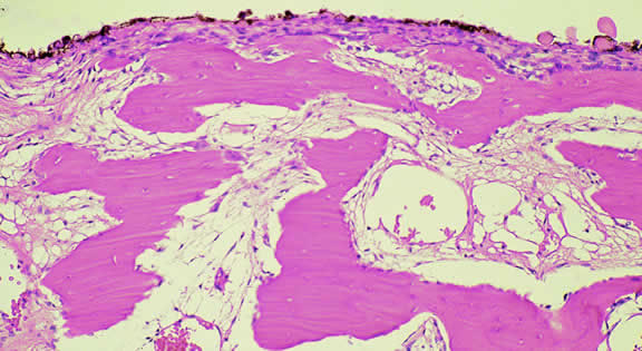
| Fig. 76. Choroidal osteoma. The tumor is composed of irregular spicules of bone surrounded by an areolar stroma containing large vascular channels. The bone is within the choroid, deep to Bruch's membrane, choriocapillaris and an intact layer of retinal pigment epithelium. The intrachoroidal location of the bone distinguishes choroidal osteoma from osseous metaplasia of the retinal pigment epithelium. (Hematoxylin-eosin, × 50.) |