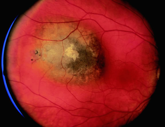
Fig. 16.
Small pigmented tumor thought to be choroidal nevus found on routine ophthalmoscopic examination. Nevus is slightly elevated and retinal pigment epithelial changes are present.