Chapter 9
The Iris and the Pupil
RANDY KARDON and JOEL WEINSTEIN
Main Menu   Table Of Contents

Search

ANATOMY AND PHYSIOLOGY OF THE PUPIL
THE IRIS TISSUE
THE LIGHT REFLEX
THE DARKNESS REFLEX
PHARMACOLOGY OF THE PUPIL
DRUG PENETRATION AND ABSORPTION
CENTRAL PUPILLARY PHARMACOLOGY
PHARMACOLOGY OF THE PARASYMPATHETIC SYSTEM
PHARMACOLOGY OF THE SYMPATHETIC INNERVATION OF THE PUPIL
THE EFFECT OF NEUROPEPTIDES ON THE PUPIL
REFERENCES

ANATOMY AND PHYSIOLOGY OF THE PUPIL
THE IRIS TISSUE
It is important to understand the normal topography of the iris and its histology in order to understand how the pupil changes size during contraction and dilation and how disorders of the iris tissue affect pupil movement. The iris can be divided into two main layers, the posterior leaf and the anterior leaf (Fig. 1). The posterior iris leaf contains the dilator muscle, the sphincter muscle, and the posterior pigmented epithelium. Both the dilator and sphincter muscles are derived embryologically from the anterior layer of the bilayered posterior pigmented epithelium. The anterior iris leaf consists of connective tissue stroma with cells, blood vessels, and nerves supplying the sphincter and dilator. The different components of the posterior and anterior iris undergo structural alterations in order to accommodate changes in pupil size during contraction and dilation.1 These changes are summarized by the series of pictures shown in Figure 2.

Fig. 1. Iris topography. A. Horizontal section through the human eye. B. Cross-sections of monkey iris in miosis (1), in moderate mydriasis (2), and in extreme mydriasis (3). C.Sagittal cross-section of human iris in mid-dilation. (B, camera obscura outlines of photomicrographs from van Alphen GWHM: The structural changes in miosis and mydriasis of the monkey eye. Arch Ophthalmol 69:802, 1963)

Fig. 2. Changing iris areas with different pupil diameter. A. This eye looked greenish hazel at a distance. The dark pupil edge is indicated by small white dots. The large white spot at 5 o'clock (marked a in B) is the corneal reflection of the electronic flash. In the ciliary portion, the anterior stroma was intact (note the shallow contraction furrows between 1 and 4 o'clock). The vessels near the iris frill were heavy and clearly marked, with prominent X formations. In the area between the pupil and iris frill, there was no anterior stroma, and many vessels could be seen to run radially toward the pupil in fairly regularly interwoven loops. The sphincter muscle was visible below these vessels as a pinkish band surrounding the pupil. B to D. Outline drawings of the same eye with different pupil sizes. The shaded areas show the inner pupillary iris ring (from pupil to collarette) and the stippled areas to the outer ciliary ring (between collarette and limbus). The fine, broken lines mark the outer edge of the sphincter muscle. The sphincter ring becomes larger and thinner when the pupil dilates, and smaller and fatter when it constricts. With large pupils, it is hidden by the iris stroma, which billows above it. The black posterior iris leaf that peeks beyond the anterior iris layers when the pupil is small becomes thinner when it enlarges. (Loewenfeld IE: Reflex integration: physiologic mechanisms. In: The Pupil: Anatomy, Physiology and Clinical Applications, Vol 1, Ch 9, pp 414–424. Detroit, Iowa State University Press, Ames and Wayne State University Press, 1993)

During pupil contraction, the outer circumference of the iris, called the outer ciliary ring (which contains the dilator), enlarges in area as the pupil becomes smaller and the iris tissue spreads out to compensate for the reduction in pupil size. The area of the inner circumference of the iris, called the inner ciliary ring, or collarette (which contains the sphincter), remains relatively constant as the pupil becomes smaller despite the increasing area taken up by iris tissue. Consequently, as the pupil becomes small, compaction of iris tissue in the inner collarette poses a mechanical limitation to iris movement. This results in a nonlinear “leveling off” of how much the pupil can contract in response to stronger light stimuli. These mechanical nonlinearities introduced by the rearrangement of iris tissue at the extremes of pupil dilation and contraction have been extensively studied by Loewenfeld and Newsome.2 The mechanical nonlinearities are important because they impose limitations on the range of pupil size at which the extent of pupil movement can be used for assessing neuronal reflexes to light stimuli or near stimuli, or for pharmacologic testing of the pupil. The limiting effect of a smaller pupil size on the degree to which it can contract to light stimuli is illustrated in the pupillograms shown in Figure 3. In this figure, a subject with equal pupils was treated with topical thymoxamine in one eye, which through its α-sympathetic blocking effect, inhibits the dilator tone and makes the treated pupil smaller. After producing unequal pupils (anisocoria) without affecting the sphincter directly, the untreated eye was stimulated with light pulses of increasing intensity while the light reflexes of both the right and left pupil were recorded pupillographically. The extent of movement of the right and left pupil in response to each light stimulus (the change from baseline size to the trough of contraction) is the same for both the treated and untreated pupils at lower stimulus intensities, where each pupil is still within its linear operating range. At brighter light stimuli, the smaller pupil contracts less because it is being driven to such a small size that it is outside of its linear response range. In the pupillograms, this results in a decrease in the degree of anisocoria during the course of contraction as the smaller, treated pupil's movement becomes impeded by the mechanical limitations on the iris movement, and the untreated pupil “catches up” to the smaller pupil at the peak of contraction. When the contraction amplitudes of the treated and untreated pupil are plotted as a function of each increasing level of light stimulus, the treated pupil starts to contract less as its excursion begins to enter the mechanical nonlinear range (Fig. 4).

Fig. 3. Pupillograms of right and left pupil after treating the right eye with thymoxamine, which blocks the adrenergic input to the eye, and thus makes the pupil smaller. Note that dimmer light stimuli produce smaller but equal contractions of the right and left pupil, as evidenced by the lack of a changing anisocoria during the pupil contraction (left). At brighter light stimuli, the contractions are greater, and as the smaller pupil becomes limited by mechanical constraints, it does not contract as much as the untreated pupil at the peak of contraction, resulting in a decreasing anisocoria (center). At the brightest stimulus light, the smaller pupil is even more limited in its contraction, resulting in a larger reduction in anisocoria (right). The straight line below each anisocoria tracing represents a level of zero anisocoria.

Fig. 4. The contraction amplitudes of the treated and untreated pupils from the pupillograms in Figure 3 are plotted as a function of stimulus light intensity. The treated pupil starts to contract less as its excursion begins to enter the mechanical nonlinear range, causing the response curves of the two pupils to come apart. The third line that decreases with stimulus intensity is the size of the smaller pupil at the peak of contraction (see Y2 axis at right). From this line, it can be seen that the two response curves of the right and left pupil start to diverge at a pupil size of 4.5 mm, where iris mechanics start to play a role in this particular subject.

CLINICAL CORRELATION: EFFECTS OF DAMAGE TO THE IRIS TISSUE ON STRUCTURAL ELEMENTS AND THEIR FUNCTIONAL CONSEQUENCES

One consequence of viral infection of the iris by herpes zoster virus is denervation of the iris sphincter and direct damage to the sphincter muscle. The sphincter muscle consists of muscle segments arranged in a series circumferentially around the pupil border. With infrared transillumination, the normal sphincter appears as a dark band as the pupil gets smaller during sphincter contraction. Figure 5 shows an unevenly reactive pupil caused by segmental atrophy of the sphincter muscle induced by previous herpes zoster iritis. In this patient, the areas of sphincter damage and loss appear as bright transillumination defects in segments of iris tissue that were damaged by the previous iritis.

Fig. 5. Asymmetric, segmental contraction of the pupil due to previous herpes zoster iritis. The top photos are slit-lamp images of the pupil in dark (left) and in light (right), showing irregular contractions. In the bottom photos, infrared transillumination of the same iris is shown in darkness and light. In areas of the iris previously damaged there is loss of tissue, which shows up as transillumination defects that appear white. In these areas, the sphincter muscle was destroyed by the inflammation and ischemia, resulting in loss of pupil contraction in the affected segment.

Back to Top
THE LIGHT REFLEX

GENERAL CONSIDERATIONS

The pupillary contraction to light is an important neuronal reflex because it gives information about the integrity of both the afferent visual system and the efferent neuronal outflow pathway to each pupil. The retina, optic nerve, chiasm, and optic tracts are composed of neurons that convey visual and pupillary afferent input, so that damage anywhere along this pathway is likely to affect both the pupillary light reflex and visual function. Because of this, physicians and investigators have used the pupillary light reflex for many years as an objective means of detecting damage along the pregeniculate afferent visual pathway.3–7 Because of similarities in neuronal input for the visual and pupillary systems, the pupillary light reflex has also been used as an investigative tool for understanding many aspects of visual function.8 The basic neuronal pathway for the pupillary light reflex is schematically depicted in Figure 6 and will be referred to throughout this chapter.

Fig. 6. Diagram of the pupil light reflex pathway. Note that the axons of ganglion cells leaving the right nasal retina cross at the chiasm to join the uncrossed, temporal retinal fibers coming from the left eye at the optic tract. From here, the afferent axons in the right and left optic tract mediating the pupillary light reflex enter the brachium of the superior colliculus on each side, before reaching the lateral geniculate body, and synapse with cells in the right and left pretectal olivary nuclei. The neurons in each pretectal nucleus send an uncrossed branch to the Edinger-Westphal nucleus on the same side and a crossed branch to the Edinger-Westphal nucleus on the opposite side. The exact site where the axons branch is still not well understood in primates, but the arrangement causes both pupils to contract regardless of which eye is stimulated. The small number of neurons in the Edinger-Westphal nucleus are preganglionic parasympathetic neurons that travel within the oculomotor nerve with axons that innervate the extraocular muscles.

Because both pupils receive an approximately equal distribution of neuronal output from the integrated afferent input of the light reflex, it is expected that in normal humans both the right and left pupils will contract with the same amplitude to a given light stimulus. In most cases, this means that either the right or left pupil movement can be used to monitor afferent input. Damage to the retina or optic nerve of one eye will therefore result in less right and left pupil contraction when a light stimulus is given to the damaged eye compared with the pupil response when the same light is given to the normal eye. Because both pupils receive approximately an equal distribution of signal from any intensity of afferent light stimuli, damage to the afferent visual input will not result in unequal pupils.

CLINICAL CORRELATION: THE RELATIVE AFFERENT PUPILLARY DEFECT

Optic nerve damage in one eye can be detected by monitoring the differences in pupil movement as a light is alternated between the normal eye and damaged eye. This provides an objective means of quantifying how much damage to the afferent arm of the light reflex is present in one eye compared with the other eye. Precise quantification of the log unit difference in sensitivity between the two eyes can be accomplished by dimming the light in the normal eye by holding up neutral density filters of known increments. For example, in the patient shown in Figure 7, who has damage to the left optic nerve, the right and left pupil do not become as small when light is shined in the left eye compared with when the light is shined in the normal right eye.

Fig. 7. Example of the swinging flashlight test in a patient with optic nerve damage in the left eye. Top. Light is shined into the normal right eye, and both pupils contract to a small size. Bottom. Light is shined into the damaged left eye, causing very little pupil contraction, and both pupils appear large.

The degree of asymmetry can be determined clinically in log units by determining the number of log units of neutral density filter needed over the right eye to balance the light reactions when the light is alternated between the two eyes. Figure 8 shows pupillographic tracings in such a patient when each eye receives the same amount of light (top) and with decreasing light intensity given to the better eye (middle), until the contractions are roughly equal no matter which eye is stimulated (bottom). The amount of log-unit neutral density filter needed to balance the reactions is known clinically as the log-unit relative afferent pupillary defect (RAPD). The clinical log-unit RAPD is important because it correlates with the amount of visual field loss.8a–12 In recent years, infrared pupillography has been used as a means of more precisely recording pupil movements during the alternating light test to quantify the log-unit RAPD more objectively and precisely.13–16

Fig. 8. Pupillograms of a patient with a left relative afferent pupillary defect. In the top tracings, it is apparent that the right and left pupil light reflexes resulting from a bright stimulus light applied to the right eye are substantial, compared with the lesser response of the two pupils when light is applied to the damaged eye. In the middle and bottom tracings, the light stimulus is reduced to the normal eye in an effort to reduce the amplitude of the pupillary contractions until they are balanced, or equal, when the light is alternated between the two eyes.

Therefore, in humans and other primates, whose two pupils react approximately the same to any given light stimulus, either pupil can be used to monitor afferent input to the eye. In other species, such as the cat, rabbit, and rat, the pupil of the eye being stimulated always reacts more (i.e., direct pupil response exceeds the consensual response). Species differences in the crossing of afferent and efferent processing of the neuronal light signal cause the direct pupil response to be greater than the consensual and results in anisocoria whenever one eye receives light. In these species, only the direct or consensual pupil response can be used to ascertain the relative afferent light sensitivity between the two eyes, rather than monitoring just the right or left pupil during the test. A greater crossing of optic nerve fibers in the chiasm and a predominantly crossed pathway in the midbrain helps to explain why the direct pupil response exceeds the consensual in lower species. The topic of species differences in the integration of the pupillary light reflex is discussed in more detail by Loewenfeld.17

In humans and other primates, anisocoria usually indicates a defect in the efferent neuronal process controlling pupil size, rather than differences in afferent input. Therefore, pupil inequality can be used to assess the integrity of the neuronal outflow and smooth muscles of the iris dilator and sphincter. Damage to the nerves supplying the pupil sphincter or direct damage to the sphincter muscle will cause the affected eye's pupil to contract less, producing unequal pupils. In this case, it would be best to observe only the undamaged pupil to monitor the afferent input to the eye. The degree of pupil inequality measured under different lighting conditions would give an indication of the extent of damage to the efferent pupil system. Examples of clinical conditions that damage the efferent pupillary system and different pharmacologic means of evaluating the cause are presented in the pharmacology section.

Some human subjects do not show equal movements of the right and left pupil in response to a light stimulus, depending on which eye receives the stimulus. This condition, known as contraction anisocoria,17–22 is observed by pupillography in up to one third of the population to some extent.23 In this condition, the pupil of the eye receiving the light stimulus contracts more than the pupil of the opposite eye (Fig. 9). In other words, the direct pupillary light reflex is greater than the consensual light reflex. The source of this condition is thought to be found at the level of the pretectal neurons in the midbrain, where afferent impulses in response to light are distributed to the right and left Edinger-Westphal nuclei. Further discussion on the pretectal integration of the pupillary light reflex in the midbrain is presented later in the chapter.

Fig. 9. Pupillograms of a subject demonstrating unilateral contraction anisocoria. Note that when the light is applied to the right eye, both the right and left pupils contract the same. When the light is applied to the left eye, however, the left pupil contracts more than the right pupil. This is also apparent by noting that the anisocoria becomes greatest at the peak of contraction.

The neuronal integration of the pupillary light reflex begins in the retina with the photoreceptors, bipolar cells, and ganglion cells. The ganglion cells, the first neurons in the chain to give rise to action potentials (the photoreceptors and bipolar cells give rise to generator potentials), are made up of different classes of neurons based on morphology, physiology, and projections. The gamma cells, a separate class of ganglion cells, appear to be primarily responsible for conveying the pupillary light reflex to the midbrain.

These ganglion cell axons synapse with the next neurons at the olivary pretectal nucleus. Neurons in the pretectum then send fibers to a small number of neurons in the Edinger-Westphal nucleus. These neurons, in turn, send preganglionic axons traveling with the oculomotor nerve to the orbital apex, where they synapse in the ciliary ganglion. From here, the last neurons in the chain pass anteriorly into the eye via the short ciliary nerves, where they innervate the iris sphincter. The properties of light that lead to pupillary movements and the site of integration of the light signal along the light reflex pathway (see Fig. 6) are discussed in the following sections.

PROPERTIES OF LIGHT AND THEIR EFFECT ON PUPIL MOVEMENT

Properties of light stimulating the retina that affect the pupil response include intensity, duration, temporal frequency, area, perimetric location, state of retinal adaptation, wavelength, and spatial frequency. There is a wealth of information on how these properties of light stimuli affect the pupillary response with regard to latency and amplitude of movement. Loewenfeld24 presented the most complete review of this topic in her book on the pupil, which should be consulted for a detailed literature review and for examples illustrating these different light effects. Table 1 summarizes the different light effects.


TABLE 1. Effect of Properties of Light Stimuli on the Pupillary Light Reflex


Stimulus PropertyEffect of Pupillary Light Reflex
Light intensityThe amplitude of contraction increases linearly over at least a 3 log-unit range with log intensity stimulus. The entire response function resembles “S” shape. Latency time (200–450 milliseconds) becomes more prolonged with dimmer light stimuli (in the range of 20–40 milliseconds further delay/log-unit decrement of light intensity).
State of light adaptationWith dark adaptation, the threshold for producing a pupil contraction decreases as rods are brought into play. However, rods do not produce as much proportional increase in pupil contraction with increases in stimulus intensity, compared with cones in the mesopic and photopic states.
DurationAt stimulus durations of less than 70 milliseconds, there is a direct inverse relationship between duration and intensity. With longer duration stimuli, the pupil contracts more, there is a shorter latency time (up to a point), and the pupillary contraction lasts longer.
AreaThe pupillary light reflex shows much greater area summation properties than visual perception (for visual threshold, summation is minimal with stimuli greater than 1°). With full-field Ganzfeld stimuli, the pupil threshold can be equal to visual threshold; with stimuli smaller than 1–2 degrees, visual threshold is usually more sensitive (by 0.5–1 log units).
Perimetric locationUnder dark adaptation, the fovea shows a decreased sensitivity compared with surrounding retinal areas due to the lack of rods here. In mesopic and photopic adaptation, the pupil responds greatest in the central field; the temporal field response is usually greater than the nasal field response.
Spectral sensitivityThe wavelength sensitivity of the pupillary light reflex follows that of visual perception, with a blue shift under dark adaptation.
Temporal frequencyThe normal pupil cannot move much faster than 4 Hz because of the relatively slow contraction of smooth muscle. Animals with striated iris muscle (pigeons) can easily follow a 10 Hz stimulus. At frequencies from 9 to 25 Hz, the steady-state pupil size increases, indicating loss of sensitivity in neuronal integration of light within this frequency range.
Spatial frequencyWhen the change in average luminance is kept constant, the pupil undergoes small contractions when a sinusoidal grating is presented. The mechanism is thought to be independent of a luminance response. The greater the spatial frequency, the less the pupil contracts to the stimulus and spatial frequency has been correlated with visual acuity.
MotionRecent evidence indicates that the pupil may respond to a motion stimulus even under conditions of isoluminance.


In the past, most investigations of the pupillary light reflex have focused on the response of the pupil to changes in light level, because the neuronal pathway for this reflex was thought to respond only to stepwise changes in light intensity. With the advent of computer graphics, more complex stimuli can be presented, allowing properties of spatial frequency, color, motion, and luminance to be more carefully controlled. A number of investigators have taken advantage of this technology to investigate whether the pupil is capable of responding to visual stimuli that change in color or spatial frequency when the average luminance does not change.25–33

The results of these studies have provided evidence that the pupil contracts to either an onset or offset of spatial frequency or color exchanges. From a practical standpoint, these responses allow the pupil response to be used as an objective indicator of visual acuity and color discrimination. From a theoretical standpoint, the pupil response to isoluminant stimuli provides a means of exploring how and where different signals are processed in the visual system.

The following sections summarize the classic concept of the integration of the pupillary afferent system in the retina, optic nerve, pretectum, and Edinger-Westphal nucleus. Although there is no question that incremental changes in intensity of diffuse light result in a neuronal pupillary light reflex that is integrated at the brain stem level, over the years clinical testing of patients with isolated damage to visual cortex has shown loss of pupillary responses to perimetric stimuli34–43 and more complex isoluminant stimuli (see earlier discussion). These unexpected results are now stimulating new experimental work to investigate the role of visual cortex in modulating the pupillary contraction to more complex stimuli and in forms of pupil perimetry.44,45

ROD AND CONE PHOTORECEPTOR INPUT

For many years, it was disputed whether rods or cones contributed to the pupillary light reflex, and whether these were the same photoreceptors as those contributing to visual perception. Loewenfeld24 summarized the extensive literature on this subject, and her book should be consulted by the reader desiring a more complete discussion of this topic. The whole topic of rod and cone input to the pupillary light reflex remained confusing for many years, because the responses measured were highly dependent on the conditions of retinal adaptation, light stimulus intensity, wavelength, size of stimulus, area of retina that was stimulated, and how sensitive a recording apparatus was available for detecting small, threshold pupil movements. By using sensitive infrared recording devices for precise measurement of small pupil movements and carefully controlled stimulus conditions, it has become apparent that both rods and cones contribute to the pupillary light reflex. The relative contribution of the rods and cones depends on the conditions under which the pupil responses are measured. In addition, evidence has overwhelmingly shown that the same rods and cones that process light input for visual perception are also used for the pupillary response, as opposed to separate photoreceptors for vision and for the pupil. Because of this, almost all modifications of stimulus condition that produce a difference in visual perception also produce a comparable change in pupillary responsiveness. These modifications include changes in retinal adaptation, wavelength of light, stimulus duration, and stimulus light intensity. In fact, in almost every way measured, the pupillary responses to light parallel those of visual perception.

For example, the wavelength sensitivity profile of pupil threshold as the color of light is changed from blue to red exactly parallels the same wavelength sensitivity of visual perception. The shift in sensitivity is also the same as the eye is changed from a condition of light adaptation to dark adaptation (Purkinje shift), providing further evidence that the same photoreceptors are used for both pupil and vision. In fact, patients with various abnormalities of either rods and cones can be shown to have the same deficits in color vision or lack of appropriate sensitivity change during dark adaptation when the results of visual threshold are compared with pupillary threshold to light stimuli.46

Under conditions of dark adaptation, low-intensity light stimuli below the level of cone threshold produce small-amplitude pupil reactions that are contributed to only by rods. The recorded pupillary waveform movements caused by rod input under stimulus conditions of dark adaptation appear indistinguishable from those produced by dim stimuli under conditions of photopic light adaptation, where cone input is the primary source of signal to the pupillary motor center. Under dark adaptation, the threshold pupillary response also favors a blue wavelength, as would be expected for a roddominated response. In fact, under these conditions there is a worsening of pupil threshold sensitivity at the fovea, causing a scotoma at the center of the visual field, where rods are lacking. This is observed when the threshold for pupil contraction is observed using low-intensity lights below the level of cone threshold. As the intensity of light stimulus is increased above the cone threshold, pupil contractions can then be recorded at the fovea, as the cone contribution comes into play. Under these stimulus conditions, both rods and cones contribute to the pupil response, a condition known as “rod intrusion.”24 If a background light is used to adapt the eye to a photopic, relatively light-adapted state, the rod contribution is suppressed and cones serve as the primary contributors to the pupillary light reflex.

Another important difference between the cone and rod contribution to the pupillary light reflex concerns the concept of pupillary threshold sensitivity versus pupillary amplitude of response. As stated earlier, the rods are far more sensitive than cones under conditions of dark adaptation. The amount of light it takes to elicit a small, threshold pupillary response is more than two log units less than what it would take to elicit a threshold pupil response from cone activation. Above the level of threshold, however, the amplitude of pupil contraction has a much greater “valence” from the contribution from cones as opposed to rods. Therefore, although the rods are more sensitive in terms of the amount of light it takes to elicit a threshold pupil response in the dark-adapted state, the amount of pupil contraction produced at light levels above threshold for each photoreceptor type is greater for cones.

With focal light stimuli given at suprathreshold intensity levels, the amplitude of the pupillary light reflex has a large cone contribution for reasons stated earlier. The pupillary light reflex is also greater in amplitude at the center of the visual field and falls off in the peripheral field (Fig. 10). The density distribution of rods and cones (and bipolar cells) accounts for only a small aspect of the pupillomotor response across the visual field. It is the ganglion cell type and their distribution within the retina that provide the primary basis for the degree of pupillary response from light stimuli at different visual field locations.

Fig. 10. Pupil perimetry response in the normal visual field compared with visual light sense perimetry in the same eye. The contraction amplitude (in millimeters excision) of the pupil is plotted as a function of perimetric location to a 1.7-degree 1000 apostilb intensity light in the pupil field. Note that the pupil contracts the most near the center of the visual field, and the response drops off in the periphery. In the visual field the threshold for light detection is given in decibels. A more sensitive area has a higher decibel threshold level.

The ganglion cells that serve the pupillomotor afferent input are densest in the central retina. The ganglion cells in the central area also have more of a one-to-one relationship with photoreceptor and bipolar cells than those in the peripheral retina, where many more photoreceptors and bipolar cells map to one ganglion cell. Therefore, it is the central density of ganglion cells and their relatively one-to-one mapping with photoreceptors and bipolar cells that account for the greater amplitude of pupillary response to stimuli in the central visual field.

GANGLION CELL INPUT

Although it appears that the pupillary and visual systems share the same photoreceptor input (and presumably bipolar cell input), the role of ganglion cell input is far less clear. Over the years, many anatomic and physiologic studies have been carried out in an attempt to classify retinal ganglion cells based on the their cell body size, dendritic field, axonal diameter, and electrical firing properties. This information has been collected in a number of species, most notably the cat and monkey, and the projections of the cell axons to the lateral geniculate body and midbrain have been studied with the use of labeling techniques. Loewenfeld24 summarized the properties of the major classes of ganglion cells in the retina and their projections. Based on these studies, there appear to be three main types of ganglion cells that make up the retina in primates: the alpha, beta, and gamma cells. In the cat, these correspond to the Y, X, and W cells, respectively (Fig. 11).

Fig. 11. Size and shape of three chief types of retinal ganglion cells (cat), as described by Boycott and Wässle in 1974. All drawings are to the same scale, shown at the lower edge. The numbers given for each cell indicate its distance from the center of the central area of the retina. Alpha cells correspond to Y or “brisk-transient” cells (left), beta cells to X or “brisk-sustained” (middle), and gamma cells to W or “sluggish” cells of other terminologies (right). Note that the dendritic trees of the alpha and beta cells increase in size with eccentricity, but those of the gamma cells do not. The dendritic configurations differ markedly among the three groups of cells and are much smaller in beta cells than in alpha and gamma cells. (Boycott BB, Wässle H: The morphological types of ganglion cells of the domestic cat's retina. J Physiol 240:397, 1974)

Table 2, from Loewenfeld,24 summarizes all of the main properties of the gamma cells (W cells). These cells appear to be the major contributor to the pretectal neurons in the midbrain serving the classic pupillary light reflex pathway. These cells may also play a role in other visually evoked reflexes (e.g., eye position control) projecting to the superior colliculus and also to the accessory optic system. The gamma cells have small cell bodies and thin, slowly conducting axons with large receptive fields. They respond primarily to incremental changes in light intensity and are relatively insensitive to movement. These cells project almost exclusively to the midbrain and not to the lateral geniculate nucleus. They have the highest density in the central field and become less dense in the periphery, which accounts in large part for the central field weighting of pupillary response. The proportion of gamma ganglion cells that serve the pupillary light reflex is not currently known, and the answer awaits specific labeling studies in primates. Such studies are technically difficult to perform, because the retrograde label must be microinjected into the specific pretectal area where light-responding neurons can be identified with the use of electrical recordings. Even then, the label reaching the retinal ganglion cells is only weakly visible; once identified in flat mount preparations, a micropipette must be used to inject these individual neurons with a more visible dye that fills each cell body, dendrites, and axon. It is currently unknown how many retinal ganglion cells project directly to the pretectal midbrain neurons serving the pupillary light reflex, but their number must be relatively small (on the order of 1%) in proportion to the total number of ganglion cells.


TABLE 2. Characteristics of the Three Chief Types of Retinal Ganglion Cells


PropertiesChief Groups of Retinal Ganglion Cells
Functional namesYXW
Anatomic namesalphabetagamma
Other terms used“Type 1” (Bishop, Fukuda), or “brisk-transient” (Cleland et al)“Type 2” (Bishop, Fukuda), or “brisk-sustained” (Cleland et al)“Type 3” (Bishop, Fukuda), or “sluggish”(Cleland et al)
Number in the retinaFew 1%–2% of population in the central area, about 10% in the periphery)Many (50%–60% of total population of retinal ganglion cells)Many (about 40% of total population of retinal ganglion cells)
Distribution in the retinaHighest density in the para-fovea, less in the center and peripheryHighest density in the central area, less in the peripheryHighest density in the central area, less in the periphery
Size of cell bodyLargest, increasing in size with eccentricityMedium, increasing in size with eccentricitySmallest; no difference with eccentricity
Axon thicknessThickestMediumThinnest
Spread of dendritic treeWidespreadSmallWidespread
Nasotemporal division of axon fibersAbout 5% of temporal fibers cross, 95% notExcept for central strip of overlap, nasal cells have crossed, temporal cells uncrossed fibersAll nasal cells and about 40%–60% of temporal cells have crossed fibers (in cats)
Type of dischargesPhasicTonicTonic and phasic
Size of receptive fieldLargeSmallLarge
Axonic conduction speedFastest (slower in attenuated branches)MediumSlowest
Latent periodShortestMediumLongest
Threshold to electric stimulationLowestMediumHighest
Time characteristics of effective stimuliFastMediumSlow
Sensitivity to anoxiaHighestMediumLowest
Resistance to barbiturate overdoseLeastMediumMost
Phylogenetic ageNewNewestOld
ProjectionTo lateral geniculate nucleus, about ½ of fibers have attenuated branches to midbrainMostly to lateral geniculate nucleus; some collateral branches to tectum, pretectumTo brain stem only (pretectum, colliculi, accessory optic accessory optic system)
Chief functional traitResponsive to fast-moving stimuliResponsive to contrast of small areas (fine gratings)Proportionately responsive to intensity but not to fast rates
Probable functionInitiate coarse, rapid eye movements (fixation reflex)Discrimination of detailPupillary and other simple reflexes, and endocrine reactions
(Loewenfeld IE: The light reflex. In: The Pupil: Anatomy, Physiology, and Clinical Applications, Vol 1, Chap 3, pp 96–135. Detroit, Iowa State University Press, Ames and Wayne State University Press, 1993)


The other two classes of ganglion cells, the alpha and beta cells in primates (or Y and X cells in cats, respectively), have different properties than the gamma cells (W cells). The alpha cells constitute only about 1% to 2% of the cells in the central retina and 10% in the periphery. They have the greatest density in the parafoveal region as well as the largest cell body size, which increases further in the peripheral retina. The axons of alpha cells are thick and have a corresponding fast conduction speed. Their receptive fields are large and have a phasic discharge. As such, these cells are tuned to respond to fast-moving stimuli and discharge in a transient fashion. The alpha cell axons project to the lateral geniculate body, but almost 50% have attenuated branches that project also to the midbrain in areas such as the superior colliculus.

The beta cells are the most numerous of all the ganglion cell classes (50% to 60% of the total ganglion cell population) and have the highest density in the central retina. They have medium-sized cell bodies and axons, and their receptive fields are small. These ganglion cells fire tonically and are most responsive to contrast of small areas such as fine grating stimuli. They are tuned toward discrimination of detail within the central field. The beta cells also project primarily to the lateral geniculate nucleus, but some axons do give rise to a collateral branch that projects to the tectum and pretectum.

From the previous description of the major classes of ganglion cells, it would appear that the gamma cells are primarily light sensitive and serve the midbrain pupillary light reflex. The alpha cells are primarily movement sensitive and provide afferent information for eye movement control and foveation of peripherally moving targets. The beta cells are mainly sensitive to high contrast detail and probably provide the visual cortex with spatial frequency and orientation information used to interpret form. In light of recent evidence of cortical mediation of the pupillary movement to perimetric stimuli and isoluminant complex stimuli (gratings and random dot patterns), it is possible that other ganglion cells besides the gamma type may play a role in mediating pupillary response to other, nontraditional types of stimuli.25–45

Clinical Correlation

Damage to the ganglion cells or their axons by optic nerve disease or damage to the inner retina causes visual field defects corresponding to the distribution of the damaged fiber bundle. There is often a corresponding pupil field defect in the same distribution as the visual field defect, as shown for a patient with anterior ischemic optic neuropathy (Fig. 12). In such cases, the focal pupil response can be used as an objective indicator of the distribution of damage within the field of vision. In other clinical examples of damage, such as glaucoma or optic neuritis (secondary to demyelinating disease), the “pupil field” may show greater damage than a person's perceptual responses, indicating that the ganglion cell response serving the pupillary light reflex was more sensitive to that specific damage (Fig. 13). Clinical examples such as this show that there may be differences between the ganglion cell integration of the pupillary response to light and the visual perception of light.

Fig. 12. Visual and pupil perimetry in anterior ischemic optic neuropathy, showing corresponding damage to the same visual field location for both pupil response and visual response.

Fig. 13. Visual and pupil perimetry in glaucoma, showing more damage to the pupil light reflex pathway.

INTEGRATION OF THE PUPILLARY LIGHT REFLEX AT THE PRETECTUM

The majority of ganglion cell axons projecting to the pretectal olivary nucleus, the next relay station for the pupillary light reflex, are from the gamma cells. Their axons leave the right and left optic tracts just prior to each lateral geniculate nucleus to travel within a small bundle of fibers on each side of the midbrain, termed the brachium of the superior colliculus. As mentioned previously, a small contribution of bifurcating fibers from the alpha and beta ganglion cell axons also travel within the brachium. Some of the fibers within the right and left brachia, the ones mediating the pupillary light reflex, synapse with the small aggregation of neurons making up the right and left pretectal olivary nucleus. Other fibers serving the near response and eye movement synapse at nearby nuclei.

As depicted in Figure 6, the ganglion cell axons from the nasal retina (temporal field) of the right eye cross at the chiasm and distribute to the contralateral (left) pretectal olivary nucleus. The ganglion cell axons from the temporal retina (nasal field) of the left eye remain on the same side and join the axons from the nasal retina of the right eye in the left optic tract to distribute to the left pretectal olivary nucleus. In this way, ganglion cell axons serving homonymous portions of the visual field distribute to the same pretectal nucleus. In other words, the axons serving the right homonymous visual field space distribute to the left pretectal olivary nucleus, and the axons serving the left homonymous visual field space distribute to the right pretectal olivary nucleus. At each pretectal olivary nucleus, a great deal of convergence takes place, with many ganglion cell axons forming connections with a relatively small number of dendritic processes of pretectal olivary neurons.

The firing properties of these neurons and their relationship to luminance level of light stimuli as well as pupil response have recently been elucidated for monkeys studied in the awake state.47 These neurons responded to a 5-second stimulus with a phasic burst of action potentials at high frequency, followed by a transient, sustained volley of action potentials at a lower frequency (Fig. 14). Of the 16 separate neurons studied, all had linear increases in the sustained component of discharge frequency as the log stimulus intensity was increased linearly, and this corresponded to linear changes in pupil diameter. However, the slope of the response relationship between firing rate and log luminance was not the same for every neuron: some had a greater gain (i.e., steeper slope of the line) than others. In fact, in cases where stimulus intensity was very low, some individual pretectal neurons showed an increase in firing frequency without any change in pupil size, demonstrating that the amount of neuronal increase in firing necessary to result in a measurable change in pupil diameter may be a summation of input from a number of pretectal neurons, especially at low light levels of stimulation. It is not known at this time whether the gain of individual pretectal neurons or the summation properties of many neurons firing in unison is regulated by higher supranuclear inputs. Perhaps it is the modulation of this firing rate and the summation properties that give rise to hippus, the random fluctuations of the pupil that are often observed under continual retinal illumination. It is also of interest that Gamlin and co-workers48 recently reported the presence of specific neuropeptides within the pretectal nucleus, which are also known to act as neuromodulators in other areas of the brain.48 The role of these neuropeptides in modulating the pretectal response, as well as the origin of these peptidergic nerve endings, is currently not well understood.

Fig. 14. A to C. Response of a pretectal luminance neuron to stimuli of 1000 trolands, 100 trolands, and 10 trolands, respectively. Note that in A, a blink occurs during the dynamic phase of pupilloconstriction. D. The change in neuronal firing rate for 16 individual pretectal luminance neurons is plotted against the logarithm of retinal illuminance. The responses of the neurons were essentially linear over this range, and linear regression analyses yielded correlation coefficients ranging from r = 0.81 to r = 0.99, with a mean correlation coefficient of r = 0.92. The solid line in D is a regression line fitted to the data from all 16 neurons. In E, pupil diameter is plotted against the logarithm of retinal illuminance. The data were obtained while recording the luminance responses of each of the 16 cells shown in D. The solid line in E is a regression line fitted to the pooled data. For the 16 luminance neurons, F shows the mean change in neuronal firing rate as a function of pupilloconstriction. The response of the neurons was essentially linear over this range, and linear regression analyses yielded correlation coefficients from r = 0.8 to r = 0.98, with a mean correlation coefficient of r = 0.9. The solid line in F is a regression line fitted to the data from all 16 neurons. Scale bar, 1-mm pupilloconstriction (Gamlin PDR, Zhang H, Clarke RJ: Luminance neurons in the pretectal olivary nucleus mediate the pupillary light reflex in the Rhesus monkey. Exp Brain Res 106:177, 1995)

The pretectal neurons also possess temporal summation properties different from neurons involved in visual perception. The intensity-dependent time delay in the pupillary light reflex is longer than occurs at other synapses. For every log unit reduction in light intensity, the latency time of the pupillary contraction may become prolonged by an additional 30 to 45 milliseconds. In order for this long delay to occur, the pretectal neuron must be capable of summating input over a relatively long period of time before having to “start over.” Therefore, the pupillary light reflex may serve as an indicator of damage to the retina or optic nerve by showing both a decrease in amplitude of pupil contraction and a prolongation of the latency time.

The receptive field properties of the pretectal olivary neurons have also been studied recently with the use of neuronal recording techniques in awake monkeys (Drs. Paul Gamlin and Robert Clarke, unpublished observations). Current evidence indicates that there are two major types of pretectal neurons, based on their firing properties to stimuli presented in different locations within the visual field:

  Type 1: This type of pretectal neuron appears to respond with a greater discharge frequency to stimuli located toward the center of the cell's receptive field, but also responds (with reduced discharge frequency) to stimuli at the periphery of its receptive field. This type has more of a center-weighted receptive field.
  Type 2: This type of pretectal neuron has a flatter response to stimuli placed in different locations within its receptive field. Its discharge frequency doesn't change substantially based on location of the light stimulus.

At this time, it is not clear how the topographic distribution of ganglion cells throughout the retina is electrically mapped to these populations of pretectal neurons, or how the simultaneous firing of these neurons is summated at the next level of integration within the Edinger-Westphal nucleus.

The integration of the discharge of the pretectal olivary neurons at the level of the Edinger-Westphal nucleus may be more complicated than previously thought. The gain control at both the pretectal and Edinger-Westphal level is not well understood, except for the fact that it is widely recognized that the latency time and responsiveness of the pupils to a given light stimulus may vary considerably over time in a given subject as well as vary widely between subjects. It is well known that some of this variability may be related to a person's state of excitation or sleepiness, which also influences pupil size. Inhibitory input to the Edinger-Westphal nucleus, arising from the reticular activating formation, tends to reduce the discharge rate of these neurons, causing pupil dilation. An excited, anxious person will usually have large pupils that may not respond well to light stimuli. In contrast, a sleepy person will demonstrate reduced inhibitory input to the Edinger-Westphal nucleus, causing fluctuations of the pupil with eventual reduction in pupil size. Narcotics and deep anesthesia have similar effects: as the inhibitory input is reduced, the pupils become small. Initially, at a stage where the pupils are still 4 to 5 mm in diameter, the reaction to light is more sensitive, but as the inhibition is further reduced, the pupils become small enough where iris excursion may become limited by mechanical constraints imposed by a small pupil size. Yet the state of a person's excitation is not the only cause of fluctuations in pupil size and response to light. Even in persons who seem to be in a stable state of wakefulness, the timing and contraction amplitude of the pupil to light also seem to vary over time. Where does this gain control reside? The answer to this important question is currently unknown. If the pupillary light reflex is to be used as an indicator of retina and optic nerve function, it will be important to understand this gain control so that reductions in pupil response due to true damage to the visual system will not be confused with nonpathologic changes in the supranuclear modulation of the pupillary light reflex.

In other words, a reduction in the pupillary response to light needs to be separated into (1) that caused by pathology to the afferent visual system, and (2) that caused by supranuclear influences on the gain control of the pupillary light reflex. Obviously, significant asymmetry in the pupillary light reflex resulting from a light alternated between the right and left eye is going to be due to pathology in the afferent input. Similarly, a defect in the topographic distribution of pupillary responses within the visual field will result only from damage to the anterior visual pathways. However, symmetric decreases in the pupillary response of each eye to global light stimuli, or symmetric decreases from focal stimuli across the visual field in both eyes, is harder to interpret until these gain control mechanisms are better understood.

As stated under the heading General Considerations, output from the pretectal neurons in the midbrain generally results in equal contractions of the right and left pupils in humans and primates. When the movements of both pupils are simultaneously recorded by infrared pupillography, however, the two pupils may not always contract to the same extent. In up to one third of persons, the direct pupil response (the pupil of the eye that is being stimulated) may exceed the consensual pupil response (the pupil of the fellow eye that is not being stimulated). This condition, termed contraction anisocoria (see Fig. 9), may be acquired as a result of viral infections that may be associated with mild encephalitis, and they may be seen with greater frequency in persons prone to infections, such as those with cystic fibrosis.23 It is possible that this condition may exist in normal persons as well. Contraction anisocoria may be present when only the right eye is stimulated or when only the left eye is stimulated, or in a smaller number of individuals it may be present when either eye is stimulated. In the latter case, the right pupil contracts more than the left when the right eye receives light, and the left pupil contracts more than the right when the left eye receives light. What might be responsible for this curious phenomenon?

According to Loewenfeld,23 contraction anisocoria may occur when asymmetries in input and output of the light signal exist both at the chiasm and at the pretectal olivary nucleus. At the pretectal level in primates, the discharge of neurons is usually distributed equally to both the right and left Edinger-Westphal nucleus, most likely via uncrossed and crossed axonal branches. The exact location where the uncrossed and crossed fibers distribute in their course to the Edinger-Westphal nuclei is currently under investigation in primates. Irrespective of the anatomic distribution of these pathways, it is known that in some persons this output may not result in an equal discharge to the right and left side. Either some of the pretectal neurons may not send an uncrossed (ipsilateral) branch to the Edinger-Westphal nucleus, or their discharge may not be equally split, with the crossed branch firing more than the uncrossed branch. This would result in a greater pupil response in the eye on the side contralateral to the pretectal nucleus. This would become apparent on full-field stimulation of the eye contralateral to the pretectal nucleus with the greater crossed output if there were also greater retinal input to this pretectal nucleus from the eye stimulated.

In other words, if the nasal retina (temporal visual field) provides greater input to the contralateral pretectal nucleus via its decussation at the chiasm compared with the uncrossed input from the temporal retina (nasal field) to the ipsilateral pretectal nucleus, then full-field stimulation of this eye would favor more input to the contralateral pretectal nucleus. If distribution of output from this nucleus were also unequal, favoring crossed output to the opposite Edinger-Westphal nucleus, then the result would be a pupillary response favoring the pupil of the eye being stimulated (i.e., the direct response would be greater than the consensual pupil response).

Clinical Correlate of Contraction Anisocoria: The Pseudo-RAPD

This condition may have clinical significance only in unilateral cases, where clinical observation of the direct pupil response during the swinging flashlight test may give the erroneous impression of a RAPD, when in reality, only contraction anisocoria may be present. Based on our own studies of pupillographic recordings of both pupils in normal subjects and in patients with organic causes of RAPDs who also happen to have unilateral contraction anisocoria, this condition may result in a log-unit “pseudo”-RAPD of as much as 0.3.

INTEGRATION OF THE PUPILLARY LIGHT REFLEX AT THE EDINGER-WESTPHAL NUCLEUS

The historical controversies surrounding the exact anatomic location of the group of preganglionic efferent neurons that give rise to pupillary constriction to light and near stimuli has been thoughtfully reviewed by Loewenfeld.24 In 1885, Edinger49 described the embryologic development of cranial nerve nuclei. In his descriptions, he noted a small column of spindle-shaped cells located dorsal to the oculomotor neurons, on each side of the oculomotor nerve complex. Two years later, Westphal50 reported the postmortem anatomic findings of a patient who had total external ophthalmoplegia and whose pupils failed to react to light, but did react to near. He found this cell group intact dorsal to the oculomotor neurons, which had degenerated, and concluded that this surviving cell group may be involved in pupillary constriction. In 1988, Spitzka51 described the midbrain anatomic findings in three similar patients and combined this with work he had done in animals; he also concluded that this small column of cells dorsal to the oculomotor neurons was involved in pupil contraction to light and near stimuli.

More detailed anatomic studies using anterograde and retrograde labels52–55 have more fully defined the location of this small group of preganglionic neurons (Fig. 15). Within this group of visceral neurons are a minority that mediate contraction of the pupillary sphincter and a majority that mediate contraction of the ciliary body for accommodation. Strictly speaking, it is probably best to reserve the term Edinger-Westphal nucleus to the small number of neurons that serve pupillary constriction. These are the same neurons that discharge when the pupil contracts to light or near stimuli; there are no separate neurons that mediate these two stimuli at the level of the oculomotor complex. The pupillary contraction to light and near stimuli is, however, mediated by separate supranuclear pathways.

Fig. 15. Visceral cells of the oculomotor nucleus, labeled by injections of horseradish peroxidase into the ciliary ganglion. The sagittal diagram E shows the levels of brain stem cross-sections A to D, where the tracer was found. The sections are arranged in rostrocaudal order. AC, anterior commissure; AM, anteromedian nucleus; CG, central gray; EW, Edinger-Westphal nucleus; FR, fasciculus retroflexus; ICA, interstitial nucleus of Cajal; IP, interpeduncular nucleus; MB, mammillary body; MLF, median longitudinal fasciculus; NP, nucleus of Perlia; PC, posterior commissure; OC, oculomotor nucleus; RN, red nucleus (Modified from Burde RM, Loewy AD: Central origin of oculomotor parasympathetic neurons in the monkey. Brain Res 198:434, 1980)

The light reflex, as discussed earlier, is mediated by input from axons of the pretectal olivary nuclei, which travel in a dorsal location in the midbrain. The near reflex is thought to originate from cortical areas surrounding visual cortex and from cortical areas within the frontal eye fields.56 The cortical neurons providing input for the near reflex are thought to synapse at least once before passing ventrally, toward the visceral neurons overlying the oculomotor complex in the midbrain. This is because a cortical lesion in this area does not produce atrophy within the oculomotor nuclear complex (it is at least one synapse removed). It is also important to realize that the near reflex consists of convergence of the eyes, accommodation, and pupil contraction, all of which should be thought of as co-movements, and not strictly dependent on one another. Any one of the three co-movements may occur in the absence of the others, as discussed by Loewenfeld.57 Because the supranuclear pathway for the near reflex passes ventrally in the midbrain and the supranuclear pathway for the light reflex passes dorsally, the two systems may be differentially affected by disease processes.

Clinical Correlation

When tertiary syphilis was common, a frequently reported finding was the presence of light-near dissociation, in which the pupil reacted poorly, if at all, to light, but would react normally to near effort. After many years of investigation, the site of damage causing this light-near dissociation was found to be in the dorsal midbrain (a location susceptible to damage from encephalitis spreading from the aqueduct to the surrounding dorsum of the brain stem), either at the pretectal olivary nucleus or at its dorsal projections.

In modern clinical practice, such a light-near dissociation caused by dorsal midbrain damage is often the result of an overlying pineal tumor compressing the dorsal fibers in transit from the pretectal olivary neurons to the Edinger-Westphal neurons. In this condition, also known as dorsal midbrain syndrome, upgaze is impaired, resulting in retraction nystagmus when the patient attempts to look up, poor reaction of pupils to light, and a near reflex that is usually better than the light reflex.

COURSE OF THE PARASYMPATHETIC PREGANGLIONIC PUPILLOCONSTRICTOR NEURONS

The preganglionic parasympathetic neurons for pupil light constriction and for ciliary body contraction during accommodation travel very close together throughout the course of the oculomotor nerve. In fact, each motor fiber destined for specific extraocular and intraocular muscles maintains a topographic distribution within the third nerve. As the third nerve fascicle exits the midbrain on the right and left ventral aspect, the pupilloconstrictor neurons maintain a medial and superior location within the oculomotor nerve. In the cavernous sinus, just before entering the orbital apex, the oculomotor nerve divides anatomically into a superior and an inferior division, which pass through the superior orbital fissure into the orbit. The accommodative and pupillary constriction fibers follow the inferior division toward the inferior oblique muscle, where they then pass into the ciliary ganglion for the final synapse in the reflex arc.

Clinical Correlation

Compression of the oculomotor nerve from a tumor or aneurysm usually causes external ophthalmoplegia, in conjunction with a reduced pupillary light reflex and reduced accommodation, as a result of the superficial location of the autonomic nerves in relation to the motor nerves. Aneurysms of the posterior communicating artery, which is located just medial to the oculomotor nerve as it exits the midbrain, commonly compress the nerve at its medial aspect, causing impairment or loss of pupillary constriction (Fig. 16). In contrast, ischemia of the oculomotor nerve at its subarachnoid location usually spares pupillary and accommodative function, but results in an external ophthalmoplegia that usually recovers within 8 to 12 weeks.

Fig. 16. Photos showing right pupil involving oculomotor nerve palsy (top) in a patient with a posterior communicating artery aneurysm (bottom).

THE CILIARY GANGLION

The preganglionic parasympathetic neurons for accommodation and pupillary constriction synapse within the ciliary ganglion, located just adjacent to the optic nerve. This ganglion contains two groups of parasympathetic neurons: one giving rise to postganglionic neurons destined to synapse in the ciliary body for accommodation, and the other destined to synapse at the pupillary sphincter. The accommodative neurons are far more numerous, outnumbering the pupilloconstrictor neurons 30:1, as would be expected on the basis of the relative mass of the ciliary muscle compared with the pupillary sphincter. In animal experiments in which an iridectomy was performed, only 3% of the neurons within the ganglion degenerated. The postganglionic neurons of the ciliary ganglion pass via the short ciliary nerves through the sclera to innervate their respective smooth muscles. The thin branches of the short ciliary nerves supply separate segments of the pupillary sphincter in a clock-hour arrangement. In anatomic investigations, it is not rare to find accessory ciliary ganglia displaced from the main ganglion that also function in a similar manner.

Clinical Correlation

In Adie's syndrome, the postganglionic axons degenerate, resulting in segmental loss of pupillary sphincter function and accommodation. With time (at least 8 to 12 weeks), surviving ganglion cells sprout new axons that find their way to the ciliary muscle and pupillary sphincter. Because the accommodative ganglion cells far outnumber the pupilloconstrictor cells, the fibers that reinnervate the ciliary muscle and pupillary sphincter muscle are almost entirely accommodative neurons. This results in a pupil whose reinnervated sphincter segments respond only to accommodation and not to light; this is the well-known light-near dissociation seen months after the acute onset of Adie's syndrome. An example of Adie's syndrome and a discussion of aberrant reinnervation will be covered later (see Pharmacology of the Pupil).

Back to Top
THE DARKNESS REFLEX
When a decrement in light level occurs, or when a light stimulus is turned off, two events must take place to allow the pupil to dilate: (1) parasympathetic relaxation, and (2) sympathetic excitation.

PARASYMPATHETIC RELAXATION

When a light stimulus is turned off, the neuronal discharge from the retina must be terminated. This occurs both from a decrease in firing of “on” ganglion cells and from an increase in firing of “off” ganglion cells in the retina. The net result is a decrement in neuronal discharge within the pretectal olivary nucleus. In addition, in a number of species, an inhibitory pathway traveling from the pretectum via the periaqueductal gray matter has been identified. When activated by darkness, its firing inhibits discharges at the Edinger-Westphal nucleus.58 The net result of these two mechanisms would be to quiet the firing of parasympathetic pupilloconstrictor neurons.

SYMPATHETIC EXCITATION

In addition to parasympathetic relaxation, the sympathetic nerves supplying the pupil dilator muscle are activated. A part of this signal for sympathetic activation results from a decrease in retinal “on” ganglion cell input to the accessory opticsuprachiasmatic system. The projections of this group of neurons, which are in close proximity to the hypothalamus, may also play an important role in light and dark cycles on neuroendocrine diurnal patterns. Loewenfeld59 also has reviewed the role of this area in the darkness reflex. It appears that when light is turned off, this area releases its inhibition on the sympathetic nerves, causing an increase in neuronal discharges recorded from the cervical sympathetics. This results in an increase in firing to the dilator muscle, helping to enlarge the pupil.

Back to Top
PHARMACOLOGY OF THE PUPIL
DRUG PENETRATION AND ABSORPTION
Although a comprehensive discussion of the physiology of drug absorption through the cornea is beyond the scope of this chapter, a few considerations are relevant. A more comprehensive review of these issues can be found elsewhere in this text and in the extremely thorough article by Loewenfeld and Newsome.2

Several mechanisms affect corneal penetration, including the kinetics of drug concentration in the precorneal tear film, the lipid solubility of the drug, and the integrity of the corneal epithelium. The precorneal tear film contains an aqueous component stabilized by mucin and a superficial lipid layer formed by meibomian secretion. Blinking mixes the instilled drugs with the marginal tear layer. Although the capacity of the adult conjunctival sac is about 7 to 9 mL, commercially available ophthalmic medications dispense a volume of about 40 to 50 mL, resulting in loss of most of the drug. Blinking also tends to pump some of the drug into the lacrimal drainage system. As a result of these factors, instillation of more than 20 mL of drug does not raise the concentration in the tear film above 50% of the instilled concentration.60 In addition, rapid consecutive instillation of two or more drugs results in mutual dilution and delivery of a diminished concentration of drug to the iris.61,62 The most effective regimen for topical instillation, therefore, consists of small volumes of drug (less than 20 mL), avoidance of blinking, and punctal occlusion. It is also important to remember that patients with diminished tear secretion will experience less dilution of drug, and other considerations being equal, a larger dose of drug will be delivered to the iris.63 Conversely, a patient who is tearing profusely (or a crying child) will receive a lower effective dose to the iris. These considerations are illustrated in Figure 17.

Fig. 17. Conjunctival sac drug kinetics. The effect of drop volume and multiple drops on absorption of pilocarpine. A. Amount of drug lost from the precorneal tear film as a function of drop volume. Note the diminishing effect of very large drop size. B. Miotic effect of 25 μL of 0.25% pilocarpine when instilled (a) alone, (b) followed by a drop of saline within 2 minutes, and (c) followed by a drop of saline within 30 seconds. (Chrai SS, Makoid MC, Eriksen ST, Robinson JR: Drop size and initial dosing frequency problems of topically applied ophthalmic drugs. J Pharmaceut Sci 63:333, 1974)

Because of the high rate of loss of aqueous solutions into the lacrimal system, a great deal of effort has been expended on the development of vehicles that maintain a higher, more prolonged concentration of topically applied drugs in the tear film. High-viscosity vehicles (e.g., methylcellulose, polyvinyl alcohol) are capable of increasing tear film saturation to approximately 80% of the instilled concentration of drug.64–67 Similar considerations apply to drugs delivered in lipid vehicles (i.e., oils and ointments).64,66,67 In addition to increasing corneal penetration (see later discussion), these vehicles contain a high concentration of drug in the lipid phase of the tear film emulsion. The lipid droplets remain in the conjunctival sac for a prolonged period, resulting in increased bioavailability of drug. Drugs delivered in a lipid base, therefore, reach a peak concentration somewhat later than those delivered in an aqueous vehicle, where peak concentration occurs soon after instillation and diminishes rapidly.64,66 Sophisticated hydrophilic release systems, such as medicated soft contact lenses (e.g., Ocusert), may permit constant delivery of drug at a controlled rate over a prolonged period of time. Such systems can deliver a lower total dose of drug compared with topical eye drops, thus minimizing unwanted side effects.68

The corneal epithelium presents little or no barrier to small molecules up to about 2.5 nanometers (25 angstroms) in size. These molecules pass between the epithelial cell junctions. Larger molecules must diffuse passively through the epithelial cell cytoplasm. Nonpolar, highly lipid-soluble molecules tend to pass through easily; in contrast, large, highly polar molecules, which are soluble in water but relatively insoluble in the lipid cell membrane, pass more slowly through the epithelial barrier. Agents that decrease surface tension (i.e., “wetting agents”) also facilitate epithelial penetration.69 This effect may be due to both the physicochemical effect on surface tension and the subclinical disruption of the epithelial barrier. The most widely used agent of this type is benzalkonium chloride, which Burstein69 has found to cause subclinical epithelial damage. Corneal permeability may be temporarily enhanced by pretreatment with an anesthetic agent, such as proparacaine.70 In general, adjustment of pH to optimize the nonpolarized form of a molecule will increase its penetration. Some compromise must often be reached, however, between the pH that optimizes penetration and the pH that optimizes drug stability. Furthermore, unwanted corneal toxicity and pain may result from drugs delivered at a very high or very low pH.

Unlike the epithelium, the corneal stroma is penetrated most easily by drugs with high aqueous solubility. Like the epithelium, the endothelium tends to accept molecules with high lipid solubility; however, these two cellular barriers may not allow passage of all molecules with the same facility. For example, the endothelium appears to act as a relative barrier to β-adrenergic blockers, which pass much more easily through the epithelium.71 For pilocarpine, however, the epithelium presents a barrier.72

Movement of drugs through the corneal barrier is not an entirely passive process. For example, Lee and co-workers72 suggested that pilocarpine may undergo considerable metabolic degradation during its passage through the cornea. Within the anterior chamber, drugs leave the eye through Schlemm's canal and the aqueous veins. Some drug may diffuse into the anterior uveal blood supply. Inactivation may also occur via binding to protein, both in the aqueous and in ocular tissues. The extent to which each of these mechanisms is important may differ from drug to drug. For example, pilocarpine seems to undergo considerable loss of activity because of protein binding in both the lacrimal fluid and the aqueous humor.73,74

Clinicians have long observed a difference between light and dark eyes in sensitivity to mydriatic agents.75 This differential sensitivity has generally been attributed to binding of mydriatic agents by melanin. Although several studies suggest that binding to melanin does occur,76,77 Loewenfeld78 found evidence that ocular pigment is not the controlling factor. Patients who responded poorly to anticholinergic agents also responded poorly to sympathomimetics and to both the miotic and the pressure-lowering effects of pilocarpine. Moreover, there was a great deal of overlap in drug sensitivity between light and dark eyes: some patients with light eyes had a poorer response than patients with very dark eyes, indicating that other factors besides pigment were important. Finally, the hyporesponsiveness of dark eyes to pilocarpine can be enhanced and prolonged by delivery in an oily vehicle, whereas the response of light eyes is unchanged.79 These observations led Loewenfeld to conclude that genetic differences in corneal permeability, coinherited with ocular pigmentation, may be the most important factor in explaining the poor response of dark eyes to various drugs. This hypothesis is also supported by observations of a gradient of corneal sensitivity to touch, with the greatest sensitivity in blue eyes and diminished sensitivity in persons with darker irides.80

Back to Top
CENTRAL PUPILLARY PHARMACOLOGY
Many centrally acting drugs produce changes in pupillary size. The actions of these drugs are mediated by specific mechanisms, generally inhibition or facilitation of neurotransmission, and by nonspecific mechanisms, which usually include changes in the level of arousal and in the activity of the reticular activating system. Pharmacology has made great advances in the past 20 years in elucidating the mechanisms of central neurotransmission, although many mechanisms are still incompletely understood. The following is a discussion of the effects of various centrally acting drugs on the pupil.

CENTRAL NERVOUS SYSTEM STIMULANTS

Amphetamine

Amphetamine consists of a racemic mixture of a d-isomer (dextroamphetamine; [Dexedrine]) and a slightly less potent l-isomer (levoamphetamine [Benzedrine]). Although amphetamine has weak peripheral sympathomimetic effects, it is among the most potent central nervous system stimulants. It releases noradrenaline and dopamine from both central (and peripheral) neurons; however, some of the physiologic effects (e.g., increased alertness) may be related to serotinergic neurons in the limbic area. Amphetamine causes mydriasis and reduced ciliary nerve impulses. Its action is probably mediated through central inhibition of the pupilloconstrictor outflow, with a minor peripheral sympathetic component.81 Several other centrally acting sympathetic stimulants, including Benzedrine, methylphenidate (Ritalin), caffeine, and theophylline, also cause pupillary dilation via inhibition of the oculomotor nucleus. For example, Loewenfeld82 found that Benzedrine, when given in very small doses to human subjects, did not affect pupillary size but did inhibit the pupillary reflex to light. A central inhibitory effect on parasympathetic outflow is also supported by the studies of Koss,81 who instilled a dextroamphetamine into the third ventricle of cats. This procedure resulted in mydriasis, which was accompanied by a reduction of action potentials in the short ciliary nerves. The effect was blocked after depletion of catecholamines with reserpine. The same effect occurs with clonidine. This effect may be inhibited by yohimbine,81 an α2-blocking agent. Evidence for central inhibition of parasympathetic outflow is also provided by the anatomic studies of Dahlstrom and associates,83 who demonstrated a network of adrenergic fibers converging upon the oculomotor nucleus of rats.

ANESTHETIC AND PSYCHOTROPIC AGENTS

Before the development of sophisticated methods for determining the depth of anesthesia, pupillary signs were widely employed as a gauge. During the early phase of excitement and delirium, the pupils were usually dilated and the light reflexes depressed. As the surgical plane of anesthesia was reached, the pupils became smaller but maintained their reaction to light, painful sensory stimuli, and loud noises. As deeper planes of surgical anesthesia were reached, the pupils became even smaller, the light reflexes became weaker, and responses to pain or sensory stimuli diminished. Deepening of anesthesia beyond this plane resulted in dilated, unreactive pupils, often accompanied by respiratory and circulatory failure. Modern anesthetic techniques, however, usually include drugs such as opiate analgesics, which render pupillary behavior clinically unreliable as an index of the plane of anesthesia. The following is a discussion of the effects of various anesthetic and psychotropic agents.

Opioids

It has been known for several hundred years that opioid drugs cause intense miosis. Lee and Wang84 demonstrated that the miosis, which persists in darkness and is inhibited by atropine, is related to increased discharge of autonomic neurons within the oculomotor nucleus. This increase in activity is presumed to be related to disinhibition of the oculomotor nucleus. The disinhibition is caused by suppression of a sympathetic inhibitory pathway that is activated by centrally acting adrenergic agents.

Sedatives, Hypnotics, and Antianxiety Drugs

Alcohol, chloral hydrate, and barbiturates are nonspecific central nervous system depressants that decrease pupillary diameter in proportion to their sedative-hypnotic effects. The mechanism of action is believed to be similar to that described above for anesthetics. The mechanism of action of diazepam and other benzodiazepines is not fully understood. In general, the pupillary reflex is depressed by these drugs, although the mechanism is not clear.85 A central inhibitory mechanism may be compounded by inhibition of GABA-ergic intraretinal pathways.

Antidepressant Drugs

Tricyclic antidepressants potentiate the central actions of adrenergic agents by a cocainelike inhibition of reuptake of neurotransmitter at adrenergic nerve endings. The tricyclic antidepressants also have anticholinergic effects. The pupillary responses reported by various investigators have therefore been somewhat ambiguous. This topic has been reviewed in detail by Loewenfeld.86

Antipsychotic Drugs

In addition to their antipsychotic effects and unwanted extrapyramidal side effects, the phenothiazines and other antipsychotic drugs have αadrenergic antagonist activity. At the doses used clinically, adrenergic effects are usually minimal; however, these drugs also have various degrees of peripheral anticholinergic activity. Common clinical side effects include mydriasis and blurred vision due to impaired accommodation. Because of the combination of central and peripheral effects of these drugs, their pupillary effects are not entirely predictable. The difficulty in separating the central and peripheral effects of these drugs is illustrated by the work of Sigg and Sigg85 on chlorpromazine, which usually produces miosis. They concluded that this drug (and perhaps others like it) does not significantly interfere with peripheral cholinergic transmission, but enhances the central cholinergic tone via diminution of inhibitory influences on the oculomotor nucleus.

Back to Top
PHARMACOLOGY OF THE PARASYMPATHETIC SYSTEM

PHARMACOLOGIC AGENTS AND THEIR MECHANISMS OF ACTION

The cholinergic innervation of the iris sphincter muscle consists of three components: (1) a peripheral parasympathetic motor nerve ending; (2) a myoneural junction consisting of presynaptic and postsynaptic components with postsynaptic muscarinic receptors; and (3) iris smooth muscle (the iris sphincter). Sphincter contraction is initiated by the release of acetylcholine, which is stored in the vesicles of all presynaptic cholinergic nerve terminals. The contents of these vesicles enter the synaptic cleft, causing stimulation of the postsynaptic cholinergic receptors. Termination of a parasympathetic action potential occurs when acetylcholine is hydrolyzed by the enzyme acetylcholinesterase. Significant levels of this enzyme are present in both iris sphincter and ciliary smooth muscle, as well as other ocular tissues, including the corneal epithelium, retina, and choroid.

Pharmacologic agents that affect the sphincter muscle can be divided into three categories: (1) direct-acting muscarinic agents (e.g., acetylcholine, pilocarpine); (2) indirect-acting muscarinic agonists (i.e., cholinesterase inhibitors, such as echothiophate); and (3) muscarinic antagonists (e.g., atropine, homatropine, tropicamide). The drugs most commonly used are listed in Table 3. The following is a discussion on their properties.


TABLE 3. Cholinergic Agents Affecting the Pupil


 Indirect-Acting 
Direct-ActingMuscarinic Agents 
Muscarinic Agonists(Cholinesterase Inhibitors)Muscarinic Antagonists
AcetylcholineDiisopropyl fluorophosphateAtropine
PilocarpineEchothiophate (phospholine)Scopolamine
Methacholine bromideNeostigmine*Homatropine
  Tropicamide
  Cyclopentolate

* Not administered topically.


Direct-Acting Muscarinic Agents

Acetylcholine is not used topically, but has been useful as an adjunct to intraocular surgery. When instilled intracamerally at a concentration of 1:1000, it results in prompt miosis to approximately 2 mm. The miotic action last approximately 10 minutes.

Pilocarpine acts directly on muscarinic receptors in the sphincter muscle. Pilocarpine is, of course, invaluable in the treatment of glaucoma through its action on the ciliary muscle. It is also the drug of choice in the diagnosis of denervation supersensitivity due to tonic pupils (see later discussion). Either a 0.08% or a 0.10% solution can be employed in the diagnosis of unilateral tonic pupils. The side effects of topical pilocarpine are related to stimulation of other systemic muscarinic receptors. These effects include lacrimation, salivation, sweating, vomiting, and diarrhea. These side effects are uncommon in the doses used either in pupillary drug testing or in the treatment of glaucoma.

Carbachol is a direct-acting cholinergic agent that actually combines the properties of acetylcholine (direct agonist) and physostigmine (cholinesterase inhibitor). It is infrequently used in clinical practice.

Methacholine bromide is similar in its pharmacologic activity to acetylcholine. This drug has also been used topically in the diagnosis of tonic pupils, but has been demonstrated to be less effective than dilute pilocarpine for this purpose. For this reason, methacholine bromide is rarely used in the diagnosis of pupillary disorders.

Indirect-Acting Muscarinic Agents (Cholinesterase Inhibitors)

The indirect agonists produce ocular effects that are similar to those of direct-acting agents, although their mechanism of action is different. These drugs (see Table 3) prevent the hydrolysis of acetylcholine by the enzyme acetylcholinesterase, thereby producing miosis, ciliary muscle contraction, and ocular hypotension. These agents are now infrequently used in the treatment of glaucoma, but are useful in the treatment of accommodative esotropia with a high accommodative convergence to accommodation ratio, as well as in the treatment of blepharitis due to lice.

Muscarinic Antagonists

These agents compete with acetylcholine for muscarinic receptor sites on the sphincter muscle. A noncompetitive antagonist effect at another receptor has also been suggested. Muscarinic antagonists are usually subdivided into naturally occurring agents (e.g., atropine, scopolamine) and synthetic agents (e.g., cyclopentolate, homatropine, tropicamide). Of these agents, atropine is the most potent and long-lasting cycloplegic, requiring an average of 12 days for full recovery. The cycloplegic effect of cyclopentolate is superior to both tropicamide and homatropine and has the advantage over atropine of being short acting. Because the antimuscarinic activity of these drugs is competitive, it can be reversed, at least in part, by muscarinic agonists. The relative mydriatic and cycloplegic potency of various muscarinic antagonists is shown in Tables 4 and 5.


TABLE 4. Comparisons of Mydriatic Effects of 0.5% Tropicamide and Homatropine 2%


 Light IridesDark Irides
 TropicamideHomatropineTropicamideHomatropine
Latency7.114.36.414.2
Maximal effect36.86842.478.4
Half recovery151.8420186.6425.4
90% recovery451.81192.84891190.4

(Average time in minutes.)
(Gambill HD, Ogle KN, Kearns TP: Mydriatic effect of four drugs determined with pupillograph. Arch Ophthalmol 77:740, 1967)


TABLE 5. Residual Accommodation (Prince Rule) After a Single Drop Instillation in Subjects 4 to 9 Years of Age


   Diopters of Residual Accommodation (Minutes After Drop)
Cycloplegic†RaceNo. of Subjects15306090120
1% TropicamideWhite224.20.43.76.45.6
 Black144.42.65.388.2
1% CyclopentolateWhite84.844.14.74.8
 Black84.84.23.33.15
5% HomatropineWhite57.66.35.95.55.4
 Black586.45.95.65.5

† Cycloplegic potency of various commonly used drugs.
(Merill DL, Goldberg G, Zavell S: bis-Tropicamide, a new parasympatholytic. Curr Ther Res 2:43, 1960)


DENERVATION SUPERSENSITIVITY

Denervation supersensitivity of the pupillary sphincter may occur after both preganglionic and postganglionic injury. A thorough review of this topic is beyond the scope of this chapter, but may be found in the text by Loewenfeld.87 As in the sympathetic nervous system, supersensitivity tends to be more pronounced after the occurrence of postganglionic lesions. Denervation supersensitivity is detectable by about 14 days after a clinical lesion involving the ciliary ganglion has occurred. Experimentally, this type of denervation supersensitivity may be produced by any mechanism that prevents stimulation of postsynaptic cholinergic receptors by neurotransmitter. These mechanisms include atropinization, nerve section, treatment with ganglionic blocking agents, and even light deprivation. In general, this form of supersensitivity is accompanied by, and presumably is at least partially due to, increased availability of postsynaptic receptor sites. However, these observations do not explain the greater intensity of denervation supersensitivity after the occurrence of postganglionic (as opposed to preganglionic) lesions.

A second mechanism, namely a marked drop in tissue cholinesterase, appears to be operative only after the occurrence of postganglionic lesions. This loss of cholinesterase results in increased concentration and prolonged action of neurotransmitter, whether occurring endogenously or applied exogenously. Neurotransmitter deactivation is also inhibited after postganglionic lesions have occurred in the sympathetic nervous system, though the mechanism is somewhat different (see Pharmacology of the Sympathetic Innervation of the Pupil later in chapter). In both cases, however, the term “supersensitivity” is inaccurate, because the hyper-responsiveness is caused by impaired deactivation of neurotransmitter, rather than increased tissue sensitivity.

CLINICAL CORRELATIONS: PARASYMPATHETIC SYSTEM

Clinical Supersensitivity Testing for Cholinergic Denervation: General Considerations

Denervation supersensitivity may be present after either preganglionic or postganglionic lesions of the parasympathetic innervation to the pupillary sphincter have occurred.

Proper technique is crucial for denervation supersensitivity testing. As noted earlier, increased corneal permeability due to mechanical injury or epithelial disease (e.g., dry eyes) may result in a false-positive test. Ideally, denervation supersensitivity testing should be carried out before applanation, testing of corneal sensitivity, or other maneuvers that might disrupt the epithelial barrier. Conversely, profuse tearing due to ocular irritation may dilute topically instilled cholinergic agents and give a false-negative result. The drug of choice for testing denervation supersensitivity is pilocarpine 0.1%.88,89 One drop is instilled in each eye and repeated 5 minutes later. Pupil diameter is evaluated photographically or by direct observation in dim illumination, with accommodation relaxed (by fixing on a distance target). Dim illumination and relaxation of accommodation are essential for two reasons. First, pupillary size is maximized, thus minimizing mechanical resistance of iris tissues to pharmacologically induced miosis. Second, cholinergic innervational input is minimized, allowing a more accurate assessment of the true pharmacologic response.

Pupil diameter should be evaluated before drops are instilled and again 45 minutes after the first instillation of drops. Two different criteria have been used to define cholinergic supersensitivity of the iris sphincter. Theoretically, a high degree of concurrence between the two criteria would be expected, although only limited comparative data exist.89,90

  First criterion: Denervation supersensitivity is considered to be present if anisocoria increases by 0.5 mm or more—that is, if the pupil of the involved eye constricts more than 0.5 mm relative to the uninvolved pupil (Fig. 18). This value represents the mean value, plus two standard deviations, of the interocular difference in pupillary constriction using the same technique.
  Second criterion: Denervation supersensitivity is considered to be present if the involved pupil is larger than the uninvolved pupil at baseline, but becomes smaller than the uninvolved pupil in response to pilocarpine.

Fig. 18. Cholinergic denervation supersensitivity in the left eye of a patient with mild Adie's syndrome. A. Prior to instillation of 0.1% pilocarpine, both pupils measure 5 mm in darkness using a Polaroid CU-5 camera with 1:1 magnification. B. Thirty minutes after instillation of 0.1% pilocarpine, the right pupil measures 5 mm and the left pupil measures 3.5 mm in darkness, indicating denervation supersensitivity of the left pupil.

The second criterion is well suited to clinical use because it does not require photographic equipment or precise measurement of pupillary size. However, it is applicable only to patients in whom the pupil of the affected eye is larger in darkness than the pupil of the fellow eye.

When bilateral denervation supersensitivity is suspected, a comparative test with 0.1% pilocarpine would not be useful. Instead, 0.0625% pilocarpine (1/16%) is instilled bilaterally (twice at 5 minute intervals). Normal pupils respond little, if at all, to this dilute solution, and greater than 1 mm of constriction of either pupil is considered a highly probable sign that denervation supersensitivity is present.*


Normative data for the pupillary response to 0.0625% pilocarpine have not been published. Therefore, 1 mm of induced anisocoria should be considered an approximate criterion for the diagnosis of bilateral cholinergic supersensitivity.{/DFN}

Tonic Pupil Versus Third Nerve Palsy

Denervation supersensitivity to dilute pilocarpine is present in 80% to 90% of patients with tonic pupils.88 Along with other signs of postganglionic denervation (see later discussion), pharmacologic testing is an important adjunct in the diagnosis of tonic pupils. Denervation supersensitivity is not, however, limited to postganglionic lesions: it may occur with either preganglionic or postganglionic third nerve injury (Fig. 19). Jacobson90 found cholinergic supersensitivity in the following classes of patients with third nerve palsy: (1) traumatic, 4 of 5 patients (80%); (2) congenital, 2 of 2 patients (100%); and (3) compressive, 5 of 11 patients (45%). Denervation supersensitivity was not present in any of 13 patients with ischemic third nerve palsy. An ischemic etiology should therefore be seriously doubted in a patient with documented cholinergic supersensitivity; this finding should prompt clinical studies to rule out a compressive lesion. In Jacobson's series, supersensitivity was not correlated with light-near dissociation, aberrant regeneration involving the lids or extraocular muscles, or Czarnecki's sign of aberrant regeneration involving the pupil.91 The degree of supersensitivity was directly correlated with the amount of baseline anisocoria. An interesting and unexpected finding in this study was subsensitivity in the involved eyes of 7 of 13 patients with ischemic third nerve palsy. The involved pupils in these seven patients tended to dilate poorly and were usually smaller than the normal pupil in darkness. These observations suggest the possibility that the clinically normal sphincter on the involved side may have a downregulated cholinergic receptor population. Perhaps the normal regenerative process results in excessive nuclear activity affecting all third nerve axons. This hypothesis is highly speculative, but it is amenable to experimental confirmation.

Fig. 19. Cholinergic denervation supersensitivity in a patient with a compressive third nerve palsy affecting the left eye. A. Before instillation of 0.1% pilocarpine, right pupil measured 5 mm and left pupil measured 6 mm in darkness using a Polaroid CU-5 (Polaroid Corp., Cambridge, MA) camera with 1:1 magnification. B. Thirty minutes after instillation of 0.1% pilocarpine, right pupil measured 2.2 mm and left pupil measured 1.8 mm in darkness, indicating denervation supersensitivity of the left pupil. The left pupil thus demonstrated denervation supersensitivity by both of the commonly used clinical criteria.

Growth and Regeneration in the Parasympathetic Nervous System

The phenomena of growth and regeneration, especially aberrant regeneration, play an important role in various clinical disorders involving the pupillary light and near reflexes. Injury to the ciliary ganglion or the short posterior ciliary nerves results in a constellation of signs, including (1) pupillary light-near dissociation; (2) segmental palsy of the iris sphincter (to both light and near); (3) tonicity of the pupillary response (i.e., slow but sometimes supranormal contraction with prolonged relaxation); and (4) denervation supersensitivity to dilute cholinergic agonists. Damage to these structures may occur in various clinical settings, including craniofacial trauma, orbital tumor, infection by neurotropic viruses (e.g., herpes zoster), and diffuse peripheral neuropathy (e.g., in diabetes mellitus). Most commonly, however, the damage is of unknown etiology and diagnosed as Adie's tonic pupil syndrome.88 Most patients with this syndrome also have deep tendon hyporeflexia for reasons that are entirely unknown.

Patients with acute injury to the ciliary ganglion or short posterior ciliary nerves do not initially manifest signs of tonicity, light-near dissociation, and cholinergic supersensitivity. Cholinergic supersensitivity occurs within several days to weeks and is in every way analogous to denervation supersensitivity, which occurs after peripheral denervation of end organs elsewhere in the parasympathetic nervous system. Light-near dissociation and tonicity, however, take longer to develop (weeks to months). These phenomena are intimately related to the regrowth and aberrant regeneration of the ciliary ganglion and short posterior ciliary nerves described later; these are also discussed in greater detail by Loewenfeld.87

The oculomotor nerves contain two types of efferent axons to the internal eye muscles: (1) fibers to the pupil, which activate the pupillary sphincter for both the light and near reflexes; and (2) accommodative fibers, which activate only the ciliary muscle as part of the near reflex. The pupillary axons are activated either by intercalated midbrain neurons after stimulation of the optic pathways, or by near vision impulses reaching the oculomotor nucleus from supranuclear sites. The ciliary ganglion cells innervating the ciliary muscle for accommodation outnumber those innervating the pupil by about 32:1, as might be expected in view of the far greater bulk of the ciliary muscle. This same proportion of ciliary muscle to sphincter axons is presumed to be present in the preganglionic (oculomotor) and postganglionic (short posterior ciliary) nerves as well.

The functional segregation of neural impulses to the ciliary muscle and sphincter is disturbed after injury to the ciliary ganglion and/or the short posterior ciliary nerves. Surviving postganglionic neurons form collateral sprouts, which attempt to reach the distal nerve stump. If successful, they may enter intact Schwann tubes destined for either the ciliary muscle or the sphincter. Injured preganglionic neurons are also apparently able to circumvent the ganglion (without synapsing) and are able to make synaptic contact with the end organ. This reinnervation process occurs in an apparently random fashion. Because axons originally destined for the ciliary muscle outnumber those destined for the pupillary sphincter by about 32:1, there is a far greater chance that the pupillary sphincter will become aberrantly reinnervated by an accommodative axon—that is, one that innervated the ciliary muscle before the injury. The chance that the sphincter will become reinnervated by an original pupillary fiber, however, is very low. The pupillary near reflex is therefore relatively preserved compared with the light reflex. In addition, the accommodative response of the ciliary muscle tends to recover in proportion to the regenerative process.

The tonic character of the light and near reflexes are also related, in part, to pathologic regrowth and reinnervation.87 The prolonged duration of contraction and the supernormal pupillary and accommodative responses have been attributed to cholinergic supersensitivity. The long latent period and the slow contraction have been attributed to (1) the decreased number of functional neuromuscular junctions; and (2) the decreased efficiency of reestablished connections, especially the aberrant ones. These conclusions are supported by the fact that the tonicity of both the pupillary and accommodative responses tends to increase with time, paralleling the regenerative process. The light reflex, however, tends to diminish with time, consistent with the ongoing destructive process that has been demonstrated in histopathologic studies of patients with Adie's syndrome.

Pharmacologic Paresis: The Fixed, Dilated Pupil

Pharmacologic sphincter paresis, inadvertent or otherwise, is common in ophthalmologic practice. This may occur when pharmacists, nurses, or other medical personnel inadvertently handle cycloplegic drops or other anticholinergic substances. Mydriatic drops may also be instilled by patients with behavioral disorders. The differential diagnosis of isolated mydriasis includes all of the causes of sphincter paresis beginning in the midbrain and ending at the pupillary sphincter. A topical neuroanatomic analysis should include each of the following: (1) dorsal midbrain compression; (2) early third nerve palsy due to a midbrain (nuclear or fascicular) or extra-axial lesion; (3) a lesion involving the ciliary ganglion or short posterior ciliary nerves; (4) pharmacologic paresis; (5) and damage to the pupillary sphincter muscle from either inflammation or trauma. Of these possibilities, dorsal midbrain lesions almost always cause bilateral pupillary abnormalities. In addition, pupillary light-near dissociation is usually present, at least in the early stages. Other signs of dorsal midbrain compression are often present, including upgaze paresis with convergence-retraction nystagmus on attempted upgaze, and pseudoabducens paresis. Compression of the third nerve rarely produces isolated pupillary paresis without extraocular muscle abnormalities.92 Even in the acute phase of transtentorial herniation, extraocular muscle abnormalities follow within hours of the pupillary abnormality, if not sooner. Patients with this problem are neurologically quite ill. Myopathic injuries can usually be ruled out with certainty by inspection of the iris and anterior segment during slit-lamp examination. If serious intracranial pathology and iris sphincter trauma or inflammation have been ruled out, the differential diagnosis reduces to tonic pupil versus pharmacologic paresis.

The pharmacologic differentiation should proceed as follows. To diagnose a tonic pupil, 0.1% pilocarpine should be used first. Denervation supersensitivity will be present in about 85% of patients with this disorder. (Other signs of postganglionic denervation should also be sought, such as segmental paresis; see Ciliary Ganglion section.) If supersensitivity is present, no further testing is needed. If supersensitivity is not present, then 1% pilocarpine should be instilled bilaterally. If both pupils react equally to this agent, then pharmacologic paresis, sphincter damage, and sphincter inflammation can be ruled out. An “equal” reaction means the same increment of change in pupillary size in each eye: for example, a change from 8 mm to 6 mm in one eye and from 5 mm to 3 mm in the other eye. A differential response of more than 1 mm after instillation of 1% pilocarpine is highly suggestive of pharmacologic paresis. If neither pupil constricts more than 1 mm to 1% pilocarpine, as may occur in dark irides, 4% pilocarpine is used and a differential response is again sought. More than 1 mm difference between the two eyes in the response to 4% pilocarpine is considered indicative of pharmacologic paresis.

Pupillary Drug Testing in the Diagnosis of Alzheimer's Disease

In 1994, Scinto and colleagues93 reported on a series of patients with Alzheimer's disease who were tested for cholinergic supersensitivity with dilute tropicamide. Their interest in this agent was prompted by consideration of various similarities between patients with Down's syndrome and patients with Alzheimer's disease. A syndrome of dementia that is clinically and pathologically indistinguishable from Alzheimer's disease often develops in Down's syndrome patients who live beyond the age of 30.94 In addition, patients with Down's syndrome (of any age) often exhibit supersensitivity to cholinergic antagonists.95 Based on these considerations, the authors compared the responses with dilute tropicamide in 14 patients with Alzheimer's disease, 40 normal controls, and 4 patients with non-Alzheimer's dementia. The authors considered this a sensitive and specific test for Alzheimer's disease.

The study contained several methodologic problems that will have to be addressed in larger series dealing with this question. First, there was no systematic control for iris color, a significant determinant of pupillary response to mydriatic agents. Second, pupillary response was calculated as the difference between pretreatment and posttreatment pupillary diameter of the treated eye, rather than the more standard scoring of change in the pretreatment and post-treatment anisocoria. The latter method uses the untreated eye as a control for changes in pupil size related to level of arousal during the testing period. Third, the authors did not report on the integrity of the corneal epithelium in their patients. For example, was there any exposure keratopathy that might have accounted for differences in corneal penetration? Fourth, the specificity of the test for Alzheimer's disease remains questionable, because the authors tested only four patients with non-Alzheimer's dementia. In addition, Marx and co-workers96 obtained responses similar to Scinto and associates' Alzheimer's subjects in a group of healthy subjects with a mean age of 32, suggesting that the test may not be valid in younger subjects.

The results of Scinto and colleagues93 were not confirmed by Newman and co-workers,96 who tested 19 patients with Alzheimer's disease and 21 controls with dilute tropicamide and found no difference between the two groups. Two studies, however, have demonstrated supersensitivity to dilute pilocarpine in patients with Alzheimer's disease.97,98 These studies leave a number of questions unanswered. It is difficult to explain supersensitivity to both cholinergic agonists and antagonists on the basis of upregulation of cholinergic receptors. A more parsimonious explanation for both phenomena may be a nonspecific increase in corneal permeability. Further investigation is clearly needed to determine the sensitivity, specificity, and mechanism of possible supersensitivity to cholinergic agents in Alzheimer's disease before the test can be employed as a clinical screening tool.

Back to Top
PHARMACOLOGY OF THE SYMPATHETIC INNERVATION OF THE PUPIL
The iris dilator muscle contains α-adrenergic receptors and hence responds most strongly to adrenergic agents with primary α-agonist properties. Among these, phenylephrine has had the most widespread clinical use. Clinical studies have demonstrated that the 2.5% solution is almost as effective as the 10% solution and produces fewer cardiovascular side effects.99 In premature neonates, in whom a single drop of the 2.5% solution can produce serious cardiovascular side effects, the 1% solution is commonly used and has been shown to be equally effective.100

Sympathetic action potentials at the neuromuscular junction are terminated by reuptake of approximately 98% of released norepinephrine back into the presynaptic nerve terminal (Fig. 20). When the entire three-neuron sympathetic pathway to the pupil is intact, the resting tonus of the dilator muscle is maintained by a continuous balance between release and reuptake of norepinephrine. Cocaine exerts its agonist effect by preventing reuptake of norepinephrine. This results in accumulation of the neurotransmitter in the synaptic cleft and continuous stimulation of the postsynaptic receptors. Cocaine will not dilate the pupils normally if a lesion at any point in the sympathetic pathway interrupts the tonic release of norepinephrine, preventing accumulation of neurotransmitter. All sympathetically denervated pupils, no matter where the site of the lesion, therefore show a relatively poor dilation to cocaine compared with the fellow eye.101

Fig. 20. Pharmacology of transmission and deactivation at an adrenergic neuromuscular junction. (Loewenfeld IE: Pupillary pharmacology. In: The Pupil: Anatomy, Physiology and Clinical Applications, Vol 1, Chap 14, p 688. Iowa State University Press, Ames and Wayne State University Press, Detroit, 1993)

Two distinct types of denervation supersensitivity occur after sympathetic denervation. For a comprehensive review of this topic, the interested reader is directed to a thorough review by Loewenfeld.102 By the second day after removal of the superior cervical ganglion in animals or humans, a profound cocainelike supersensitivity develops and persists indefinitely unless the damage is repaired by regeneration. This form of supersensitivity is thought to result from increased concentration of drug in the synaptic cleft due to loss of reuptake by intact adrenergic nerve endings. This mechanism does not, therefore, represent a true increase in tissue sensitivity to neurotransmitter. As expected, this mechanism is inconspicuous after central or preganglionic lesions. Over the next 2 to 3 weeks, there is a gradual rise in sensitivity to exogenously applied neurotransmitter analogs. During this second phase of supersensitivity, denervated muscles show an enhanced reaction not only to adrenergic mediators, but also to nonspecific stimuli, such as calcium ions and even mechanical stretch. Current evidence suggests that this second phase is related to increased availability of postsynaptic receptor sites.

CLINICAL CORRELATION: DIAGNOSIS AND NEUROANATOMIC LOCALIZATION OF HORNER'S SYNDROME

Clinical testing for sympathetic denervation (Horner's syndrome) is performed in room light by instilling two sets of 10% cocaine, 5 minutes apart, in each eye. The results are interpreted at 45 minutes. More than 0.8 mm of post-test anisocoria (regardless of pretest pupillary size) is considered a positive result (mean odds ratio of 1050:1 that Horner's syndrome is present; lower 95% confidence limit equals 37:1).101

Hydroxyamphetamine aids in the localization of the lesion causing oculosympathetic paresis. It dilates the pupil by releasing norepinephrine from intact postganglionic neurons. A lesion of the sympathetic pathway affecting the postganglionic neuron will therefore impair pupillary dilation by hydroxyamphetamine. Lesions of the central or preganglionic neuron, however, have little or no effect on the activity of topically applied hydroxyamphetamine. Hydroxyamphetamine therefore differentiates between postganglionic lesions, which dilate poorly in response to hydroxyamphetamine, and central or preganglionic lesions, which dilate normally to hydroxyamphetamine. No pharmacologic test is available that can differentiate a central from a preganglionic lesion. This must be done on the basis of other clinical data: for example, the presence of hypothalamic, brain stem, or spinal cord signs and symptoms in the case of a central lesion; or chest or neck signs or symptoms in a preganglionic lesion. This clinical differentiation is usually not difficult.

On the basis of clinical data and pharmacologic testing in 54 patients with Horner's syndrome, Cremer and associates103 concluded that after instillation of 1% hydroxyamphetamine, a difference in dilation of 1 mm indicates an 85% probability of a postganglionic lesion.103 A difference of 1.5 mm would correspond to a 96% probability of a postganglionic lesion. In children younger than 4 years of age, hydroxyamphetamine testing may indicate a postganglionic lesion regardless of the site of damage to the sympathetic pathway. This phenomenon of postganglionic insensitivity to hydroxyamphetamine after preganglionic damage is presumed to reflect trans-synaptic changes that may occur in the developing neonatal or infantile sympathetic nervous system.104

The hydroxyamphetamine and cocaine tests should not be done on the same day. Cocaine may inhibit uptake of hydroxyamphetamine by postganglionic nerve endings, yielding spurious results. In addition, although some degree of denervation supersensitivity due to dilute adrenergic agents is found in patients with postganglionic lesions, preganglionic lesions can also display this property. This overlap precludes the clinical use of adrenergic supersensitivity testing, which has been largely abandoned in favor of the hydroxyamphetamine test.

Back to Top
THE EFFECT OF NEUROPEPTIDES ON THE PUPIL
Various neuropeptides have been shown to affect the pupillary sphincter. These effects have been discussed at length in two articles.105,106 Various neuropeptides have been reported to be present in capsaicin-sensitive sensory neurons and are believed to produce iris sphincter contraction after iris injury or inflammation. In vitro testing with monkey iris sphincter by Almegaard and colleagues105 demonstrated a very high degree of sensitivity to cholecystokinin 8s (CCK-8s). This activity was inhibited by the cholecystokinin antagonists lorglumide and loxiglumide. These investigators screened eight other neuropeptides for in vitro activity and found significant (but weak) activity only with vasopressin.

In another set of experiments, Almegaard and Bill106 investigated the in vivo activity of the Cterminal calcitonin gene-related peptide (CGRP) fragments somatostatin-28 and vasopressin. The interaction of these agents with cholecystokinin A receptors was also evaluated. Several CGRP fragments were found to have low potency miotic activity, which was blocked by the cholecystokinin (CCK) receptor antagonist loxiglumide. Somatostatin-28 did not demonstrate in vivo activity. Vasopressin did cause a small reduction in pupil size, which was not blocked by loxiglumide but was partially inhibited by a vasopressin receptor antagonist.

Back to Top
REFERENCES

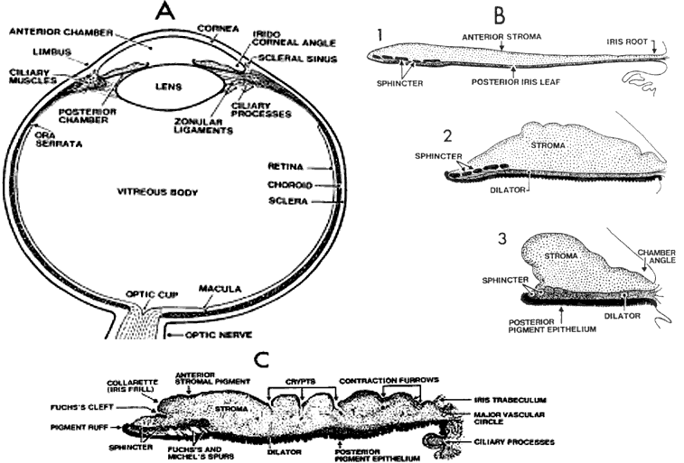
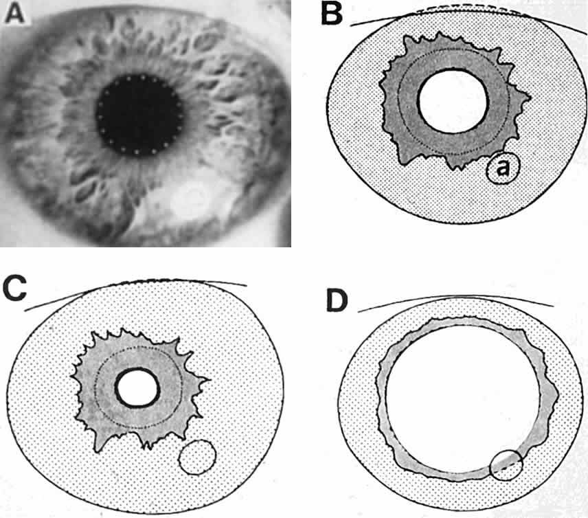
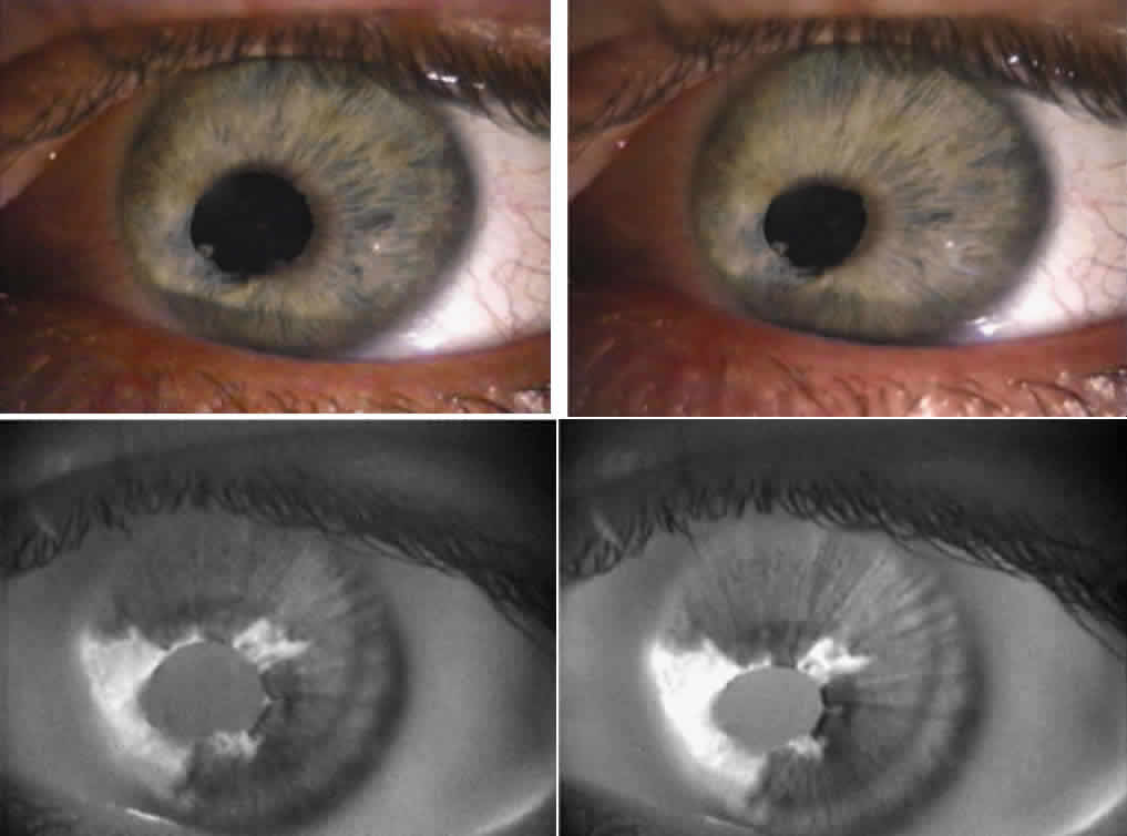
1. Newsome DA, Loewenfeld IE: Iris mechanics: II. Influence of pupil size on details of iris structure. Am J Ophthalmol 71:553, 1971

2. Loewenfeld IE, Newsome DA: Iris mechanics: I. Influence of pupil size on dynamics of pupillary movements. Am J Ophthalmol 71:347, 1971

3. Kestenbaum A: Clinical methods of neuro-ophthalmological examination, p 289. New York, Grune & Stratton, 1946

4. Levatin P: Pupillary escape in disease of the retina or optic nerve. Arch Ophthalmol 62:768, 1959

5. Fineberg E, Thompson HS: Quantitation of the afferent pupillary defect. In Smith JL (ed): Neuro-ophthalmology Focus, pp 25–29. New York, Masson, 1980

6. Thompson HS, Corbett JJ: Asymmetry of pupillomotor input. Eye 5:36, 19916a.Thompson HS: Putting a number on the relative afferent pupillary defect. In Thompson HS (ed): Topics in Neuro-ophthalmology, pp 157–158. Baltimore, Williams & Wilkins, 19796b.Thompson HS, Corbett JJ, Cox TA: How to measure 
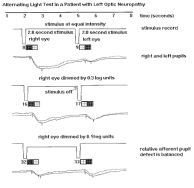
the relative afferent pupillary defect. Surv Ophthalmol 26:39, 1981

7. Rosenberg ML, Oliva A: The use of crossed polarized filters in the measurement of the relative afferent pupillary defect. Am J Ophthalmol 110:62, 1990

8. Lowenstein O, Kawabata H, Loewenfeld I: The pupil as indicator of retinal activity. Am J Ophthalmol 57:569, 1964

8a. Cox TA, Thompson HS, Hayreh SS, Snyder JE: Visual evoked potential and pupillary signs: a comparison in optic nerve disease. Arch Ophthalmol 100:1603, 1990

9. Thompson HS, Montague P, Cox TA, Corbett JJ: The relationship between visual acuity, pupillary defect, and visual field loss. Am J Ophthalmol 93:681, 1982

10. Brown RH, Zillis JD, Lynch MG, Sanborn GE: The afferent pupillary defect in asymmetric glaucoma. Arch Ophthalmol 105:1540, 1987

11. Johnson LN, Hill RA, Bartholomew MJ: Correlation of afferent pupillary defect with visual field loss on automated perimetry. Ophthalmology 95:1649, 1988

12. Kardon RH, Haupert C, Thompson HS: The relationship between static perimetry and the relative afferent pupillary defect. Am J Ophthalmol 115:351, 1993

13. Fison PN, Garlick DJ, Smith SE: Assessment of unilateral afferent pupillary defects by pupillography. Br J Ophthalmol 63:195, 1979

14. Cox TA: Pupillography of a relative afferent pupillary defect. Am J Ophthalmol 101:320, 1986

15. Cox TA: Pupillographic characteristics of simulated relative afferent pupillary defects. Invest Ophthalmol Vis Sci 30: 1127, 1989

16. Kawasaki A, Moore PA, Kardon RH: Variability of the relative afferent pupillary defect. Am J Ophthalmol 120: 622, 1995

17. Loewenfeld IE: The light reflex. In: The Pupil: Anatomy, Physiology and Clinical Applications, Vol 1, Chap 3, Section V, Pupillary reflex pathways, pp 198–240. Detroit, Iowa State University Press, Ames and Wayne State University Press, 1993

18. Lowenstein O: Alternating contraction anisocoria. Arch Neurol Psychiatry 72:742, 1954

19. Cox TA, Drewes CP: Contraction anisocoria resulting from half-field illumination. Am J Ophthal 97:577, 1984

20. Smith SA, Ellis CJK, Smith SE: Inequality of the direct and consensual light reflexes in normal subjects. Br J Ophthalmol 63:523, 1979

21. Smith SA, Smith SE: Contraction anisocoria: nasal versus temporal illumination. Br J Ophthalmol 64:933, 1980

22. Wyatt HJ, Musselman JF: Pupillary light reflex in humans: evidence for an unbalanced pathway from the nasal retina, and for signal cancellation in brainstem. Vision Res 21: 513, 1981

23. Loewenfeld IE: Damage to the intercalated pretectal neuron: consensual deficit. In: The Pupil: Anatomy, Physiology and Clinical Applications, Vol 1, Chap 18, pp 945–955. Detroit, Iowa State University Press, Ames and Wayne State University Press, 1993

24. Loewenfeld IE: The light reflex. In: The Pupil: Anatomy, Physiology and Clinical Applications, Vol 1, Chap 3, Section III, pp 96–135. Detroit, Iowa State University Press, Ames and Wayne State University Press, 1993

25. Young RSL, Han B, Wu P: Transient and sustained components of the pupillary responses evoked by luminance and color. Vision Res 33:437, 1993

26. Young RSL, Kennish J: Transient and sustained components of the pupil response evoked by achromatic spatial patterns. Vision Res 33:2239, 1993

27. Barbur JL, Harlow AJ, Sahraie A: Pupillary responses to stimulus structure, colour, and movement. Ophthalmic Physiol Opt 12:137, 1992

28. Barbur JL, Forsyth PM: Can the pupil response be used as a measure of visual input associated with the geniculo-striate pathway? Clin Vision Sci 1:107, 1986

29. Barbur JL, Keenleyside MS, Thomson WD: Investigation of central visual processing by means of pupillometry. In Kulikowski JJ, Dickinson CM, Murray IJ (eds): Seeing Colour and Contour, pp 431–451. Oxford, Pergamon Press, 1989

30. Slooter JH, van Noren D: Visual acuity measured with pupil responses to checkerboard stimuli. Invest Ophthalmol Vis Sci 19:105, 1980

31. Ukai K: Spatial pattern as a stimulus to the pupillary system. J Opt Soc Am 2:1094, 1985

32. Barbur JL, Thomson WD: Pupil response as an objective measure of visual acuity. Ophthalmic Physiol Opt 7:425, 1987

33. Cocker KD, Moseley MJ: Visual acuity and the pupil grating response. Clin Vision Sci 7:143, 1992

34. Harms H: Grundlagen, Methodik und Bedeutung der Pupillenperimetrie fur die Physiologie und Pathologie des Schorgans. Graefes Arch Klin Exp Ophthalmol 149:1, 1949

35. Cibis G, Campos E, Aulhorn E: Pupillary hemiakinesia in suprageniculate lesions. Arch Ophthalmol 93:1322, 1975

36. Hellner K, Jensen W, Muller-Jensen A: Video-processing pupillography as a method for objective perimetry in pupillary hemiakinesia: Proceedings of the Second International Visual Field Symposium, Tubingen, 1976. Doc Ophthalmol 14:221, 1977

37. Hellner KA, Jensen W, Muller A: Video processing pupillographic perimetry in hemianopsia. Klin Monatsbl Augenheilkd 172:731, 1978

38. Alexandridis E, Krastel H, Reuther R: Disturbances of the pupil reflex associated with lesions of the upper visual pathway. Graefes Arch Klin Exp Ophthalmol 209:199, 1979

39. Reuther R, Alexandridis E, Krastel H: Disturbances of the pupil reflex associated with cerebral infarction in the posterior cerebral artery territory. Arch Psychiatr Nervenkr 229:249, 1981

40. Reuther R, Krastel H, Alexandridis E: Disturbances of the pupil reflex associated with homonymous hemianopic paracentral scotoma. Arch Psychiatr Nervenkr 229:259, 1981

41. Hamann K, Hellner K, Muller-Jensen A, Zschocke S: Videopupillographic and VER investigations in patients with congenital and acquired lesions of the optic radiation. Ophthalmologica 178:348, 1979

42. Narasaki S, Kawai K, Kubota S, Noguchi J: Videopupillographic perimetry and its clinical application. Jpn J Ophthalmol 18:253, 1974

43. Yoshitomi M: The examination of pupillary light reflex with a specific pupillary paralysis. Acta Soc Ophthalmol Jpn 9: 853, 1955

44. Kardon RH, Kirkali PA, Thompson HS: Automated pupil perimetry. Ophthalmology 98:485, 1991

45. Kardon RH: Pupil perimetry. Curr Opin Ophthalmol 3: 565, 1992

46. ten Doesschate J, Alpern M: Response of the pupil to steady state retinal illumination: contribution by cones. Science 149:989, 1965

47. Gamlin PDR, Zhang H, Clarke RJ: Luminance neurons in the pretectal olivary nucleus mediate the pupillary light reflex in the rhesus monkey. Exp Brain Res 106:177, 1995

48. Reiner A, Williams J, Gamlin PDR: Characterization of neuropeptides, calcium binding proteins and glutamate receptors found in the olivary pretectal nucleus of the Rhesus monkey. Invest Ophthal Vis Sci 36(suppl):S292, 1995

49. Edinger L: Ueber den Verlauf der centralen Hirnnervenbahnen mit Demonstration von Praparaten. Arch Psychiatr Nervenkr 16:858, 1885

50. Westphal C: Ueber einen Fall von chronischer progressiver Lahmung der Augenmuskeln (ophthalmoplegia externa) nebst Beschreibung von Ganglienzellengruppen im Bereiche des Oculomotoriuskerns. Arch Psychiatr Nervenkr 18:846, 1987

51. Spitzka EC: The oculo-motor centres and their co-ordinators. J Nerv Ment Dis 13:413, 1988

52. Warwick R: The ocular parasympathetic nerve supply and its mesencephalic sources. J Anat 88:71, 1954

53. Carpenter MB, Harbison JW, Peter P: Accessory oculomotor nerve nuclei in the monkey: projections and effects of discrete lesions. J Comp Neurol 140:131, 1970

54. Akert K, Glicksman MA, Lang W et al: The Edinger-Westphal nucleus in the monkey: a retrograde tracer study. Brain Res 184:491, 1980

55. Burde RM, Loewy AD: Central origin of oculomotor parasympathetic neurons in the monkey. Brain Res 198:434, 1980

56. Jampel RS: Representation of the near-response on the cerebral cortex of the macaque. Am J Ophthalmol 48: 473, 1959

57. Loewenfeld IE: The reaction to near vision. In: The Pupil: Anatomy, Physiology and Clinical Applications, Vol 1, Chap 5, pp 295–317. Detroit, Iowa State University Press, Ames and Wayne State University Press, 1993

58. Loewenfeld IE: The light reflex. The Pupil: Anatomy, Physiology and Clinical Applications, Vol 1, Chap 3, Section V, pp 256. Detroit, Iowa State University Press, Ames and Wayne State University Press, 1993

59. Loewenfeld IE: Reactions to darkness. In: The Pupil: Anatomy, Physiology and Clinical Applications, Vol 1, Chap 4, p 282. Detroit, Iowa State University Press, Ames and Wayne State University Press, 1993

60. File RR, Patton TS: Topically applied pilocarpine. Pupillary response as a function of drop 
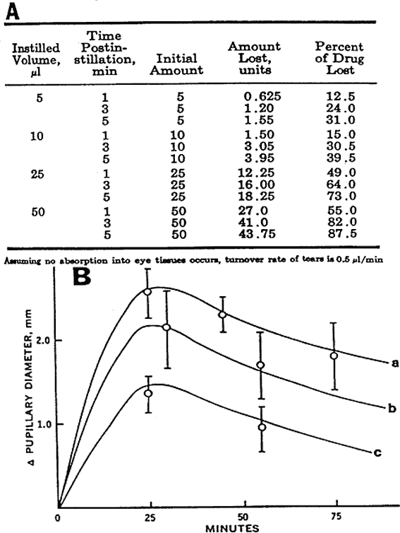
size. Am J Ophthalmol 98:112, 1980

61. Chrai SS, Makoid MC, Eriksen ST, Robinson JR: Drop size and initial dosing frequency problems of topically applied ophthalmic drugs. J Pharm Sci 63:333, 1974

62. Apt L, Henrick A: Pupillary dilation with single eye drop combinations. Am J Ophthalmol 89:553, 1980

63. Korczyn AD, Rubenstein AE, Yahr MD, Axelrod FB: The pupil and familial dysautonomia. Neurology 31: 628, 1981

64. Sugaya N, Nagataki S: Kinetics of topical pilocarpine in the human eye. Jpn J Ophthalmol 22:127, 1978

65. Duffin RM, Camras CB, Gardener SK, Pettit TH: Inhibitors of surgically induced myosis. Ophthalmology 89:966, 1982

66. Settone MF, Glannaccini B, Savigni P, Wirth A: The effect of different ophthalmic vehicles on the activity of tropicamide in man. J Pharm Pharmacol 32:519, 1980

67. Nagataki S, Sugaya N: Methylcellulose and ointment vehicles: their effects on ocular pharmacokinetics. Nippon Ganka Gakkai Zasshi 82:127, 1978

68. Zimmerman TJ, Leader B, Kaufman HE: Advances in ocular pharmacology. Ann Rev Pharmacol Toxicol 20:415, 1980

69. Burstein NL: Preservative cytotoxic threshold for benzalkonium chloride and chlorhexidine digluconate and cat and rabbit corneas. Invest Ophthalmol Vis Sci 19:308, 1980

70. Lyle WM, Bobier WR: Effective topical anesthetics on phenylephrine-induced mydriasis. Am J Optometry 54: 276, 1977

71. Araie M, Takase M, Yokoyama Y, Kitagawa M: Timolol maleate: pharmacokinetic analysis of ocular penetration in the rabbit eye and effects on human aqueous humor dynamics. Acta Soc Ophthalmol Jpn 84:2139, 1980

72. Lee VHL, Hui HW, Robinson JR: Corneal metabolism of pilocarpine in pigmented rabbits. Invest Ophthalmol Vis Sci 19:210, 1980

73. Mikkelson TJ, Chrai SS, Robinson JR: Altered bioavailability of drugs in the eye, due to drug-protein interaction. J Pharmacol Exp Ther 62:1648, 1973

74. Mikkelson TJ, Chrai SS, Robinson JR: Competitive inhibition of drug-protein interaction in the eye fluids and tissues. J Pharm Sci 62:1942, 1973

75. Loewenfeld IE: Pupillary Pharmacology. In: The Pupil: Anatomy, Physiology and Clinical Applications, Vol 1, Chap 14, Section IV-C2, pp 809–816. Detroit, Iowa State University Press, Ames and Wayne State University Press, 1993

76. Patil PN: Cocaine binding by the pigmented and the nonpigmented iris and its relevance to the mydriatic effect. Invest Ophthalmol 11:739, 1972

77. Mishima S: Clinical pharmacokinetics of the eye. Invest Ophthalmol Vis Sci 21:504, 1981

78. Loewenfeld IE: Pupillary Pharmacology. In: The Pupil: Anatomy, Physiology and Clinical Applications, Vol 1, Chap 14, Section IV-C3, pp 817–822. Detroit, Iowa State University Press, Ames and Wayne State University Press, 1993

79. Borgmann H, Worster W: Der Einfluss unterschiedlicher Konzentrationen Und Vehikel auf die Pilocarpin-Miosis: II. Unterschiedliche Vehikel. Klin Monatsbl Augenheilkd 163:51, 1973

80. Millodot M: Do blue-eyed people have more sensitive corneas than brown-eyed people? Nature 235:151, 1975

81. Koss MC: Studies on the mechanism of amphetamine in the cat. J Pharmacol Exp Ther 213:49, 1980

82. Loewenfeld IE: Pupillary Pharmacology. In: The Pupil: Anatomy, Physiology and Clinical Applications, Vol 1, Chap 14, Section III-B, pp 774–775. Detroit, Iowa State University Press, Ames and Wayne State University Press, 1993

83. Dahlstrom A, Fuxe K, Hillarp NA, Malmfors T: Adrenergic mechanisms in the pupillary light reflex path. Acta Physiol Scand 62:119, 1964

84. Lee HK, Wang SC: Mechanism of the morphine-induced miosis in the dog. J Pharmacol Exp Ther 192:115, 1975

85. Sigg EB, Sigg TD: The modification of the pupillary light reflex by chlorpromazine, diazepam and pentobarbital. Brain Res 50:77, 1973

86. Loewenfeld IE: Pupillary Pharmacology. In: The Pupil: Anatomy, Physiology and Clinical Applications, Vol 1, Chap 14, Section III-C, pp 788–791. Detroit, Iowa State University Press, Ames and Wayne State University Press, 1993

87. Loewenfeld IE: Lesions in the Ciliary Ganglion and Short Ciliary Nerves: The tonic pupil (“Adie's” syndrome). In: The Pupil: Anatomy, Physiology and Clinical Applications, Vol 1, Chap 24, Section III-C, pp 1111–1116. Detroit, Iowa State University Press, Ames and Wayne State University Press, 1993

88. Thompson HS: Adie's syndrome: some new observations. Trans Am Ophthalmol Soc 75:587, 1977

89. Jacobson DM, Olson KA: Influence of pupil size, anisocoria, and ambient light on pilocarpine miosis: implications 
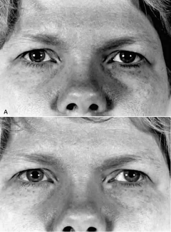
for supersensitivity testing. Ophthalmology 100:275, 1993

90. Jacobson DM: A prospective evaluation of cholinergic supersensitivity of the iris sphincter in patients with oculomotor nerve palsies. Am J Ophthalmol 118:377, 1994

91. Czarnecki JC, Thompson HS: The iris sphincter in aberrant regeneration of the third nerve. Arch Ophthalmol 96: 1606, 1978

92. Burde RM, Savino PJ, Trobe JD: Clinical Decision in Neuro-Ophthalmology, pp 178–185. St. Louis, CV Mosby, 1985

93. Scinto LFM, Kirk RD, Dressler D et al: A potential noninvasive neurobiological test for Alzheimer's disease. Science 266:1051, 1994

94. Wisniewski HM, Rabe A, Wisniewski KE: In Davies P, Finch C (eds): Molecular Neuropathology of Aging, Banbury Report, pp 399–413. Cold Spring Harbor, NY, Cold Spring Harbor Laboratory, 1988

95. Sacks B, Smith S: People with Down's syndrome can be distinguished on the basis of cholinergic dysfunction. J Neurol Neurosurg Psychiatry 52:1294, 1989

96. Loupe DN, Newman NJ, Green RC et al: Pupillary responses to tropicamide in patients with Alzheimers disease. Ophthalmology 103:495, 1996

97. Katz B: Letter to the Editor. Science 267:1578, 1995

98. Sitaram N, Pomara N: IRCS Med Sci 9:409, 1981

99. Forman AR: A new low-concentration preparation for mydriasis and cycloplegia. Ophthalmology 87:213, 1980

100. Sindell BD, Baker MD, Maisels MJ, Weinstein JM: A comparison of the pupillary and cardiovascular effects of various mydriatic agents in preterm infants. J Pediatr Ophthalmol Strabismus 23:273, 1987

101. Kardon RH, Denison CE, Brown CK, Thompson HS: Critical evaluation of the cocaine test in the diagnosis of Horner's syndrome. Arch Ophthalmol 108:834, 1990

102. Loewenfeld IE: Injury and repair in the nervous system. In: The Pupil: Anatomy, Physiology and Clinical Applications, Vol 1, Chap 11, Section II-D, pp 540–545. Detroit, Iowa State University Press, Ames and Wayne State University Press, 1993

103. Cremer SA, Thompson HS, Digre KB, Kardon RH: Hydroxyamphetamine mydriasis in Horner's syndrome. Am J Ophthalmol 110:66, 1990

104. Weinstein JM, Zweifel TJ, Thompson HS: Congenital Horner's syndrome. Arch Ophthalmol 98:1074, 1980

105. Almegaard B, Stjernschantz J, Bill A: Cholecystokinin contracts isolated human and monkey iris sphincters: a study with CCK receptor antagonists. Eur J Pharmacol 211: 183, 1992

106. Almegaard B, Bill A: C-terminal calcitonin gene-related peptide fragments and vasopressin but not somatostatin-28 induce miosis in monkeys. Eur J Pharmacol 250:31, 1993

Back to Top