|
|
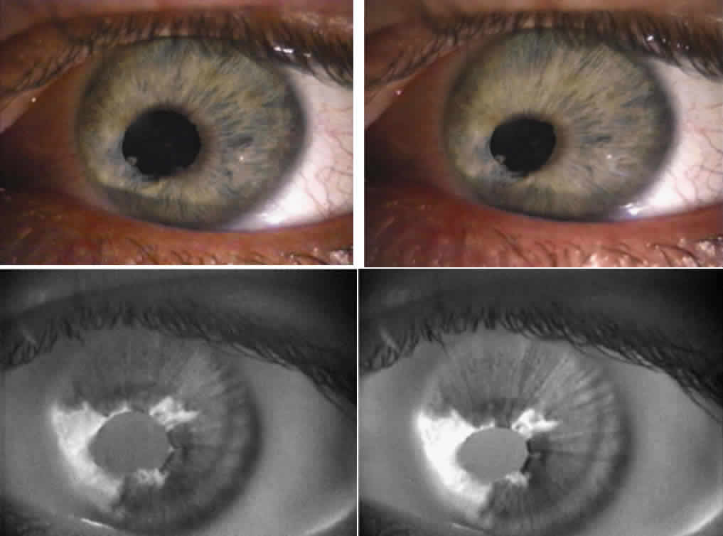
| Fig. 5. Asymmetric, segmental contraction of the pupil due to previous herpes zoster iritis. The top photos are slit-lamp images of the pupil in dark (left) and in light (right), showing irregular contractions. In the bottom photos, infrared transillumination of the same iris is shown in darkness and light. In areas of the iris previously damaged there is loss of tissue, which shows up as transillumination defects that appear white. In these areas, the sphincter muscle was destroyed by the inflammation and ischemia, resulting in loss of pupil contraction in the affected segment. |