|
|
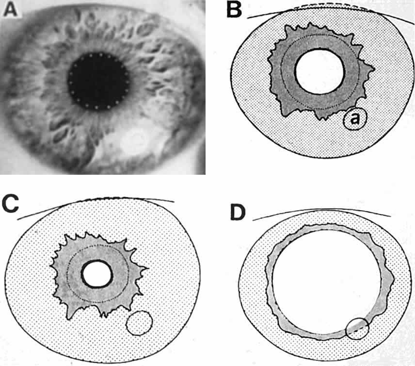
| Fig. 2. Changing iris areas with different pupil diameter. A. This eye looked greenish hazel at a distance. The dark pupil edge is indicated by small white dots. The large white spot at 5 o'clock (marked a in B) is the corneal reflection of the electronic flash. In the ciliary portion, the anterior stroma was intact (note the shallow contraction furrows between 1 and 4 o'clock). The vessels near the iris frill were heavy and clearly marked, with prominent X formations. In the area between the pupil and iris frill, there was no anterior stroma, and many vessels could be seen to run radially toward the pupil in fairly regularly interwoven loops. The sphincter muscle was visible below these vessels as a pinkish band surrounding the pupil. B to D. Outline drawings of the same eye with different pupil sizes. The shaded areas show the inner pupillary iris ring (from pupil to collarette) and the stippled areas to the outer ciliary ring (between collarette and limbus). The fine, broken lines mark the outer edge of the sphincter muscle. The sphincter ring becomes larger and thinner when the pupil dilates, and smaller and fatter when it constricts. With large pupils, it is hidden by the iris stroma, which billows above it. The black posterior iris leaf that peeks beyond the anterior iris layers when the pupil is small becomes thinner when it enlarges. (Loewenfeld IE: Reflex integration: physiologic mechanisms. In: The Pupil: Anatomy, Physiology and Clinical Applications, Vol 1, Ch 9, pp 414–424. Detroit, Iowa State University Press, Ames and Wayne State University Press, 1993) |