Chapter 41
Congenital Craniofacial Anomalies
STEVEN J. COVICI, PETER D. FRIES and JAMES A. KATOWITZ
Main Menu   Table Of Contents

Search

EMBRYOLOGY
CRANIOSYNOSTOSIS
CLEFTING SYNDROMES
BASELINE OPHTHALMIC EVALUATION
SURGICAL MANAGEMENT
SUMMARY
REFERENCES

Congenital abnormalities of the cranium and face present complex diagnostic and therapeutic challenges to the ophthalmologist. Patients with craniofacial anomalies should be managed by a multidisciplinary team including specialists from plastic surgery, neurosurgery, ophthalmology, otolaryngology, oral surgery, orthodontics, anesthesia, and genetics as well as specialists in the disciplines of psychiatry, social work, and nutrition. All these specialists must work together to provide for the overall well-being of the patient. To assist in the evaluation and management of these patients, the ophthalmologist must possess a basic understanding of craniofacial syndromes as well as the necessary medical and surgical interventions required to improve ocular and adenexal problems. This chapter systematically reviews the major craniofacial anomalies of ophthalmic importance as well as highlights salient management issues. Craniofacial syndromes may be conveniently divided into two broad categories: craniosynostosis or premature closure of the cranial sutures and clefting syndromes

To facilitate understanding of the congenital craniofacial anomalies, a review of the normal embryogenesis of the human head is needed. Although a more complex review of embryology is beyond the scope of this chapter, essential morphogenetic events are presented here to help the reader visualize the formation of the craniofacial anomalies.

Back to Top
EMBRYOLOGY
The fourth to eighth weeks of gestation are referred to as the embryonic period. The face develops mainly between the fifth and eighth weeks and by the end of the eighth week the embryo has an unquestionably human appearance. Early in the fourth week, the embryo is a straight flat structure with a dorsal neural tube (Fig. 1). Formation of the neural tube begins in the middle of the embryo and progresses toward the cranial and caudal ends. As the neural tube separates to a more ventral position from the surface ectoderm, a subset of neuroectodermal cells migrate to the dorsolateral aspects of the tube to become the neural crest. Neural crest cells ultimately give rise to the connective tissue elements of the eye and adnexa including corneal keratocytes, iris stroma, corneal stroma, ciliary smooth muscle, sclera fibroblasts, optic nerve meanings, orbital fibroadipose tissue, orbital nerve Schwann's cells, and orbital bone. In addition, early in the fourth week of gestation the paraxial mesoderm forms into longitudinal columns of paired cuboidal bodies called somites that laterally flank the neural tube. These somites are destined to form most of the paraxial skeleton and associated musculature.

Fig. 1. Early fourth-week embryo demonstrating cephalad open neural groove, closing neural plate, and flattened structure. (Fries PD, Katowitz JA: Congenital craniofacial anomalies of ophthalmic importance. Surv Ophthalmol 35:87, 1990, with permission from Elsevier Science.)

As the rostral neural groove closes, the first branchial arch begins to appear along the lateral portion of the neural tube. The first branchial arch, or mandibular arch, forms two paired mandibular processes, which will eventually fuse to become the mandible. At the superior lateral portion of each mandibular process, a small bud develops into the maxillary process, which eventually forms the maxilla, zygomatic bone, and squamous part of the temporal bone (Fig. 2). Thus, at the midfourth week, after neural tube invagination, the primitive cavity or stomodeum is flanked by five distinct facial processes: two paired mandibular processes, two paired maxillary processes, and a central frontonasal process. Most congenital malformations of the head and neck originate as the branchial arches transform into their adult derivatives.

Fig. 2. Midfourth week embryo after neural tube invagination. Maxillary, mandibular, and frontocranial processes are beginning growth into intended facial position. (Fries PD, Katowitz JA: Congenital craniofacial anomalies of ophthalmic importance. Surv Ophthalmol 35:87, 1990, with permission from Elsevier Science.)

The branchial arches are initially composed of an external layer of ectoderm and an internal core of endoderm. It is the migration of neural crest cells into the branchial arches that causes them to enlarge. The ectodermal furrows dividing the facial processes are eventually obliterated by proliferating neural crest cells and mesenchyme. Fusional failure of the facial processes caused by the absence of neural crest cell migration is one of the proposed etiologies of “true” facial clefts and are in more detail in later sections. At the beginning of the fourth week, the developing eye appears as a placode on the lateral forebrain and by the end of the fourth week has become an easily identifiable optic vesicle.

During the fifth week of gestation, the maxillary processes continue to enlarge and laterally border the recently perforated stomodeum (Fig. 3). The nasal placodes, which appear late in the fourth week, transform into olfactory grooves. Horseshoe-shaped medial and lateral nasal prominences develop and the nonfused inferior portion of the olfactory groove remains continuous with the stomodeum. Also during the fifth week, six swellings, called auricular hillocks, develop from the first and second branchial arches and represent the rudimentary external ear. As the optic vesicle continues to grow, the overlying surface ectoderm differentiates into the lens placode. The optic vesicle invaginates to form the double layered optic cup and the overlying surface ectoderm, with mesenchymal contributions from the neural crest cells, which will later form the eyelids (Fig. 4). The eyelids fuse at the ninth week and at the 25th to 26th weeks, the fused margins separate.

Fig. 3. Frontal view, late fifth week embryo demonstrating prominent anteriorly located nasal pits with the nonfused inferior portion connecting to the recently perforated stomodeum. (Fries PD, Katowitz JA: Congenital craniofacial anomalies of ophthalmic importance. Surv Ophthalmol 35:87, 1990, with permission from Elsevier Science.)

Fig. 4. Late fifth week demonstration of ocular development showing lens vesicle, two-walled optic cup and optic stalk with choroidal fissure incorporating the ophthalmic vessels. (Fries PD, Katowitz JA: Congenital craniofacial anomalies of ophthalmic importance. Surv Ophthalmol 35:87, 1990, with permission from Elsevier Science.)

In the sixth week, the medial nasal prominences fuse with each other and with the maxillary prominence (Fig. 5). Failure of the medial nasal prominence to merge with the maxillary prominence results in a cleft lip, with or without cleft palate; the cleft lip may be unilateral or bilateral. Continued growth of the maxillary processes promotes the formation of the nasolacrimal groove along the lateral border of the nasal processes. The nasolacrimal groove will incorporate neuroectodermal cells, which will eventually become the nasolacrimal drainage apparatus. The mandibular process continues its growth and fuses at the midline by the end of the fifth week.

Fig. 5. Sixth week embryo shows fusion of the maxillary process to the lateral nasal groove forming the nasal pit and nasolacrimal furrow. Note lateral position of developing eye. (Fries PD, Katowitz JA: Congenital craniofacial anomalies of ophthalmic importance. Surv Ophthalmol 35:87, 1990, with permission from Elsevier Science.)

During the seventh week of gestation, the maxillary processes fuse at midline, separating the nares from the mouth (Fig. 6). The enlargement of the maxillary process and expansion of the lateral head, combined with the horizontal collapse and the vertical elongation of the frontonasal process, allows for anterior rotation of the developing eyes. This anterior rotation of the eyes is not be fully achieved until approximately 4 months of gestation and abnormalities in this process may result in hypertelorism (Fig. 7). By the seventh week, nearly all lower facial fissures and furrows have completely closed.

Fig. 6. Seventh week embryo with completed maxillar and mandibular midline fusion, continued frontal rotation of the orbits and prominent frontonasal overhang. (Fries PD, Katowitz JA: Congenital craniofacial anomalies of ophthalmic importance. Surv Ophthalmol 35:87, 1990, with permission from Elsevier Science.)

Fig. 7. Separation of globes at 2 months' gestation, 3 months' gestation, birth, and adulthood. (Fries PD, Katowitz JA: Congenital craniofacial anomalies of ophthalmic importance. Surv Ophthalmol 35:87, 1990, with permission from Elsevier Science.)

By the eighth week of gestation, the embryonic face has distinct human characteristics despite the obvious differences in facial proportions. By the end of the embryonic period, external ears begin to assume their final shape despite being set low on the head. The eyes assume a more almond shape and continue their anterior rotation. The cranium loses most of its overhang while the chin and nose achieve a more pronounced profile.

In addition to an appreciation of the development of facial features, a fundamental understanding of the development of the bony skull is critical to understanding of congenital craniofacial anomalies. The calvaria and cranial base represent the two major portions of the developing skull and have distinct embryonic etiologies. The calvaria arises from neural crest cell migration into cartilaginous condensations at the site of the future crista galli, lesser wings of the sphenoid, and petrous ridges. Primitive dura matter arises from these sites and is first observed at 5 to 6 weeks' gestation. The dural sheaths expand to accommodate the rapidly enlarging brain and the junctions at which these dural sheaths appose determine the sites of the cranial sutures (Fig. 8). Intramembranous ossification of mesenchyme forms the bones of the cranial vault. Portions of the dura prevent closure of the cranial sutures and allow for expansion of the intracranial contents during fetal and infant growth. Premature fusion of one or more of the cranial sutures, termed craniosynostosis, produces various abnormal head shapes, which are discussed in more detail in later sections.

Fig. 8. Normal sutures and fontanels of fetal skull. (Fries PD, Katowitz JA: Congenital craniofacial anomalies of ophthalmic importance. Surv Ophthalmol 35:87, 1990, with permission from Elsevier Science.)

At 4 to 5 weeks' gestation, the neural crest cells responsible for formation of the cranial base first flank and later surround the notochord (Fig. 9). These include the alisphenoid, orbitosphenoid, and prechordal cartilage, which develop into the sphenoid, zygoma, temporal, and ethmoid bones. Premature fusion of these sutures, especially the sphenozygomatic and sphenoethmoidal sutures can lead to shallow orbits and poor growth of the midface. The prechordal cartilage becomes the nasal septum, which, with ethmoid development, plays a vital role in the collapse of the wide embryonic interpalpebral distance. Variations in development of the ethmoid sinus and nasal septum may lead to hypertelorism.

Fig. 9. Schematic representation of cranial base cartilage and growth at weeks five and eight of gestation. The alisphenoid, orbitosphenoid, and prechordal cartilages develop into the sphenoid, zygoma, temporal, and ethmoid bones in the adult skull. (Fries PD, Katowitz JA: Congenital craniofacial anomalies of ophthalmic importance. Surv Ophthalmol 35:87, 1990, with permission from Elsevier Science.)

Most congenital craniofacial anomalies arise from developmental abnormalities before the eighth week of gestation. Striking similarities between characteristics of the developing embryonic face and certain craniofacial deformities should now be readily apparent. It is almost as if growth and differentiation progress normally to a certain stage and then are abruptly interrupted. The following sections review in detail the craniofacial deformities most frequently encountered by the ophthalmology consultant.

Back to Top
CRANIOSYNOSTOSIS
Craniosynstosis comprises a major group of congenital malformations in which one or more of the cranial sutures are closed prematurely. In 1851, the German pathologist Virchow first described how premature closure of one cranial suture promotes growth parallel to that suture and inhibits growth perpendicular to it.1 Although the terms craniosynostosis and craniostenosis are often used interchangeably, the preferred, more accurate term is craniosynostosis. The word craniosynostosis describes the process of premature sutural fusion, craniosynostosis is simply the result.2

There are several different types of craniosynostosis. First, craniosynostosis may be defined as either simple or compound. Simple craniosynostosis refers to the premature closure of a single suture whereas a compound craniosynostosis involves two or more sutures. The most prevalent type of craniosynostosis is scaphocephaly, in which the sagittal suture is synostosed, which results in a boat-shaped skull. Boys are affected much more commonly than girls, whereas sagittal synostosis occurs in girls more often than boys.2

Second, craniosynostosis may be designated as either primary or secondary. In primary synostosis, the most common type, the cranial sutures are fused prematurely because of a genetic predisposition. In secondary craniosynostosis, the premature sutural fusion is secondary to another known disorder. Examples include hematologic disorders (thalassemia and sickle cell disease), metabolic disorders (hyperthyroidism), metabolic disorders (mucopolysaccharoidosis), and malformations (microcephaly).2

Finally, craniosynostosis may be either isolated or syndromic. In the isolated form, the patient has no other abnormalities except those directly related to early sutural obliteration. In contrast, patients with the syndromic craniosynostosis have other primary morphogenic alterations. For example, in Apert's syndrome, syndactyly of the hands and feet occur frequently whereas congenital heart defects accompany the craniofacial malformation.

Specific terms, such as scaphocephaly, have been used to describe the typical skull deformities associated with various synostoses (Fig. 10). Single coronal sagittal, metopic, or lambdoidal synostosis, respectively, leads to various degrees of brachycephaly, scaphocephaly, trigonocephaly, or plagiocephaly. Oxycephaly (tower head) is used to describe multiple synostosis involving at least the coronal and sagittal sutures, with compensatory upward growth of the cranium. Triphyllocephaly3 or kleeblattschädel describes a cloverleaf-shaped skull, which is clinically characterized by the synostosis of the lambdoid and coronal suture with herniation of the cerebrum through a patent sagittal suture. The general term plagiocephaly refers to closure of a single cranial suture, whether sagittal, coronal, or lambdoidal and may lead to wide variability in cranial morphology. Although descriptive, these terms may be confusing and it is probably simpler to classify these patients by the sutures involved.4

Fig. 10. Frontal and lateral views of fetal skulls demonstrating more common calvarial abnormalities resulting from suture closure. (Fries PD, Katowitz JA: Congenital craniofacial anomalies of ophthalmic importance. Surv Ophthalmol 35:87, 1990, with permission from Elsevier Science.)

Traditional classification of craniosynostosis has been divided into three main groups: (1) craniofacial dystosis or Crouzon's syndrome, (2) acrocephalosyndactyly or “Apert's syndrome,” and (3) simple or single craniosynostosis. This classification system has largely been replaced by Cohen's5–7 more extensive classification.8 Cohen's classification of craniosynostosis categorizes these anomalies based on clinical similarities and genetic transmission5–7 (Table 1).


TABLE 1. Craniosynostosis Syndromes


NameOphthalmic Involvement
Crouzon's syndromeShallow orbits, proptosis, hypertelorism, exotropia, exposure keratitis, luxation of globe
Apert's syndromeHypertelorism, shallow orbits, strabismus, proptosis
Pfeiffer syndromeHypertelorism, proptosis, strabismus
Saethre-Chotzen syndromeHypertelorism, ptosis, strabismus, nasolacrimal duct obstruction, optic atrophy
Carpenter syndromeHypertelorism, epicanthal folds, medial canthal dystopia, corneal opacities
Kleeblattschadel anomaly: Cloverleaf skullVariable: proptosis, hypertelorism, exposure keratitis, iris coloboma, orbital hypoplasia, nasolacrimal duct obstruction
Christian syndromeHypertelorism, ophthalmoplegia, lateral canthal dystopia
Summitt syndromeEpicanthal folds, strabismus
Baller-Gerold syndromeHypertelorism, epicanthal folds
Lowry syndromeProptosis, strabismus
Gorlin-Chaudhry-Moss syndromeLid colobomas, ptosis, microphthalmia
Hermann-Opitz syndromeHypertelorism, proptosis, exotropia
Hermann-Pallister-Opitz syndromeHypertelorism, exotropia
Sakati-Nyhan-Tisdale syndromeHypoplastic orbits, proptosis
(Adapted from Cohen MM Jr: An etiologic and nosologic overview of craniosynostosis syndromes. Birth Defects 11:137, 1975.)


Three classic theories of pathogenesis explain craniosynostosis. Virchow1 believed that premature fusion of the cranial sutures were the primary dysgenesis and that the cranial base abnormalities were secondary to the craniosynostosis. Moss,8 conversely, postulated that the cranial base malformation was the primary event, resulting in secondary obliteration of the cranial sutures. Moss speculated that spatially malformed lesser sphenoidal wings in coronal synostosis, and a malformed cribriform plate in sagittal synostosis transmit aberrant tensile forces at points of dural attachments leading to premature fusion of the overlying sutures. Park and Powers9 implicated a primary defect in the mesenchymal blastema that produced both an abnormal cranial base and craniosynostosis.

There are chromosomal and environmentally induced etiologies for the craniosynostosis syndromes. The specific inheritance patterns and genetic anomalies of the individual craniosynostosis syndromes of ophthalmic importance are individually discussed in later sections. For many dysmorphic syndromes, it is possible to find families that display a particular phenotype in either an autosomal dominant, recessive, or x-linked manner.10 In addition, patients with the same genetic condition may have fusion of completely different sutures.11 Nonsyndromic synostosis may be sporadic or familial, with similar suture involvement in several members of the same family.12 With the recent advancements in gene mapping and cloning techniques, investigation of the genetic elements of craniofacial dysmorphologies has developed rapidly. It is critical for geneticists to be intimately involved with other craniofacial medical specialists so that they may provide genetic counseling to the patient's family as well as further research, which may ultimately lead to possible genetic therapy.

CROUZON'S SYNDROME

Crouzon's syndrome, which he first described in 1912, is characterized by the triad of craniosynostosis, midface retrusion, and proptosis (Fig. 11).13 Crouzon's syndrome, originally termed hereditarily craniofacial dysostosis, has a birth prevalence of 16.5 per million and counts for 4.8% of all cases of craniosynostosis, making it the most common of the craniosynostosis syndromes.14 Inheritance is autosomal dominant with variable expression and complete penetrance. Approximately 50% of cases represent a new familial mutation.15,16 Crouzon's syndrome has been linked to the long arm of chromosome 10, specifically 10q25-q26.17–20 Apert's, Jackson-Weiss, and Pfeiffer's syndrome have also been genetically linked to chromosome 10q25-q26.19,20 Variations in the B exon of the fibroblast growth factor receptor 2 gene (FGFR2) has been linked as a cause of Crouzon's syndrome in one published study.21 FGFR2 is in the family of tyrosine kinase receptors and is involved in the signal transduction pathways for embryogenesis and cellular differentiation.22 Crouzon's syndrome is seen in many different ethnic groups and is associated with advanced paternal age.16

Fig. 11. Crouzon's syndrome showing typical midfacial findings including prominent exophthalmos, hypertelorism, and recessed supraorbital and lateral orbital rims.

Calvarial suture synostosis in Crouzon's syndrome varies, which is why no head shape is characteristic of this syndrome.23 The premature and progressive craniosynostosis is often observed at birth and is usually completed by 2 to 3 years of age.24 Despite this variable presentation, between 96% and 98% of patients with this syndrome display radiographic evidence of fusion of the coronal and sagittal sutures whereas 79% have lambdoidal synostosis.18 This pattern of fusion leads to the somewhat more predicable skull shape, the features of which include a shortened calvaria, steep forehead, and flattened occiput.3,25–30

Early fusion of the cranial sutures imposes physical restraints on the growing brain, which results in neurologic complications. These included headaches (29% to 50%), mental retardation (13%), and seizures (11.5%).7,16,23 In addition to synostosis of the calvarial sutures, the cranial base sutures and midface sutures of the orbits and maxilla display premature fusion.16,31–37 This may lead to hypoplasia of the ethmoid, frontal, maxillary, and sphenoid bones, which contributes to the midfacial retrusion and shallow, widespread orbits characteristics of the “toadlike” facies associated with Crouzon's syndrome.16,30 Other consistent facial features include a parrot-beak nose, hypoplastic cheekbones, flattened nasal bridge, hypoplastic supraorbital ridge, hypertelorism, and shallow orbits with resulted proptosis.24

Hypertelorism and exophthalmos are universal features in Crouzon's syndrome.16 Complications arising from the proptosis include exposure keratopathy, exposure conjunctivitis, and more rarely, globe subluxation anterior to the eyelids (Fig. 12).15,16 These patients also exhibit exorbitism, which is an increased angle of divergence of the orbits in excess of the normal 90 degrees. Other ocular anomalies that have been sporadically reported in Crouzon's syndrome include aniridia, anisocria, blue sclera, cataracts, compound astigmatism, corectopia, ectopia lentis, glaucoma, iris coloboma, keratoconus, megalocornea, microcornea, nystagmus, and optic nerve hypoplasia.30,38,39 Adenexal abnormalities may include canthal dystopia, ectropion, entropion, nasolacrimal duct obstruction, and ptosis.40–46

Fig. 12. A. Child with Crouzon's syndrome and severe orbital hypoplasia with resultant proptosis anterior to eyelids. B. Axial computed tomogram of child in Figure 12A demonstrating extreme orbital shallowness.

Strabismus has been described in as many as 92% of patients with Crouzon's syndrome, with V-pattern exotropia the most common variety. Esotropia, exotropia, hypertropia, and vertical and horizontal phorias have also been reported.7,23,47,48 The frequently encountered V-pattern exotropia has been postulated to occur secondary to an underaction or absence of the superior oblique muscle with a relative overreaction of the inferior oblique.48 In fact, absent or abnormally inserting extraocular muscles may contribute to the aforementioned patterns for strabismus.49

Optic nerve dysfunction may be present in up to 80% of patients with Crouzon's syndrome.23 Optic nerve involvement may present clinically as either optic atrophy or papilledema and its etiology remains elusive. This papilledema, in a study by Bentelson, was not correlated to hydrocephalus, increased intracranial pressure or narrow optic canals.23

Nonophthalmic findings in Crouzon's syndrome include crowding of the maxillary teeth, lateral palatal swellings, and jaw malocclusion secondary to maxillary hypoplasia.16 Of these patients, 55% acquire conductive hearing loss while 30% have atretic external auditory canals.16 Obstructive sleep apnea is unfortunately common and is related to a constricted nasopharynx and midfacial retrusion.15,16,18 Cervical spine anomalies occur in 30% of patients; most of these abnormalities involve fusion of the C2 and C3 vertebrae.16 Radiographic evidence of calcification of the stylohyoid ligament has also been reported in 88% of patients. As mentioned previously, headaches, seizures, and hydrocephalus may occur; however, unlike the case of Apert's syndrome, mental retardation is rare.16

APERT'S SYNDROME

Apert's syndrome (acrocephalosyndactyly type I) was described by Apert in 1906,50 although others had reported similar cases prior to his publication.50,52 Apert's syndrome is characterized by craniosynostosis with symmetric syndactyly of the hands and feet. Two clinical categories of acrocephalosyndactyly exist: typical (or Apert) variant and the atypical variant. Typical acrocephalosyndactyly is clinically distinguishable by complete distal fusion of the soft tissues of digits of 2, 3, and 4 with variable degrees of bony fusion in both the hands and feet.53 There is the presence of a middigital hand mass with a common single nail to digits 2 through 4. Despite a range of craniofacial variability, it is the clinical reproducibility of the hand anomaly that distinguishes these individuals from those with the atypical acrocephalosyndactyly.

Apert's syndrome has an estimated frequency of 1 per 160,000 live births53 and comprises approximately 4.5% of all cases of craniosynostosis. Although this syndrome is inherited in an autosomal dominant pattern with complete penetrance, most reported cases are sporadic.54–58 Advanced paternal age appears to be a risk factor for the fresh sporadic mutations53,59,60 and the syndrome has been observed with equal incidence across all populations.7 As mentioned previously, the genetic defect associated with Apert's syndrome has been linked to chromosome 10q25-q26.19,61,62 Apert's, Crouzon's, Jackson-Weiss, and Pfeiffer's syndromes are all craniosynostoses genetically linked to chromosome 10q25-q26 and are associated with point mutations in the extracellular domain of FGF2.19,61,62

The characteristic limb malformations differentiate Apert's syndrome from other craniosynostosis associated with syndactyly.50,63 Based on craniofacial dysmorphology alone, Apert's syndrome can be confused with Crouzon's, Pfeiffer's and, less often, Saethre-Chotzen syndromes. Despite multiple and variable suture involvement in Apert's syndrome, craniosynostosis usually affects the coronal sutures, thereby limiting the anterior and posterior growth of the cranium.15 The skull contour is typically tall, with a steepened forehead and a flattened occiput. Similar to Crouzon's syndrome, there is hypoplasia of the midface, hypertelorism, and shallow orbits with proptosis, which may lead to exposure keratopathy. Patients with Apert's syndrome also may possess parrot-beak nose, low-set ears, and a horizontal groove above the supraorbital ridge (which diminishes with age) as well as down-slanting (antimongoloid) palpebral fissures (Fig. 13).

Fig. 13. Apert's syndrome showing frontal overhang, “parrot-beak” nose, mild exophthalmos, and exorbitism.

The hypertelorism, decreased orbital volume, and proptosis in Apert's syndrome are secondary to the midface retrusion and dysmorphic cranial base and are generally less severe than in Crouzon's syndrome.15 Strabismus occurs in 70% to 100% of patients and is usually of the exotropic variety. V-pattern exotropia predominates, although V-pattern esotropia may be observed. Absence of the superior rectus muscle, absence of the superior oblique muscle, and structural abnormalities of the extraocular muscles have been reported as well.48,64 Refractive errors such as high astigmatism and anisometropia are common in this syndrome. Other rare ophthalmic manifestations include keratoconus, ectopia lentis, congenital glaucoma, and optic atrophy.9,36,37

Apert's syndrome does not necessarily include mental retardation, although it is often pessimistically presented by clinicians to patients' families in this way.65 Intelligence is often subnormal and in one study of 15 patients with Apert's syndrome, the average IQ was 72.5.66 Development may be adversely affected by progression of increased intracranial pressure, although early craniotomy does not appear to ameliorate development.67 Other neurologic anomalies associated with Apert's syndrome include megalocephaly, as well as defects of the corpus callosum, brain, and gyral anomalies.66,68

Oral manifestations of Apert's syndrome include crowded teeth, impaction, and thickened gingiva.15,69 Clefting of the soft palate and bifid uvula occur in 30% of patients and malocclusion, usually class 3, related to the maxillary hypoplasia is seen. Many cardiac and visceral anomalies, such as coarctation of the aorta, ventricular septal and atrial septal defects, patent ductus arteriosus, and tracheoesophageal and pyloric stenosis have been reported, but such findings are relatively uncommon.70 In adolescent patients, 70% display peculiar acneiform eruptions on the trunk and forearms.71 Conductive hearing loss, often from chronic otitis media, is unfortunately extremely common with the incidence of deaths reported as high as 30%.72–74

Syndactyly of the hands and feet is a major feature of Apert's syndrome (Fig. 14). As mentioned previously, a middigital hand mass involving the second, third, and fourth digits is observed. The first and fifth digits may be adjoined, but when the thumb is separate it is usually broad. There is symmetric syndactyly of the toes with medial deviation of the great toe and progressive fusion of the midfoot and hindfoot in the supinated position.75 Radiologic studies show some degree of brachydactyly in all five digits, synphalangism of the interphalangeal joints, and eventual fusion of the carpal, tarsal, and interphalangeal joints over time.7,76 Other orthopedic anomalies in Apert's syndrome include ankylosis of the elbow, shoulder, and hip and cervical stenosis.77

Fig. 14. Hand of child with Apert's syndrome showing proximal syndactyly of digits 2 and 3, 4 and 5 and near-total fusion of digits 3 and 4. (Fries PD, Katowitz JA: Congenital craniofacial anomalies of ophthalmic importance. Surv Ophthalmol 35:87, 1990, with permission from Elsevier Science.)

Several additional cranial synostoses share common craniofacial features with Crouzon's syndrome but occur less frequently; these include Carpenter's, Pfeiffer's, and Sathre-Chotzen syndrome.

CARPENTER'S SYNDROME

Carpenter's syndrome (acrocephalopolysyndactyly type II) is a rare entity first described by Carpenter in 1901. It was finally recognized as a distinct entity in 1966 when Tentamy reported 1 case and studied 12 cases previously described in the literature under the name of Laurence-Moon-Biedl syndrome.78,79 Carpenter's syndrome is clinically characterized by craniosynostosis, preaxial polydactyly of the feet, variable brachydactyly, syndactyly, and obesity. Inheritance is autosomal recessive, but many cases represent new spontaneous mutations.72,79

Ocular findings are less distinct due to the rarity of the disorder. Hypertelorism with relative telecanthus and epicanthal folds has been described in most cases.80 Shallow orbits with proptosis and mild down-slanting of the palpable fissure may also be observed. Corneal opacity, microcornea, optic atrophy, and pseudopapilledema occur sporadically.78–81

The craniosynostosis of Carpenter's syndrome is characterized by multisutural involvement. Variable premature fusion of the coronal, lambdoidal, and sagittal sutures normally result in a grossly normal calvaria, which is usually shortened in all directions.15,79,82 Most affected patients are mentally retarded; however, those with normal intelligence have been reported.82,83 Short stature and obesity are usually present. Between 30% and 50% of patients have congenital heart defects including atrial and ventricular septal defects, patent ductus arteriosus, and tetralogy of Fallot.79,84

Patients with Carpenters' syndrome may also have low-set ears, a hypoplastic mandible, flattened nasal bridge, and high arched palate.7,80,81 Other anomalies include pes varus, genu valgum, laterally displaced patella, ilial flaring, and decreased hip mobility.15,74,79,82–85 Preaxial polysyndactyly of the feet, soft tissue syndactyly of the hands, and brachydactyly and duplication of the second toe may be seen in many of these patients.

PFIEFFER'S SYNDROME

The combination of craniosynostosis, broad thumbs and toes, and partial soft tissue syndactyly of the fingers and toes was described by Rudolf Pfieffer in 1964 in eight members of one family affected over three generations (Fig. 15).86 Pfeiffer's syndrome is an autosomal dominant disorder with complete penetrance and variable expression.87,88 The genetic mutations in Pfeiffer's syndrome have been identified as FGF2 on chromosome 10 and FGF1 on chromosome 8.89 Interestingly, identical mutations of FGF2 have been identified in Crouzon's and Pfieffer's syndrome despite their phenotypic differences in appearance. Cohen classified Pfeiffer's syndrome into three subtypes:

Fig. 15. A. Clinical photograph of Pfeiffer's syndrome demonstrating exophthalmos, hypertelorism, small mandible, “parrot-beak” nose, recessed supraorbital ridges, and increased forehead height. B. Enlarged thumb of same patient. C. Broad, long great toe. (Fries PD, Katowitz JA: Congenital craniofacial anomalies of ophthalmic importance. Surv Ophthalmol 35:87, 1990, with permission from Elsevier Science.)

Patients with type I have normal intelligence whereas types II and III have developmental retardation.87 The usefulness of this classification lies chiefly in genetic counseling, given that much clinical overlap exists among the different subtypes.

The ophthalmic manifestations in Pfeiffer's syndrome closely parallel those found in Crouzon's and Apert's syndromes. Hypertelorism, ocular proptosis, oxybrachycephaly (often secondary to bilateral coronal synostosis), strabismus, flattened nasal bridge with beaked nose, maxillary hypoplasia with a relative mandibular prognathism, and malocclusion are typical findings in this syndrome. Other abnormalities include high arched palate, crowded teeth, hydrocephalus, seizures, bifid uvula, low-set ears, and conductive hearing loss.90

The distinguishing features of this syndrome are the broad, medially deviated great toes and thumbs. In fact, the ratio of the width of the great toe to the second toe may be useful in diagnosing this syndrome.87,91 Brachydactyly, clinodactyly, and partial soft tissue syndactyly may be present whereas radiographic studies of the hand and foot show the occasional absence of the middle phalanges, broad distal thumb phalanges, triangulation of the proximal thumb, and the unusual appearance or the duplication of the first metatarsal bone.92 Associated abnormalities include lumbar hyperlordosis, fusion of lumbar or cervical vertebrae, shortened humerus, umbilical hernia, preauricular skin tags, and supernumerary teeth.

SAETHRE-CHOTZEN SYNDROME

Saethre-Chotzen syndrome is characterized by craniosynostosis, low-set frontal hairline, facial asymmetry, deviated nasal septum, and partial simple syndactyly especially involving the middle and index fingers (Fig. 16).93,94 This syndrome, as described by Saethre and Chotzen, is an autosomal dominant disorder with high penetrance and variable expression. The genetic defect has been mapped as a distal part of the short arm of chromosome 7.95,96 Advanced paternal age is a risk factor for new mutations and translocation defect of chromosome 7 have been noted in certain families with the syndrome.7,97–99

Fig. 16. Frontal (A) and lateral (B) view of patient with Saethre-Chotzen syndrome with low-set hairline, ptosis, strabismus, mild facial asymmetry and low set ears. (A, Fries PD, Katowitz JA: Congenital craniofacial anomalies of ophthalmic importance. Surv Ophthalmol 35:87, 1990, with permission from Elsevier Science.)

Saethre-Chotzen syndrome is often underdiagnosed because craniosynostosis is diagnostic for the syndrome and cosmetic abnormalities are usually mild. Variability in head shape is usually seen as plagiocephaly. Scaphocephaly, brachycephaly, and oxycephaly have also been reported.100,101 The midface is spared the retrusion seen in the other craniosynostosis. Frontal bossing with a high forehead, which is often slightly concave, and low-set frontal hairline are fairly distinctive features. The ears are small, misshapen, and often set low.102 Hearing impairment has also been observed. Dental anomalies occur in approximately 50% of patients and include malocclusion, high-arched palate, and cleft palate.102–105

Although most patients with Saethre-Chotzen syndrome have normal intelligence, seizures and schizophrenia have been reported.7,93,94 Hand anomalies are frequent and most commonly include simple partial syndactyly, clinodactyly, and simian creases.101

Ophthalmic features of Saethre-Chotzen differ from the other craniosynostoses. Ptosis is far more common and strabismus is seen in over 50% of cases.100,101 Proptosis and hypertelorism are less common than in Apert's and Crouzon's syndromes, presumably because of the relative absence of midface hypoplasia. Nasolacrimal drainage abnormalities with epiphora occurs in 50% of patients whereas optic atrophy and amblyopia complicate approximately 24%.101

Back to Top
CLEFTING SYNDROMES
The second major category of craniofacial abnormalities are the clefting syndromes. As a group, craniofacial clefting is both etiologically and pathogenetically heterogeneous.106,107 The meaning of the word cleft is easily understood when one is faced with a clinical example such as a cleft lip or palate. However, defects in opposition of junctional structures are not always so obvious and easy to classify.

Historically, many attempts have been made to classify the clefting and craniofacial anomalies. Efforts put forth by Morin and coworkers,108 Lund,109 Burian,110 Sanvenero-Rosselli,111 Franceschetti and associates,112 and Karfik113 have enjoyed only brief popularity, as there were numerous shortcomings and overlaps, as well as a poor understanding of the underlying pathogenesis.

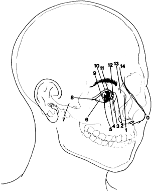
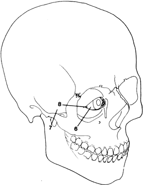
Paul Tessier, in 1976, presented an orderly classification of the clefting syndromes that has been universally accepted due to its simplification of nomenclature106,113 (Fig. 17). Tessier's system assigns each craniofacial cleft a number from 0 to 14, beginning at 0 in the midline of the lower face and progressing clockwise around the right orbit ending at 14 in the upper facial midline. The left side of the face is organized in the same manner except the designation numbers run counterclockwise. Tessier's classification scheme is purely descriptive and provides no pathogenetic or etiologic information. Nevertheless, this is the most widely accepted system because it facilitates recording and communication among observers.106,113

Fig. 17. Oblique view of skull with adnexal soft tissue demonstrating positions of facial clefts. (Fries PD, Katowitz JA: Congenital craniofacial anomalies of ophthalmic importance. Surv Ophthalmol 35:87, 1990, with permission from Elsevier Science.)

The morphogenesis of facial clefting is not well understood and is a topic of much debate. To explain the clefting phenomenon, Dursy in 1869114 and His in 1892115 proposed the classic theory of fusion. They suggested that clefts develop as a result of the failure of fusion of the normal embryonic facial processes. In contrast to the classic theory, Johnston116 and Wetson117 subsequently implicated either (1) the failure of neural crest cell migration, which prevents the mesenchyme responsible for skeletal and connective tissue formation of the face or (2) the degeneration of these cells before their migration as the source of clefts. More recently, studies by Vermiej-Keers and coworkers suggests that neural crest cell migration may not be the source of all facial clefting.118,119 the group led by Vermiej-Keers identified four primary sites in embryonic facial development that may lead to facial clefts: (1) between the lateral and medial facial processes, (2) between the lateral nasal and maxillary processes, (3) between the maxillary and mandibular processes, and (4) between the palatine processes.119 These sites are identified as “true clefts” because they are associated with normal sutures of embryologic development. True clefts tend to be well-defined and occur within the seventh week of gestation.

Within each of the facial embryonic bony plates there are multiple ossification centers, which vary in location and number, and which arise to complete bone development. Failure of these ossification centers to produce bone may cause bony defects and soft tissue abnormalities forming “pseudoclefts.” These are termed pseudoclefts because they lack association with true facial sutures. Most clefts would thus be considered pseudoclefts using this classification.

Clefting phenomena have both genetic and environmental influences. Genetics appears to play only a limited role, the major exception being mandibulofacial dysostosis (Treacher-Collins syndrome), which is transmitted as an autosomal dominant trait and is linked to chromosome 5q31.1-q33.3.120,121 Both trisomy 13 and trisomy 18 are associated with facial clefting at incidences of 40.7% and 6.9%, respectively.

Radiation exposure in Japanese survivors of the atomic bombs has been implicated in inducing clefts.122 Following the 1985 earthquake in Santiago, Chile, the rate of facial clefts locally rose from 1.6 to 2.01 per 1000 with a peak incidence 7 months after the quake.123 Infectious agents such as influenza A2 virus and Toxoplasma have been implicated as causative factors as well.124,125

Anticonvulsant drugs taken during pregnancy have been shown to increase the risk of craniofacial clefts six times over the risk of children born to mothers without seizure disorders.126 Maternal use of cocaine and benzodiazepines has also been reported to be associated with cleft lip, cleft palate, and atypical facial clefts.127 Dilantin, corticosteroids, and retinoids have also induced clefting in animal models.128 Certainly the variety of potential causal agents identified and the early period of facial embryogenesis, when the mother may be unaware of her pregnancy, creates a situation for unforeseen environmental induction unpreventable by parents.

Amniotic band syndrome, which is reviewed in depth later in this chapter, is another cause of clefting phenomenon. Amniotic bands result from ruptures in the amniotic membrane, which then form into tissue chords, which may wrap around or be swallowed by the fetus. These bands lead to facial clefts presumably by causing pressure necrosis. Amniotic bands may actually be the most frequent cause of congenital clefts.

REGIONAL CLASSIFICATION OF ORBITAL AND ADENEXAL CLEFTS

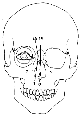
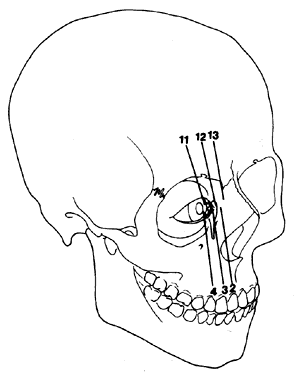
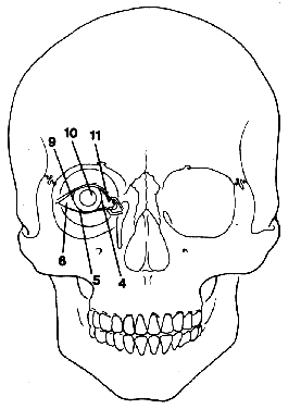
Fries and Katowitz grouped the orbital clefts into four regional categories that stress the ophthalmic features associated with the clefts4 (Figs. 18 to 21). This system was not intended to replace Tessier's classification but rather to emphasize the clinically involved ophthalmic adnexa affected by these clefts. The four categories, from medial to lateral, are (1) median facial clefts (Tessier's cleft numbers 0,1, 13, 14), which primarily present with hypertelorism; (2) medial canthal and nasolacrimal system clefts (Tessier's cleft numbers 2, 3, 4, 11, 12, and 13), which primarily present with medial canthal dystopia and nasolacrimal abnormalities; (3) central eyelid clefts (Tessier's cleft numbers 4, 5, 6, 9, 10,and 11), which primarily present with colobomatous defects; and (4) lateral canthal clefts (Tessier's cleft numbers 4, 5, 6, 9, 10, and 11), which primarily present with lateral canthal dystopias.

Fig. 18. Median facial cleft position. (Fries PD, Katowitz JA: Congenital craniofacial anomalies of ophthalmic importance. Surv Ophthalmol 35:87, 1990, with permission from Elsevier Science.)

Fig. 19. Medial canthal and nasolacrimal cleft positions. Note relation to punctum and infraorbital foramen for cleft number 4. (Fries PD, Katowitz JA: Congenital craniofacial anomalies of ophthalmic importance. Surv Ophthalmol 35:87, 1990, with permission from Elsevier Science.)

Fig. 20. Eyelid clefts positions. (Fries PD, Katowitz JA: Congenital craniofacial anomalies of ophthalmic importance. Surv Ophthalmol 35:87, 1990, with permission from Elsevier Science.)

Fig. 21. Lateral canthal and facial cleft positions. (Fries PD, Katowitz JA: Congenital craniofacial anomalies of ophthalmic importance. Surv Ophthalmol 35:87, 1990, with permission from Elsevier Science.)

The soft tissue and bony tissue relationships in the craniofacial clefts are often important. The medial clefts typically have more soft tissue involvement than bony abnormality except for cleft number 3, which may be one of the more severe facial clefts. The more lateral clefts typically display more bony deformity than overlying soft tissue deformity.

The Fries and Katowitz classification4 includes some overlap between the regional groups. For instance, cleft number 4 is included in both the medial canthal/nasolacrimal and the first groups. Its position just lateral to the punctum can lead to proximal punctal or canalicular involvement, resulting in epiphora in addition to medial lower lid clefts or colobomas. Similar “borderline” or overlapping clefts include numbers 6, 11, and 13.

Although the variety of clefting combinations is wide, syndromes exist that involve a distinct clefting pattern with consistent ophthalmic features. These syndromes are discussed later, along with some of the rarer clefts of specifically ophthalmic importance.

Regional Classification—Area I Midline Craniofacial Clefts (Tessier's Cleft Numbers 0, 1, 13, and 14)

The midline craniofacial clefts vary in severity. Mild involvement includes a minor vermilion notch of the upper lid and a bifid nose. More severe deformities include a true median cleft lip and a nasal duplication. Perhaps the most striking anomaly in this group is a frontal encephalocele resulting from a Tessier's number 14 cleft (Fig. 22).

Fig. 22. Midline facial cleft, Tessier's cleft numbers 0 and 14, demonstrating hypertelorism, frontal encephalocele, and bitid nose.

The primary manifestation of these clefts is orbital hypertelorism. Orbital hypertelorism is defined as an increased distance between the medial orbital walls and therefore this classification is based on bony anatomy. Telecanthus, conversely, is a soft tissue measurement referring to an increased distance between the medial canthi. Radiographic studies have shown that average intraorbital distance at birth is 16 mm.108,129 In adults, the average female measurement is 25 mm and, in men 28 mm. In infants and children, the upper limit of normal is less distinct but is generally 20 mm in infants and 24 mm in children.108,129 The relationship of the intercanthal distance to the interpupillary distance is a clinically helpful measurement is assessing telecanthus and hypertelorism. The intercanthal distance is normally about one half the interpupillary distance.

Two main developmental failures can cause hypertelorism in the midline craniofacial clefts. Premature fusion of the cranial base sutures slows or stunts midfacial orbital migration when the orbits are still widely separated.130 The second major cause is related to the normal development of the nasal cartilage, which promotes vertical expansion of the face, allowing the orbits to further approach midline. Failure in septal growth not only produces hypertelorism but also forces the developing forebrain anteriorally, thus producing frontal encephaloceles.131

Kawamoto132 classified the relationship of congenital hypertelorism to specific clefting patterns into five basic types. This classification begins medially and proceeds laterally. Kawamoto Group I includes isolated hypertelorism in the craniosynostosis and has previously been discussed. Midline clefts associated with hypertelorism is known as Kawamoto Group II, median craniofacial dysraphia, the median cleft face syndrome, or Tessier's 0 to 14 clefting syndrome. The clinical manifestations of the syndrome include hypertelorism, widened dorsum of the nose, bifid anterior cranium, midline facial cleft involving the nose, lip, and sometimes the palate, hypoplastic nasal tip, and a widow's peak. Kawamoto Group III is the frontal encephalocele or giant pneumatization of the frontal sinus. This cleft is synonymous with Tessier's number 14 cleft and involves the midline of the cranium. As already mentioned, with its facial counterpart, the number 0 cleft, it is termed the median craniofacial dysrhaphia. In Kawamoto Group III, the crista galli may be widened and the cleft at number 14 position allows the frontal lobes to herniate through the frontal bone in the area of the frontal sinus. The clinical features of this syndrome spare the lower face but involve the upper midface with hypertelorism and widened forehead with frontal encephalocele. The fourth group in Kawamoto's classification is due to the unilateral paramedian facial clefts typically involving cleft numbers 1 and 13. The fault in this syndrome lies medial to the junction of the nasal bone and the frontal process of the maxilla. This cleft, just lateral to the facial midline, displaces the ipsilateral orbit laterally, producing an asymmetric hypertelorism. Kawamoto Group V comprises the naso-ocular clefts, which include Tessier's cleft numbers 2, 3, 11, and 12. The more lateral the cleft is, the less hypertelorism will be produced. However, more lateral clefts produce more medial canthal and nasolacrimal abnormalities than the other hypertelorism-producing clefts. From an ophthalmic perspective, naso-ocular clefts are best grouped functionally with the medial canthal/nasolacrimal group despite overlapping characteristics with the midline craniofacial clefts.

Regional Classification—Area II: Medial Canthal/Nasolacrimal Clefts

The medial canthal/nasolacrimal clefts correspond to Tessier's cleft numbers 2, 3, and 4, as well as their superior counterparts, cleft numbers 10, 11, and 12. The lower face clefts (numbers 2, 3, and 4) are much more common than upper face clefts (numbers 10, 11, and 12), which generally occur paired with their lower face counterparts to form clefting pairs comprising 4 and 10, 3 and 11, and 2 and 12.

Cleft number 2, the most medially located in this group, is relatively rare.106,113 Nearly 50% of all cleft numbers 1 and 2 present bilaterally, whereas the more oblique clefts 3, 4, and 5 are bilaterally only 20% to 30% of the time.133,134 Its medial location causes hypertelorism but rarely involves the medial canthus and palpebral fissure. Although the upper nasolacrimal system is spared, maxillary bone involvement may cause distal lacrimal duct obstruction.

Cleft number 3 is more common and typically more severe than cleft 2. It begins inferiorly as a cleft lip and runs cephalad through the nasal alar-cheek junction and continues to the medial aspect of the lower eyelid, just medial to the inferior punctum. Thus, the lower canalicular system is severely deformed and affected patients demonstrate epiphora and recurrent infections. The nasolacrimal system in these patients is extremely difficult to reconstruct surgically. Clinically, the medial canthus is inferiorly displaced whereas the canthal tendon is hypoplastic. There also may be nasal alar colobomas, a vertically shortened alar-medial canthal distance, and occasionally microphthalmos.

If paired with cleft number 11, the medial one third of the upper lid is usually colobomatous with inferior displacement of the medial eyebrow and dysplasia at the ipsilateral forehead.

Cleft number 4 is one of the “borderline clefts” and has clinical features of both regional area 2 (the medial canthal/nasolacrimal group) and area 3 (the lid group) (Fig. 23). Cleft number 4 begins midway between the phitral column and the commissure, continues laterally past the nasal ala staying medial to the infraorbital foramen but lateral to the lower lid punctum.106,113 Severe clefting usually produces canalicular disfunction, medial canthal dystopia, and severe lower lid deformities. If the clefting is mild, medial canthal and nasolacrimal abnormalities may be absent with only a medial lower lid coloboma present.

Fig. 23. Tessier's number 4 cleft involving left maxilla, orbits and oral cavity. Patient also has Tessier's number 7 cleft on right. (Fries PD, Katowitz JA: Congenital craniofacial anomalies of ophthalmic importance. Surv Ophthalmol 35:87, 1990, with permission from Elsevier Science.)

Regional Classification—Area III: Eyelid Clefts

The group of clefts that primarily affect the eyelids include cleft numbers 4, 5, and 6 inferiorly and numbers 9, 10, and 11 superiorly (Fig. 24). Cleft number 4 was discussed previously. Cleft number 5 begins just medial to the oral commissure and runs laterally across the cheek and approaches the orbit just lateral to the infraorbital foramen to terminate within the region of the middle and lateral third portions of the lower lid. Cleft number 5 rarely occurs in isolation and is normally found paired with upper cleft number 9.135 In the number 5 cleft, there is vertical shortening of the tissue between the lateral lower lid and the lateral upper lip. This absence of tissue pulls the lower lid inferiorly while displacing the ipsilateral upper lid superiorly. Bony growth from the surrounding ossification centers accentuates the soft tissue deformity along the cleft site. This creates a deep V-shaped lower lid coloboma which points towards the oral component. The skin component is affected more than the underlying bone, however defects in the superior maxillary sinus may be large enough to permit herniation of orbital contents. There may be microphthalmia or anophthalmia. Moreover, even if the globe itself is normal there is often severe ocular exposure secondary to the absence of lower lid tissue.

Fig. 24. Tessier's number 9 cleft demonstrating superior lid, orbital rim and frontal bone abnormalities with encephalocele. (Fries PD, Katowitz JA: Congenital craniofacial anomalies of ophthalmic importance. Surv Ophthalmol 35:87, 1990, with permission from Elsevier Science.)

Unlike clefts number 4 and 5, cleft number 6 does not have an oral component. It runs from the junction of the middle and lateral third of the lower eyelid and extends inferiorly towards the angle of the mandible. The skin defect is usually mild causing a lower lid pseudocoloboma with lateral canthal dystopia. The underlying osseous defect is characterized by zygomatic hypoplasia with the cleft coursing through the zygomaticomaxillary suture. Cleft number 6 may be grouped with both the regional eyelid clefts on the lateral canthal group. In fact, several sources believe that an isolated cleft number 6 may be a part of the Treacher-Collins syndrome.139,140

Regional Classification—Area IV: Lateral Canthal Defects

The lateral canthal region involves clefts numbers 6, 7, and 8. Cleft number 6 has already been reviewed. The Tessier number 7 cleft is the most common and probably the earliest craniofacial cleft recorded, having been traced to cuneiform inscriptions left by the Chaldean civilization of Mesopotamia in 2000 BCE (Fig. 25). The number 7 cleft fault line runs from the corner of the mouth laterally across the lower cheek and crosses the zygomatic arch to terminate near the auricular tragus. The hypoplastic zygoma may cause drooping of the superolateral orbital angle resulting in lateral canthal dystopia. The mandibular coronoid process, condyle, and ramus are hypoplastic as well. Soft tissue abnormalities include macrostomia, auricular tags, and hypoplasia of the parotid gland and duct, tongue, and soft palate. Severe ear malformations include the external auditory canal and the middle ear ossicles.

Fig. 25. Lateral view of Tessier's number 7 cleft demonstrating lateral angle of mouth involvement, pretragal skin tags, and low set but otherwise normal pinna. (Fries PD, Katowitz JA: Congenital craniofacial anomalies of ophthalmic importance. Surv Ophthalmol 35:87, 1990, with permission from Elsevier Science.)

Tessier's cleft number 8 runs laterally from the lateral canthal angle posteriorly toward the frontozygomatic suture. The isolated form of cleft number 8 is rarely seen. However, when present it produces a lateral canthal coloboma with fusion of the lid defect to the underlying bulbar conjunctiva with disruption of the lateral canthal tendon. When present, the osseous component tends to involve the frontozygomatic suture and the greater wing of the sphenoid.

The relationship of the lateral canthal cleft numbers 6, 7, and 8 with other distinguishing abnormalities form the basis for several specific clinical syndromes that represent some of the most common clefting syndromes. These include Treacher-Collins syndrome, Goldenhar's syndrome, and hemifacial microsomia.

TREACHER-COLLINS SYNDROME

Treacher-Collins syndrome, otherwise known as mandibulofacial dystosis or Berry's syndrome, is transmitted in an autosomal dominant fashion with variable expression (Fig. 26). New mutations account for between 50% and 60% of cases136–138 and the syndrome has been described in several racial groups.137,138 Advanced paternal age is a risk factor for new spontaneous mutations.

Fig. 26. A. Treacher-Collins syndrome demonstrating malar hypoplasia and micrognathia. B. Lateral view of patient with Treacher-Collins syndrome showing malar hypoplasia and micrognathia. C. Waters' view of patient with Treacher-Collins syndrome demonstrating “tear-drop” orbital rim representing inferior orbital cleft. (B and C, Fries PD, Katowitz JA: Congenital craniofacial anomalies of ophthalmic importance. Surv Ophthalmol 35:87, 1990, with permission from Elsevier Science.)

The features of this condition are secondary to abnormal development of the first and second branchial arches. Clinical features may include hypoplasia of the mandible and zygoma, antimongoloid palpebral fissure obliquity, pseudocolobomas or true notching at the lateral lower lid (and sometimes the upper lid), hypoplasia of the lashes over the medial two thirds of the lower lid, external and middle ear malformations, deafness, macrosomia with malocclusion and abnormal dentition, pretragal fistula, and inferior extension of the sideburns onto the upper cheek.139 Various other skeletal and facial anomalies have been reported associated with Treacher-Collins syndrome, but they are not consistent features. These include microphthalmos, orbital hypoplasia, cataract, lacrimal duct atresia, pupillary ectopia, strabismus, distichiasis, various ocular colobomata, sinus abnormalities, nasal atresia, cleft lip and palate, enlarged sphenoid bone fissures, and skeletal and cranial synostosis.42,139–142 It is the combination of the flattened nasofrontal angle combined with the macrostomia and receding chin that produces the characteristic birdlike or fishlike facies.

The most salient ophthalmic features of this syndrome are the lid and adenexal abnormalities.143 The horizontal palpebral length is shortened whereas the lateral canthus is displaced inferomedially. The intelligence of the patient is usually normal although cases of mental retardation have been reported.144,145 Patients with this syndrome may be falsely presumed to be retarded secondary to severe hearing impairment and are often socially impaired due to their appearance.139,141

In 1949, Franceschetti and Kline extensively reviewed this syndrome and classified it into five clinical forms: (1) the complete form, encompassing nearly all of the abovementioned features; (2) the incomplete form, presenting variably with less severe and less extensive auricular, ocular, zygomatic, and mandibular abnormalities; (3) the abortive form, in which only the lower lid pseudocoloboma and zygomatic hypoplasia are present; (4) the unilateral form, with the anomalies limited to only one side of the face regardless of severity; and (5) the atypical variant, an incomplete form combined with other abnormalities not usually part of the complete form listed above.140

The most common forms are the complete and incomplete varieties whereas the typical and abortive forms are rarer.146 Many experts have disputed the actual occurrence of the unilateral form; early reports may actually represent another entity, hemifacial microsomia, which shares many features with Treacher-Collins syndrome.139,140,147

Tessier was the first to recognize the relationship between Treacher-Collins syndrome and complete or incomplete cleft numbers 6, 7, and 8.113 The incomplete form of Treacher-Collins syndrome is represented by a number 6 orbital cleft whereas the complete form involves cleft numbers 6, 7, and 8. The variable involvement of the severity and extent of each cleft contributes to myriad clinical manifestations.

Genetic studies have mapped Treacher-Collins syndrome to the long arm of chromosome 5, specifically 5q32-5q33.2.120,148–150 Despite the genetic demonstration of true nonpenetrance, radiologic studies of patients with a positive family history have demonstrated subtle degrees of zygomatic hypoplasia.151 Multiple theories of pathogenesis have been presented to explain this syndrome. These include delayed or failed ossification of the facial bones derived from the first branchial arch, defects in neural crest cell migration, and ingestion of retinoic acid (in nonhuman animal models).140,152–155

Other work in nonhuman animal models have implicated abnormalities in the stapedial arch, which supplies the first branchial arch during ebryogenesis.156 In fact, McKenzie and Craig demonstrated the absence of the incus and stapes with a coexistent stapedial artery anomaly in a child with Treacher-Collins syndrome.157 An interesting study by Sulik and coworkers has demonstrated the premature death of ectodermal placode cells with resultant mandibular, maxillary, and auricular abnormalities suggestive of Treacher-Collins syndrome.158

GOLDENHAR'S SYNDROME

Oculoauriculovertebral dysplasia or Goldenhar's syndrome was first described by Van Duise in 1882 and later reported by Goldenhar in 1952.159 Similar to Treacher-Collins syndrome, Goldenhar's involves abnormalities of the first and second branchial arches. In fact, some authors like to lump Goldenhar's, Treacher-Collins, and hemifacial microsomia together for this reason. The clinical features of Goldenhar's syndrome include corneoscleral dermoids, subconjunctival or anterior orbital dermoids (usually lipodermoids), eyelid colobomas, unilateral aplasia or hypoplasia of the mandibular ramus, small size or abnormal shape of one side of both ears, prearicular skin tags, and auricular anomalies (Fig. 27).160

Fig. 27. A. Frontal view of patient with Goldenhar's syndrome showing asymmetry of mandible. B. Lateral view of same patient with ear abnormality and micrognathia.

Most authors require the presence of periocular choristomas to establish the diagnosis of oculoauriculovertebral dysplasia. The incidence of epibulbar and orbital choristomas ranges from 32% to 92% depending on which study is referenced and how the author chose to define the inclusion criteria.161,162 Upper eyelid colobomas occur in between 12% and 20% of cases and are usually ipsilateral to the side exhibiting the choristema.161 In Mansour and associates' classic study,162 12% of patients had ptosis and 11% had nasolacrimal anomalies including nasolacrimal duct obstruction and lacrimal fistulas. Other ocular findings include horizontal palpebral fissure shortening, eyelid skin tags, and iris colobomas. Strabismus is noted in from 10% to 19% of patients and include Duane's syndrome, esotropia, exotropia, and palsy affecting cranial nerve VI.161–163 Rare occurrences include microphthalmia, anophthalmia, and cataracts.74,162,164–167

Extraocular anomalies in the syndrome are numerous and varied. Middle ear abnormalities, agenesis of the external auditory canal, and preauricular fistulae may be found. Other associated features include micrognathia, macrostomia, microcephaly, hydrocephaly, plagiocephaly, intracranial dermoids, various cranial asymmetries, and various palatal and facial clefts.159,162,165,168,169 Vertebral anomalies include cervical vertebral fusion primarily at the C2-C3 level, hemivertebrae, butterfly vertebrae, and pericervical cartilaginous tags, spina bifida, scoliosis, and hypoplastic or fused ribs.106,109,112,115,116 Cardiopulmonary anomalies include atrial septal, ventricular septal defects, pulmonary stenosis, Tetralogy of Fallot and tracheoesophageal fistula.139,160,170–173 Genitourinary problems, such as horseshoe kidney, have been reported.164 Clubfoot, forearm hypoplasia, and digital anomalies have also been documented.162,165,168,169

The etiology of Goldenhar's syndrome is unknown although an early cell-induction abnormality due to environmental, vascular, or topographic insult has been postulated.127,153 Most cases occur sporadically although both autosomal recessive and autosomal dominant inheritance patterns have been hypothesized in certain cases.74,169,174–176

HEMIFACIAL MICROSOMIA

Hemifacial microsomia is a highly variable craniofacial disorder, which, like Goldenhar's and Treacher-Collins syndromes, is related to an abnormal morphogenesis of the branchial arches (Fig. 28).155 This is one of the more common craniofacial abnormalities with an estimated frequency of 1 in 4000 live births.155 The term itself was popularized by Gorlin and Pindborg.177 Other terms include craniofacial microsomia, otomandibular dystosis, and auricular branchiogenic dysplasia. There is a male predominance178 and a positive family history may be seen in 50% of cases.179,180

Fig. 28. Clinical photo of left hemifacial microsomia showing severe mandibular hypoplasia with jaw shift, left maxillary hypoplasia with nasal shift, low set ears and lateral canthal dystopia.

The clinical spectrum of hemifacial microsomia is highly variable with only slight microtia and asymmetric mandible at the least severe end of the spectrum. Severely affected patients may present with absence of the ear, lateral orofacial clefts (Tessier's cleft 7), orbital dystopia and hypoplasia, mandibular dysplasia, and corresponding cleft tissue abnormalities. Although this condition is predominantly unilateral, 10% of cases may be asymmetrically bilateral.181 Of note is that when this syndrome is unilateral, it is more common on the right side.174,182–185 Lauritzen and associates186 proposed a classification scheme for microsomia based primarily on clinical features (Table 2).


TABLE 2. Classification of Hemifacial Microsomia


Type IComplete facial skeleton, intact intramuscular joint, asymmmetric mandible
Type IIAplasia of the condylar head, hypoplastic ascending ramus, ear deformities
Type IIIType II defects and hypoplastic zygomatic arch, rudimentary gelnoid fossa
Type IVType III defects and hypoplastic lateral and inferior orbital rims
Type VType IV defects and orbital hypoplasia and dystopia, flattened temporal fossa


In a study of 49 patients by Hertle and coworkers178 67% of patients had ocular findings. In addition, 16% had visual loss secondary to amblyopia caused by uncorrected refractive errors (27%), anisometropia (8%), strabismus (22%), and nystagmus and ptosis (12%). Limbal dermoids and orbital dermoids are frequently observed in this syndrome158 whereas microphthalmos and anopthalmos are rarely present. Varying degrees of canthal dystopia occur. The orbit is frequently decreased in size and volume. The most common adenexal abnormalities are dacryostenosis and blepharoptosis. Colobomas of the upper eyelid may be found as well as iris colobomas. Common findings include esotropia, exotropia, Duane's syndrome, and several cranial nerve palsies affecting the motility.185 Cataracts, glaucoma, and keratitis sicca also occurred frequently.

Many theories have been advanced to explain the etiology of this syndrome. Several features overlap Goldenhar's and Treacher-Collins syndromes such as upper eyelid colobomas and even more importantly, the morphogenic abnormalities of the first and second branchial arches. However, certain skeletal and suspended manifestations help to differentiate hemifacial microsomia from the other first and second branchial arches. These include cerebral, malar, mandibular, temporal, and zygomatic hypoplasia; various cerebral anomalies; a wide variety of cranial nerve abnormalities with the facial nerve most commonly involved; and rare neural and cardiac anomalies.111,186,187

AMNIOTIC BAND SYNDROME

Amniotic band syndrome, also known as constriction ring syndrome, is another cause of facial clefting. The clefts in this syndrome are variable and do not conform to any specific pattern. Incidence of this syndrome varies from 1:1,200 to 1:15,000 live births.188 Nearly all cases are sporadic, although a few instances of familial transmission have been reported.189 Distribution by gender and race is equal. Any understanding of the pathogenesis of this syndrome should begin with a review of the normal development of the amniotic sac, which contains amniotic fluid that bathes the developing embryo. The amniotic fluid provides the embryo with constant temperature in a nonrestricted environment as protection against trauma.190 As normal gestation progresses, the amnion thickens, as does the chorion or the semipermeable membrane, which lies deep to the amnion. The fetus begins swallowing amniotic fluid at approximately 3 to 6 months of embryogenesis.188

The etiology of amniotic bands is subject to wide debate. The most likely explanation is related directly to the formation of amniotic bands. Early in the middle trimester, the amnion may separate from the chorion and produce thin free-floating tissue strips. These amniotic bands may wrap around parts of the embryo in a bandlike manner, restricting growth or even causing full structural defects, such as amputation of arms or limbs. Rupture of the amnion not only produces amniotic bands but also reduces levels of amniotic fluid temporarily. As already mentioned, as gestation proceeds the amnion thickens. Thus, separation of the amnion from the chorion at later stages of gestation promotes thicker bands, which are capable of producing more deformities. These amniotic bands may also be swallowed, becoming anchored in the mouth. If this occurs, the bands may stretch across the face and cranium and can interrupt facial fusional processes or create disruption in tissues already formed creating cranial deformities of alarming magnitude (Fig. 29).191 If the band encircles the umbilical chord, it may lead to fetal hypoxia and death. Pericranial constrictions can cause anencephaly or encephaloceles. Clefting defects may be caused by pressure necrosis or interruption of normal fusion in facial processes. Any combination of clefts may arise in this syndrome. Larger bands may also cause intrauterine amputations. In some cases, the amniotic band may be found in the cleft or peripheral constriction defect thus providing some insight regarding the etiology of the deformity.

Fig. 29. Amniotic band syndrome producing craniofacial defects, including large cranial encephalocoele, nasal clefts, and medial orbital clefts with severe upper lid deformities, and globe exposure. (Fries PD, Katowitz JA: Congenital craniofacial anomalies of ophthalmic importance. Surv Ophthalmol 35:87, 1990, with permission from Elsevier Science.)

An alternative theory suggests that extrinsic band formation does not occur and that such constrictions, compressions, and clefts might result from focal fetal dysplasia.192 This theory survives because it is difficult to explain certain internal malformations. Associated with this syndrome (dysplastic kidneys, imperforate anus, and ectopic gallbladder).193

As expected, ophthalmic complications may arise from the amniotic band syndrome.194–196 The most common anomaly is colobomas of the eyelids (upper, lower, or both). Associated colobomas tend to be large and irregular and often lead to corneal exposure and other related complications. Symblepharon from the lid to the cornea can be associated with these colobomas. Paramedian clefts (Tessier's numbers 3 and 4) frequently have nasolacrimal system abnormalities, including nasolacrimal duct obstruction, punctal or canalicular agenesis, or even complete absence of the nasolacrimal system. Hypertelorism occurs in approximately 45% of these cases and is often associated with frontal encephalocele in the median or perimedian cleft (Tessier's cleft numbers 0, 1, 2, 12, 13, and 14) Canthal dystopia and ptosis may also be observed.

Extraocular muscle abnormalities may include esotropia, exotropia, oblique muscle overreaction, and paralytic strabismus. These strabismus patterns may be primary or secondary to the cranial nerve abnormalities. Less common abnormalities include iris and choroidal coloboma and entropion, ectropion, megalocornea, and varying degrees of myopia and hypertropia.

MANAGEMENT OF CRANIOFACIAL DEFORMITIES

As mentioned previously, patients with craniofacial deformities require management by a team of specialists, which will usually include consultants from genetics, plastic surgery, neurosurgery, oral surgery, orthodontics, ophthalmology, and otolaryngology, as well as psychiatry, nutrition, speech therapy, and social work. Team management begins after each member has evaluated the patient and family. Ideally, the patient is examined by each specialist on a designated day at the treatment facility. At the end of the day, the entire team meets to discuss the diagnosis as well as short- and long-term management goals. This coordinated effort is more convenient for the families and facilitates interdisciplinary communication and management.

Back to Top
BASELINE OPHTHALMIC EVALUATION
A systematic and ophthalmic evaluation should be performed as soon as possible after the diagnosis of any craniofacial abnormality is made and before any surgical intervention. Use of a preprinted examination sheet of the globe and adnexa helps ensure detection of subtle as well as major abnormalities. For example, a child with a number 3 craniofacial cleft has an obvious lower lid and medial cheek defect with tears exiting along the lower lid deformity. A second cause for the epiphora may also be present, namely, an abnormal nasolacrimal system created by this same cleft.4

VISION

Amblyopia is responsible for significant visual loss in patients with craniofacial disorders.197 In infancy, visual acuity is estimated by using preferential fixation testing (as with Teller acuity cards). At approximately 3 years of age, HOTV Allen cards and Snellen's visual acuity tests may be obtained. The many causes of amblyopia in these children include corneal opacification, ptosis, strabismus, refractive error, postoperative swelling, occlusive dressing, and tarsorrhaphy, among others. Prompt initiation of therapy for amblyopia is of utmost importance in this patient population. Thus, each initial examination should include a cycloplegic refraction with correction of the refractive error and penalization therapy of the better eye.

GLOBE

A complete ocular examination including pupillary assessment, slit-lamp examination, cycloplegic refraction, tonometry (if possible) and dilated funduscopic examination must be performed on every patient.

Pupillary evaluation helps identify any afferent pathway abnormalities that may accompany the associated optic nerve atrophy seen in many of the craniosynostoses. Slit-lamp examination helps identify any opacifications of the cornea or lens. In addition, cobalt blue illumination of florescence on the corneal surface may be performed, at the slit lamp or remote distance to evaluate possible exposure keratopathy. Dilated funduscopic examination may identify any optic nerve, retinovascular, pigmentary, or choroidal disturbances.

LIDS AND CANTHUS

The eyelids and canthal regions are frequently involved in many craniofacial syndromes and are often the sites of involvement of the clefting syndromes. Furthermore, craniofacial surgical techniques, which involve mobilization and shifting of the orbits, commonly alter the position of the lids and canthi.

The intercanthal distance (distance between the medial canthi) and the interpupillary distances are recorded. A good clinical guide to the degree of hypertelorism is the intercanthal distance to interpupillary distance ratio, which normally is approximately 50%. Amounts greater than this may indicate telecanthus with or without underlying hypertelorism. The clinician must attempt to estimate the interpupillary distance accurately when an esotropia or exotropia is present. This ratio also may be indirectly determined from photographs taken in the office when the child will not tolerate direct measurements. The normal distance of soft tissue between the medial canthi is generally less than 20 mm in infants, less than 24 mm in older children, and less than 30 mm in adults.

The lateral canthus is normally positioned 1 to 2 mm superior to the medial canthus. Any upward or downward translocation of either canthi is termed canthal dystopia and should be recorded and quantified in millimeters of vertical elevation or depression. The eye should be carefully observed for ectropion, entropion, colobomas, trichiasis, and epicanthal folds. Ptosis requires documentation of vertical fissure height, position of the lid crease and fold, margin-reflex distance, and levator muscle function. These measurements, although only gross approximations in infants, ought still be recorded in millimeters as accurately as possible. Terms such as poor, fair, good, and excellent levator functions are somewhat ambiguous and should be avoided. Horizontal fissure lengths should also be recorded.

NASOLACRIMAL SYSTEM

A careful history of tearing problems and any associated infections should be obtained. Nasolacrimal outflow abnormalities are common in the craniofacial syndromes and may be congenital or acquired from several surgical repair maneuvers. Periosteal stripping, bony cuts through the nasolacrimal area, and failure to reattach the medial canthal tendon, if disinserted, can produce anatomic or functional failure of the lacrimal pump. Fracture through the lateral nasal wall or medial maxilla can sever the nasolacrimal duct or collapse the bony nasolacrimal canal. Transnasal wiring for telecanthus may also disrupt the medial canthal tendon, lacrimal sac, and canaliculus.

It is difficult to perform nasolacrimal and tearing tests intended for adults (e.g., Jones' or Schirmer's tests) on children. The dye-disappearance test is a relatively easy way to evaluate the function of the nasolacrimal system and is well suited for infants and children. Ancillary studies such as dacryocystography and lacrimal scintigraphy may also be useful assess lacrimal outflow disruption but commonly are not required.46 Irrigation of the nasolacrimal system may be possible in older, more cooperative children.

Failure of the nasolacrimal duct to canalize is probably no more common in newborns than in the general population.46 Certain anomalies that may contribute to epiphora in patients with craniofacial disorders include lateral displacement of the puncta, absence of the puncta, elongation of the canaliculi, absence of the nasolacrimal duct or sac, and bony abnormalities of the lacrimal sac fossa or nasolacrimal duct.

EXOPHTHALMOS

Preoperative and postoperative measurements of proptosis in the craniofacial patient is important. Accurate measurement of proptosis before surgery is essential to evaluate the effect of orbital advancement surgery. Hertel's exophthalmometry gives an accurate measurement; however, due to anatomic variation of the lateral orbital rims, it may yield misleading information in craniofacial patients. Luedde's exophthalmometer also measures the distance from the lateral orbital rim to the corneal apex but may be more valuable in children because it is less threatening.

STRABISMUS AND BINOCULAR SENSORY STATUS

Extraocular motility disturbances, common in many craniofacial syndromes, need to be managed appropriately to prevent amblyopia. This may entail spectacle correction, occlusion therapy, or surgical therapy. Evaluation of strabismus and the binocular sensory status of the child with craniofacial disorders is the same as in the nonaffected child and includes the prism-cover test, Worth's four-dot test, and the Titmus stereo acuity. The aforementioned tests are valuable in addition to a careful motility examination. Abnormal or absent extraocular muscles, orbital anomalies, cranial nerve disruption, and craniofacial facial surgery may all contribute to the presence of strabismus.

Back to Top
SURGICAL MANAGEMENT

PREOPERATIVE COMPLICATIONS

In the early era of craniofacial reconstruction, surgical repair was delayed until the child was older. It was originally thought that surgery at an early age could have deleterious effects on craniofacial growth and was too risky for such young patients. Now surgery may be performed between 6 and 12 months of age thus permitting use of full-thickness cranial bone graphs. These donor sites will regenerate bone at this early age and cranial bone has a much lower resorption rate than rib grafts, although it cannot be bent or molded so easily.

Surgery is often performed in stages to take advantage of different craniofacial growth patterns. For example, a typical staging for Apert's syndrome may involve craniovault surgery in infancy, followed by midfacial advancement in childhood to increase the intracranial and intraorbital volume further and improve the nasal airway and self-image; orthognathic surgery in adolescence can improve occlusion and speech.34 Severe synostosis may require craniovault remodeling within the first few months of life, whereas less severe cases are treated between 6 to 12 months of age.198 One must also consider positive contributions to psychosocial development that early surgery may provide. Studies suggest that unattractiveness and infant facial deformities may affect the quality of parental-infant interactions as well as produce emotional stresses as the child interacts with other children outside the household.199

As previously emphasized, the ophthalmologist must manage two major areas of importance: amblyopia prevention and corneal protection. Other members of the craniofacial team may be more concerned with more urgent concerns and may inadvertently overlook these issues. Thus, the ophthalmologist must continually monitor these patients to prevent any potential loss of vision.

The ophthalmologist's expertise may also be called on for intraoperative advice. The anterior two thirds of the orbit are frequently dislocated and mobilized in craniofacial procedures and may fortunately lead to severe intraorbital complications. These surgical techniques are performed using a bicoronal approach, often combined with an anterior craniotomy, to gain access to the medial, lateral, and superior orbits. The orbital floor is approached through a transconjunctival or subciliary route. Mobilization of the orbital contents is achieved through a subperiosteal dissection and by fracturing all four orbital walls approximately 15 mm anterior to the orbital apex. This allows the orbit to be shifted anteriorally or in any needed direction. Perhaps the most devastating intraoperative complication is intraorbital hemorrhage with increasing orbital pressure, optic nerve compromise with pupillary dilation, and potential central retinal artery occlusion and subsequent visual loss. Bone grafts or sharp surgical instruments may inadvertently perforate the globe or injure the optic nerve. The ophthalmologist should be prepared to perform intraoperative pupillary examinations, ophthalmoscopy and drain or manage intraorbital hematomas. Fortunately, intraoperative complications are rare.

SURGICAL TIMING

The types of surgical procedures for the various ophthalmic problems encountered in congenital craniofacial syndromes generally follow the basic tenets that govern their use in other patients. Early surgical intervention is indicated in conditions that may lead to visual or ocular loss such as severe corneal exposure, optic atrophy, entropion, or severe ptosis. In the following discussions of these principles, little time is spent on the specific operative techniques. Instead, emphasis is directed at the general principles of surgical management.

Corneal Exposure

Corneal exposure in craniofacial patients is common and may be severe enough to compromise the integrity of the globe and the vision itself. The two most common conditions responsible for this are (1) lid colobomas that fail to cover the cornea and (2) exophthalmos severe enough to prevent closure of the eyelids. Either situation necessitates immediate medical therapy with aggressive lubrication and if necessary, occlusive devices such as plastic food wrap. Although medical therapy may be adequate, it is normally used only as a temporizing measure until definitive surgical therapy can be instituted. In addition, the clinician ought be conscious of causing amblyopia with prolonged administration of ointments and protective dressings.

Shallow orbits associated with Crouzon's and Apert's syndromes may cause severe exophthalmos. If lid closure is prevented by severe proptosis and corneal exposure threatens the integrity of the globe, orbital advancement surgery to enlarge the bony orbit may be performed within the first few weeks of life. Fortunately, the globe is usually not immediately threatened and alternative measures are sufficient. These include ocular lubrication, moisture chambers, temporary suture tarsorrhaphy, or large lateral tarsorrhaphy. When the globe is severely proptotic, upper and lower lid retractors may need to be disinserted to ensure success of the tarsorrhaphy.

In the case of lid colobomas, surgical intervention is delayed until at least 6 months of age if the integrity of the cornea is not threatened. Sliding or rotational flaps are preferable to lid-sharing techniques in the pediatric population because of the concern of deprivation amblyopia. Similarly, use of postoperative occlusion dressings should be kept to a minimum. The primary goal of initial surgery is to restore function whereas secondary procedures should address cosmetic concerns such as lid contour on ptosis.

Canthal Deformities

Canthal dystopias can usually be repaired at the time of initial craniofacial surgery. Medial canthopexy to address medial canthal dystopias and transnasal wiring to correct telecanthus are readily permitted at this time because of the excellent exposure and easy access to the nasal area. Lateral canthal dystopias may need to be repaired in stages when bony support is lacking, such as in Treacher-Collins syndrome. Several surgical techniques have been described to achieve this.137,171,200,201

Nasolacrimal Anomalies

The nasolacrimal system is frequently dysfunctional in craniofacial patients with midfacial involvement.42 In such instances, correction of epiphora should be delayed until all other bony reconstruction in the area has been completed. The necessary manipulation of bony elements and soft tissue in craniofacial surgery of the midface may disrupt a well-functioning nasolacrimal system or cause previously successful nasolacrimal surgery to fail. If craniofacial surgery can be delayed until the patients has become a teenager, conservative nasolacrimal repair such as probing and irrigation with or without Silastic tubing may be performed when indicated. Dacryocyst or amniotocele should be managed in the same manner as in noncraniofacial patients. Dacryocystorhinostomy, canaliculodacryo-cysto-rhinostomy, and conjuctivorhinostomy should be deferred until all work on osseous structures has been completed unless frequent infections force earlier intervention.

Ptosis

The timing of ptosis repair should allow a minimum number of procedures and increase the likelihood of definitive repair. Deferring ptosis repair is preferred because craniofacial surgery can worsen the ptotic lid. However, if the ptosis is so severe that there is a risk of occlusion amblyopia, then the lid should be elevated. A frontalis suspension procedure using Supramid or Silastic tubing may be used as reversible means of ptosis correction.

Amblyopia

Recognition of amblyopia and treatment should be pursued in all craniofacial patients. In a review of 322 patients with major craniofacial abnormalities presenting to the craniofacial clinic at the Children's Hospital of Philadelphia between 1979 and 1991, 20% of all eyes had visual acuity less than 20/40.202 This amblyopia was secondary to strabismus, refractive error, anisometropia, and deprivation (corneal opacities, cataracts, and ptosis). Moreover, findings included anomalies affecting primarily the extraocular muscles, especially in Apert's and Crouzon's syndromes.203 The superior rectus and superior oblique muscles may be hypoplasic or missing and preoperative computed tomography scans may be helpful in accessing these variations.

Motility

One would expect that impaired vision would be primarily due to structural problems in craniofacial deformities. In fact, our experience has demonstrated that only 10% of visual degeneration is due to actual structural causes and that 90% is due to amblyopia that resulted primarily from strabismus ad refractive errors. Occlusive amblyopia secondary to ptosis can also occur. Use of Teller's visual acuity cards is of value in assessing these types of amblyopia.

Back to Top
SUMMARY
Congenital craniofacial syndromes present uncommon and often perplexing problems. Proper evaluation and management of these patients require understanding specific syndromes and their ophthalmic features as well as providing a means to classify them into manageable and clinically useful categories. Classification of the many syndromes into the two major categories of craniosynostosis and clefting syndromes focuses attention on different mechanisms of abnormal morphogenesis. Subsequent categorization of the clefting syndromes into four anatomic/functional categories4 provides the ophthalmologist with a more comprehensible order to this confusing subject. In the repair of ocular and adenexal deformities, the concept of timing underscores the importance of the interaction of the clinicians who comprise the craniofacial team. Preservation and development of vision are the primary goals of the ophthalmologist and may require early and persistent intervention.
Back to Top
REFERENCES

1. Virchow R: Über den Crestinismus, Manenlick in franken, and über pathologische Schadelformen. Ver Phys Med (Warsburg) 2:230, 1851

2. Jane JA, Edgerto MT, Futrell JW et al: Immediate correction of sagittal synostosis. J Neurosurg 49:705, 1978

3. David JD, Poswillo D, Simpson D: The Craniosynostosis: Causes, Natural History and Management. New York: Springer, 1982

4. Fries PD, Katowitz JA: Congenital craniofacial anomalies of ophthalmic importance. Surv Ophthalmol 35:87, 1990

5. Cohen MM Jr: Perspectives on craniosynostosis. West J Med 132:507, 1980

6. Cohen MM Jr: Craniosynostosis and syndromes with craniosynostosis: Incidence, genetics, penetrance, variability and new syndrome updating. Birth Defects 15:13, 1979

7. Cohen MM Jr: An etiologic and nosologic overview of craniosynostosis syndromes. Birth Defects 11:137, 1975

8. Moss ML: The pathogenesis of premature cranial synostosis in man. Acta Anat 37:351, 1959

9. Park EA, Powers GF: Acrocephaly and caphocephaly with symmetrically distributed malformations of the extremities. Am J Dis Child 20:235, 1920

10. Moore GE: Molecular genetic approaches to the study of human craniofacial dysmorphologies. Int Rev Cytol 158: 215, 1995

11. Herman J, Opitz JM: An unusual form of acrocephalosyndactyly. Birth Defects 5:39, 1969

12. Le Merrer M, Ledinot V, Renier D et al: Genetic counseling in craniostenosis: Results of a prospective study performed with a group of studies on craniofacial malformations. J Genet Hum 36:295, 1988

13. Crouzon O: Dystostose craniofaciale herediture. Bull Mem Soc Med Hosp Paris 33:545, 1912

14. Cohen MM Jr: Frieborg S: Birth prevalence studies of Crouson syndrome: Comparison of direct and indirect methods. Clin Genet 41:12, 1992

15. Cohen MM: Syndrome with craniosynostosis. In Cohen MM (ed): Craniosynostosis: Diagnosis, Evaluation and Management, pp 413–590. New York: Raven Press, 1986

16. Kreiborg S: Crouzon syndrome: A clinical and roentgen cephalometric study. Scand J Plast Reconstr Surg (Suppl) 18:1, 1981

17. Li X, Lewanda AF, Eluma F et al: Two craniosynostotic syndrome loci, Crouzon and Jackson-Weiss, map to chromosome 10q23-q26. Genomics 22:418, 1994

18. Losken HW, Preston RA, Post JC et al: The gene for Crouzon craniofacial dysostosis maps to chromosome 10q25-q26. Proceedings of the 63rd Annual Meeting of the American Society of Ophthalmic Plastic and Reconstructive Surgery, San Diego, CA, September 24–30, 1994

19. White RA, Dowler LL, Angeloni SV et al: Assignment of FGF* to human chromosome 10q25-q26: Mutations in FGF8 may be responsible for some types of acrocephalosyndactyly linked to this.

20. Preston RA, Post JC, Keats BJ et al: A gene for Crouzon craniofacial dysostosis maps to the long arm of chromosome 10. Nat Genet 7:149, 1994

21. Reardon W, Winter RM, Rutland P et al: Mutations in the fibroblast growth factor receptor 2 gene cause Crouzon syndrome. Nat Genet 8:98, 1994

22. Jabs EW, Li X, Scott AF et al: Jackson-Weiss and Crouzon syndromes are allelic with mutations in fibroblast growth factor 2. Nat Genet 8:275, 1994

23. Bentelson TI: The premature synostosis of the cranial sutures. Arch Ophthalmol (Copenh) 5(Suppl):47, 1958

24. Waardenburg J, Franceschetti A, Klein D: Genetics and Ophthalmology, p 222. Vol 1. Springfield, IL: Charles C Thomas, 1961

25. Mermann J, Pallister PD, Opitz JM: Craniosynostosis and craniosynostosis syndromes. Rocky Mt Med J 66:45, 1969

26. Marhac D, Renier D: Craniosynostosis. World J Surg 13:358, 1989

27. Parks MM, Costenbader FO: Craniofacial dysostosis (Crouzon's disease). Am J Ophthalmol 33:77, 1950

28. Anderson FM, Geiger L: Craniosynostosis: A survey of 204 cases. J Neurosurg 22:229, 1965

29. Anderson H, Gomes SP: Craniosynostosis: Review of the literature and indications for surgery. Acta Paediatr Scand 57:47, 168

30. Atkinson FRB: Hereditary craniofacial dysostosis or Crouzon's disease. Med Press 195:118, 1937

31. Shiller JG. Craniofacial dystosis of Crouzon: A case report and pedigree with emphasis on heredity. Pediatrics 23: 107, 1959

32. Kreiborg S: Postnatal growth and development of the craniofacial complex in premature craniosynostosis. In Cohen MM (ed): Craniosynostosis: Diagnosis, Evaluation and Management, pp 157–189. New York: Raven Press, 1986

33. Kreiborg S, Pruzandky S: Craniofacial growth in patients with premature craniosynostosis. Scand J Plast Reconstr Surg 15:171, 1981

34. Posnick JC. Craniofacial dysostosis: Staging of reconstruction and management of the midfacial deformity. Neurosurg Clin North Am 2:683, 1991

35. Kolar JC, Munro IS, Farkas LG: Patterns of dysmorphology in Crouzon syndrome: An anthropometric study. Cleft Palat J 25:235, 1988

36. Bergstrom L, Weblett LM, Hemenway WG: Otologic manifestations of acrocephalosyndactyly. Arch Otolaryngol 96: 177, 1972

37. Mathur JS, Nema HV, Metra KS et al: Apert's anomaly a transition. Acta Ophthalmol 46:1041, 1968

38. Waardenburg J, Franceschetti A, Klein D: Genetics and Ophthalmology, p 123. Vol 1. Springfield, IL: Charles C Thomas, 1961

39. Perlman JM, Zaidman GW: Bilateral keratoconus in Crouzon's syndrome. Cornea 13:80, 1994

40. Miller MT: Ocular abnormalities in craniofacial malformations. Int Ophthalmol Clin 24:143, 1984

41. Hertle RW, Wuinn GE, Minguini M et al: Visual loss in patients with craniofacial synostosis. J Pediatr Ophthalmol Strabismus 28:344, 1991

42. Whitaker LA, Katowitz JA: Facial anomalies involving the nasolacrimal apparatus. In Tessier P, Callahan A, Mustarde J et al (eds): Symposium on Plastic Surgery in the Orbital Region. St Louis: CV Mosby, 1974

43. Whitaker LA, Katowitz JA: Canthal and nasolacrimal apparatus problems. In Tessier P, Callahan A (eds): Symposium on Pediatric Surgery. St Louis: CV Mosby, 1974

44. Whitaker LA, Katowitz JA, Randall P: the nasolacrimal apparatus in congenital facial anomalies. J Maxillofac Surg 2:59, 1974

45. Francois J, Bacskulin J: External congenital fistula of the lacrimal sac. Ophthalmologica 159:249, 1969

46. Whitaker LA, Katowitz JA, Jacobs WE: Ocular adenexal problems in craniofacial deformities. J Maxillofac Surg 7:55, 1979

47. Choy AE, Margolis S, Breinin GM et al: Analysis of preoperative and postoperative extraocular muscle function in surgical translocation of bony orbits: A preliminary report. In Converse JM (ed): Symposium on Diagnosis and Treatment of Craniofacial Anomalies, p 128. St Louis: CV Mosby, 1979

48. Diamond GR, Whitaker LA: Ocular motility in craniofacial reconstruction. Plast Reconstr Surg 73:31, 1984

49. Diamond GR, Katowitz JA, Whitaker LA et al: Ocular alignment after craniofacial reconstruction. Am J Ophthalmol 90:248, 1980

50. Apert E: De l'acrocephalosyndactylie. Bull Mem Soc Med Hop Paris 243:1310, 1906

52. Weaton SW: Two specimens of congenital cranial deformities in infarcts associated with fusion of fingers and toes. Trans Pathol Soc Lond 45:238, 1894

53. Blank CE: Apert's syndrome (a type of aerocephalosyndactyly): Observations on a British series of thirty-nine cases. Ann Hum Genet 24:151, 1960

54. Bull MJ, Escobar V, Bixler D et al: Phenothpe definition and recurrence risk in the acrocephalosyndactyly syndrome. Birth Defects 15:65, 1979

55. Eastman JR, Escobar V, Bixler D: Linkage analysis in dominant acrocephalosyndactyly. J Med Genet 15:292, 1978

56. Gramey D, Farriaux JP: A dominant syndrome associated with polysyndactyly, spatula thumb, facial abnormalities, and mental retardation: A particular form of Noack's acrocephalodacrylia. J Genet Hum 19:299, 1971

57. Lemaitre A, Van Reck J: Genetic aspects of craniofacial dystosis, the Crouzon disease and the Apert-Crouzon disease. Acta Stomatol Belg 73:279, 1976

58. Sanchez HM, De Negrotti TC: Variable expression in Pfeiffer syndrome. J Med Genet 18:73, 1981

59. Erickson JD, Cohen MM Jr: A study of parental age effects on the occurrence of fresh mutations for the Apert syndrome. Ann Hum Genet 38:89, 1974

60. Smith CAB: Note on the estimation of parental age effects. Ann Hum Genet 35:337, 1972

61. Wilkie AO, Starey SF, Oldridge M et al: Apert syndrome results from localized mutations of FGFR2 and is allelic with Crouzon syndrome. Nat Genet 9:165, 1995

62. Park WJ, Theda C, Maestri Ne et al: Analysis of phenotypic features and FGFR2 mutations in Apert syndrome. Am J Hum Genet 57:321, 1995

63. Diamond GR, Katowitz JA, Whitaker LA et al: Variations in extraocular muscle and structure in craniofacial dystosis. Am J Ophthalmol 90:416, 1980

64. Cuttone JM, Brazis PT, Miller MT et al: Absence of the superior rectus muscle in Apert's syndrome. J Pediatr Ophthalmol Strabismus 16:349, 1979

65. Galli Ml: Apert syndrome does not equal mental retardation. J Pediatr 89:691, 1976

66. Murcovic JA, Posnick JC, Drake JM et al: Hydrocephalus in the Apert syndrome: A retrospective review. Pediatr Neurosurg 1963:151, 1993

67. Nowtzel MJ, Marsh JL, Palkes H et al: Hydrocephalus and mental retardation in craniocynostosis. J Pediatr 107:885, 1985

68. Cohen MM, Kreigorg S: Unusual cranial aspects of Apert syndrome. J Craniofac Genet Dev Bus 14:48, 1994

69. Ferraro NF: Dental, orthodontic, and one/maxillofacial evaluation and treatment in Apert syndrome. Clin Plast Surg 18:291, 1991

70. Cohen MM Jr: Cardiovascular anomalies in Apert type acrocephalosyndactyly. Birth Defects 8:239, 1982

71. Stegger C: The acneiform eruption of Apert's syndrome is not acne vulgaris. Am J Dermatopathol 6:213, 1984

72. Marden PM, Smith DW, McDonald MJ: Congenital anomalies in newborn infant including minor variations. J Pediatr 64:358, 1964

73. McGill T: Otolaryngologic aspects of Apert syndrome. Clin Plast Surg 18:309, 1991

74. Gorlin RJ, Cohen MM, Levin LS: Syndromes of the Head and Neck. New York: Oxford University Press, 1990

75. Mah J, Kasser J, Upton J: The foot in Apert syndrome. Clin Plast Surg 18:391, 1991

76. Green SM: Pathological anatomy of the hands in Apert's syndrome. J Hand Surg 7:450, 1982

77. Fogh-Anderson P: Rare clefts of the face. Acta Univ Scand 129:275, 1965

78. Carpenter G: Two sisters showing malformations of the skull and other congenital abnormalities. Rep Soc Study Dis Child Lond 1:110, 1901

79. Temtamy SA: Carpenter's syndrome: Acrocephalopolysyndactyly. An autosomal recessive syndrome. J Pediatr 69: 111, 1964

80. Eaton AP, Sommer A, Kontres SB et al: Carpenter's syndrome: Acrocephalopolysyndactyly type II. Birth Defects 10:249, 1974

81. Der Kaloustain VM, Sinno AA, Nassar SI: Acrocephalopolysyndactyly type II (Carpenter's syndrome). Am J Dis Child 124:716, 1972

82. Robinson LK, James HE, Mubarak SJ et al. Carpenter syndrome: Natural history and clinical spectrum. Am J Med Genet 20:461, 1985

83. Frias JC, Felman AH, Rosenbloom AL et al. Normal intelligence in two children with Carpenter's syndrome. Am J Med Genet 2:191, 1978

84. Cohen DM, Green JG, Miller J et al: Acrocephalopolysyndactyly type II: Carpenter syndrome: Clinical spectrum and attempt at unification with Goodman and Summit syndromes. Am J Med Genet 28:311, 1987

85. Nissen SJ, Turner S, Ray S: An unusual presentation of Carpenter syndrome. Contemp Orthop 27:129, 1993

86. Pfeiffer RA: Dominant erbliche Akrocephalosyndaktylie. Z Kinderheilk 90: 301, 1964

87. Cohen MM: Pfeiffer syndrome update, clinical subtypes, and guidelines for differential diagnosis. Am J Med Genet 45:300, 1993

88. Vanes J, Logan F: Pfeiffer's type acrocephalosyndactyly in two families. J Med Genet 19:289, 1982

89. Schell B, Hehr A, Feldman GJ et al: Mutations in FGFR1 and FGFR2 cause familial and sporadic Pfeiffer's syndrome. Hum Med Genet 4:323, 1995

90. Navey Y, Freedman A: Pfeiffer's syndrome: Report of a family in review of the literature. J Med Genet 13:227, 1976

91. Kreiborg S, Cohen MM: A severe case of Pfeiffer syndrome associated with stub thumb on the maternal side of the family. J Craniofac Genet Dev Biol 13:73, 1993

92. Saldino RM, Seinbech HL, Epstein CJ: Familial acrocephalosyndactyly (Pfeiffer syndrome). AJR Am J Roentgenol 116:609, 1972

93. Chotzen F: Eine eigenartige familiars Entwicklungsstorung (Akrocephalosyndaktylie: Dystosis cranifacialis und Hyperteloismus). Monatsschr Kinderheilk 55:97, 1932

94. Saethre H. Ein Beitrag zum turmschadel Problem. Pathogenese, Erblichkeit und Symptomatologie. Stsch Z Nervenheilk 117:533, 1931

95. Lewanda AF, Green ED, Weissenbach J et al: Evidence that the Saethre-Chotzen syndrome locus lies between D75664 and D75507, by genetic analysis and detection of a microdeletion in a patient. Am J Hum Genet 55:1195, 1994

96. Van Merwerden L, Rose CS, Reardon W et al: Evidence for locus heterogeneity in acrocephalosyndactyly: A refined localization for the Saethre-Chotzen syndrome locus on distal chromosome up—and exclusion of Jackson-Weiss syndrome from craniosynostosis loci on up and 5q. Am J Hum Genet 54:669, 1994

97. Reid C, McMorrow LE, McDonald-McGinn DM et al: Saethre-Chotzen syndrome with familial translocation at chromosome 7, p. 22. Am J Med Genet 47:637, 1993

98. Rearden W, McManus SP, Summers D et al: Cytogenetic evidence that the Saethre-Chotzen gene maps to 7p.212. Am J Med Genet 47:633, 1993

99. Franks V et al: Saethre-Chotzen syndrome with additional abnormalities in a Mexican family. Birth Defects 13:241, 1977

100. Evans CA, Christiasen RL: Cephalic malformations in Saethre-Chotzen syndrome. Radiology 121:399, 1976

101. Pantke OA, Cohen MM Jr, Witkop CJ et al: The Saethre-Chotzen syndrome. Birth Defects 11:190, 1975

102. Kopysc Z, Stanska M, Ryzko J et al: The Saethre-Chotzen syndrome with partial bifid of the distal phalanges of the great toes. Hum Genet 56:195, 1980

103. Bianchi E, Arico M, Podesta AF et al: A family with the Saethre-Chotzen syndrome. Am J Med Genet 22:649, 1985

104. Prudzansky S, Pashayan H, Kreiborg S et al: Roentgen cephalometric studies of the premature craniofacial synostosis: Report of a family with the Saethre-Chotzen syndrome. In Bergsma (ed): Malformation Syndromes. Amsterdam: Excerpta Medica for the National Foundation—March of Dimes. Birth Defects 11:226, 1975.

105. Aase JM, Smith DW: Facial asymmetry and abnormalities of palms and ears: A dominantly inherited developmental syndrome. J Pediatr 76:928, 1970

106. Kawamotu MK: The kaleidoscopic world of rare craniofacial clefts: Order out of chaos (Tessier classification). Clin Plast Surg 3:529, 1976

107. Cohen MM: Syndromes with cleft lip and cleft palate. Cleft Palate J 15:306, 1978

108. Morin J, Hio J, Anderson J et al: A study of growth in the interorbital region. Am J Ophthalmol 152:13, 1966

109. Lund OE: Combination of ocular and cranial malformations with craniofacial dysplasia. Ophthalmologica 152: 13, 1966

110. Burian F: Median clefts of the nose. Acta Chir Plast 2: 180, 1960

111. Savenero-Rosselli G: Developmental pathology of the fact and the dysraphic syndromes: An essay of interpretation based on experimentally produced congenital defects. Plast Reconst Surg 11:36, 1953

112. Franceschetti A, Their CJ: Symptomenkomplex in Rahmen der mandibulofazialen Dysplazion. Klin Monatsbl Augenheilkd 135:391, 1959

113. Tessier P: Anatomical classification of facial, craniofacial, laterofacial clefts. J Maxillofac Surg 4: 69, 1976

114. Dursy E. Zur entwicklungs Geschichte des Kopfes des Menschen und der heheren Wirbelt geire. Tubingen: Verlag der H. Lauppschen-Buchhandlung, 1869

115. His W:. Die Entwicklung der Menschiliche und Thierischer Physiognomen. Arch Anat Entwicklungs Gesch 5:384, 1892

116. Johnston MC: The neural crest in abnormalities of the face and brain. Birth Defects 11:416, 1975

117. Wetson JA: A radioautographic analysis of the migration and localization of trunk neural crest cells in the chick. Dev Biol 6:279, 1963

118. Vermeij-Keers C, Mazzola RF, Van der Meulen JC et al: Cerebrocraniofacial and craniofacial malformations: An embryologic analysis. Cleft Palate J 20:128, 1983

119. Vermeij-Keers C, Poelmann RE: The neural crest: A study in cell degeneration and the improbability of cell migration in mouse embryos. Neth J Zool 30:74, 1990

120. Dixon MF, Read AP, Donnai D et al: The gene for Treacher-Collins syndrome maps to the long arm of chromosome 5. Am J Hum Genet 49:17, 1991

121. Dixon MJ, Dixon J, Houseal T et al: Narrowing the position of the Treasher-Collins syndrome locus to a small interval between three new microsatellite markers at 5q32-33, 1. Am J Hum Genet 52:907, 1993

122. Neel JV: A study of major congenital defects in Japanese infants. Am J Hum Genet 10:398, 1958

123. Montnenegro MA, Palomino H et al: The influence of earthquake-induced stress on human facial clefting and simulation in mice. Arch Oral Biol 40:33, 1995

124. Erdelyi R: The influence of toxoplasmosis on the incidence of congenital facial malformations: Preliminary report. Plast Reconstr Surg 20:306, 1957

125. Leck I, Hay S, Witte JI et al: Malformations recorded on birth certificates following A2 influenza epidemics. Public Health Rep 84:971, 1969

126. Erickson JD, Oakley JP: Seizure disorder in mothers of children with orofacial clefts: A case-control study. J Pediatr 84:244, 1974

127. Fries MTL, Kuller JA, Norton T et al: Facial features of infants exposed prenatally to cocaine. Teratology 48:413, 1993

128. Granstrom G, Kullaa-Mikkohen A: Experimental craniofacial malformations induced by retinoids and resembling branchial arch syndromes. Scand J Plast Reconstr Surg Hand Surg 24:3, 1990

129. Hansman C: Growth of intraorbital distance and skull thickness as observed in roentgenographic measurements. Radiology 86:87, 1966

130. Mazzola RF: Congenital malformations in the frontonasal area: Their pathogenesis and classification. Clin Plast Surg 3:573, 1976

131. David DJ, Mahatumarat C, Cooter RD: Hemifacial microsomia: A multisystem classification. Plast Reconstr Surg 80:525, 1987

132. Kawamoto HF Jr: Incidence, pathology and classification of orbital clefts and the pathology of orbital hypertelorism. In Converse JM et al (eds): Symposium or Diagnosis and Treatment of Craniofacial Anomalies, p 164. St Louis: CV Mosby, 1979.

133. Ortiz-Monasterio F, Fuente Del Campo A, Dimopulos A: Nasal Clefts. Ann Plast Surg 18:377, 1987

134. Kawamoto HF: Rare craniofacial clefts. In McCarthy J (ed): Plastic Surgery. Vol 4. Philadelphia: WB Saunders, 1990

135. Mann I: A theory of the embryology of oxycephaly. Trans Ophthalmol Soc UK 55:279, 1935

136. Fuente del Campo A, Elizendo MM, Arnaud E. Treacher-Collins syndrome (mandibulofacial dysostosis). Clin Plast Surg 21:613, 1994

137. Rogers BO: The surgical treatment of mandibulofacial dystosis (Berry syndrome, Treacher-Collins syndrome, Franceschetti-Zwahlen-Klein syndrome). Clin Plast Surg 3:653, 1976

138. Rovin S, Dachi SF, Borenstein DB et al: Mandibulofacial dysostosis, a familial study of five generations. J Pediatr 65:215, 1964

139. Rogers BO: Berry-Treacher-Collins syndrome: A review of 200 cases. Br J Plast Surg 17:109, 1964

140. Franceschetti A, Kline D: The mandibulofacial dysostosis: A new hereditary syndrome. Acta Ophthalmol 27:144, 1949

141. Hurwitz P: Mandibulofacial dysostosis. Arch Ophthalmol 57:69, 1954

142. Rogers BO: Mandibulofacial dysostosis: Treacher-Collins syndrome. In Converse JM et al (eds): Symposium on Diagnosis and Treatment of Craniofacial Anomalies, pp 423–432. St Louis: CV Mosby, 1979

143. Hertle RW, Ziylan S, Katowitz JA: Ophthalmic features and visual prognosis in the Treacher-Collins syndrome. Br J Ophthalmol 77:642, 1993

144. Harrison SH: Treacher-Collins syndrome. Br J Plast Surg 3:282, 1951

145. Stovin JJ, Lyon JA, Clemmens RL: Mandibulofacial dysostosis. Radiology 74:225, 1960

146. Berry GA: Note on a congenital defect (?coloboma) of the lower lid. R Lond Ophthalmol Hosp Rep 12:255, 1889

147. Wilson TG: A case of unilateral mandibulofacial dystosis associated with agenesis of the homolateral lung. J Laryngol Otol 72:238, 1959

148. Dixon MJ, Dixon J, Raskova D et al: Genetic and physical mapping of the Treacher-Collins syndrome locus: Refinement of the localization to chromosome 5q 32-332. Hum Molec Genet 1:249, 1992

149. Loftus SK, Edwards SJ, Scherpbier-Heddema T et al: A combined genetic and radiation hybrid map surrounding the Treacher-Collins syndrome locus on chromosome 5q. Hum Mol Genet 2:1785, 1993

150. Jabs EW, Lix F, Lovett M et al: Genetic and physical mapping of the Treacher-Collins syndrome locus with respect to loci in the chromosome 5q3 region. Genomics 18:7, 1993

151. Marres HA, Cremers CW, Dixon MJ et al: The Treacher-Collins syndrome. A clinical, radiological and genetic linkage study on two pedigrees. Arch Otolaryngol Head Neck Surg 121:509, 1995

152. Harstadius S: The Neural Crest, p 176. London: Oxford University Press, 1950

153. Morels O: Zur Pathogenese der Missbildugen des 1 Visceralbogens unter desunderer Berucksichtigung der Dysostosis mandibulofacialis. Z Kinderheilkd 73:532, 1953

154. Nucci P, Brancato R, Carone F et al: Mandibulofacial dysostosis and corneal guttata. Am J Ophthalmol 108:204, 1989

155. Poswillo D: The pathogenesis of the Treacher-Collins syndrome. Br J Oral Surg 13:1, 1975

156. Keerl G: Zurformalen und causalen Genese der Dysostosis mandibulofacialis. Ophthalmologica 143:5, 1962

157. McKenzie J, Craig J: Mandibulofacial dysostosis (Treacher-Collins syndrome). Arch Dis Child 30:391, 1955

158. Sulik KH, Johnston MC, Smiley SJ et al: Mandibulofacial dysostosis (Treacher-Collins syndrome): A new proposal for its pathogenesis. Am J Med Genet 27:359, 1987

159. Goldenhar M: Associations malformations de l'oeil et de l'oreille, en particular le syndrome dermoide epibulbaire-appendices. Auricularies-fistula congenita et ses relations avec la dysostose mandibulo-faciale. J Genet Hum 1:243, 1952

160. Pruzandky S, Miller MT: Ocular defects in craniofacial syndrome. In Goldberg MF (ed): Genetic and Metabolic Eye Disease, p 245. 2nd ed. Boston: Little, Brown, 1986

161. Baum JL, Feingold M: Ocular aspects of Goldenhar's syndrome. Am J Ophthalmol 75:250, 1973

162. Mansour AM, Wang F, Henkind P et al: Ocular findings in facioauriculovertibral sequence (Goldenhar-Gorlin syndrome). Am J Ophthalmol 100:555, 1985

163. Feingold M, Gellis SS: Ocular abnormalities associated with first and second arch syndromes. Surv Ophthalmol 14:30, 1968

164. Rollnick BR, Kaye CI, Nagatoshi K et al: Oculoauriculovertebral dysplasia and variants: Phenotypic characteristics of 294 patients. Am J Med Genet 26:361, 1987

165. Wilson GN: Cranial defects in the Goldenhar syndrome. Am J Med Genet 14:435, 1983

166. Buick V: Genetic aspects of hemifacial microsomia. Hum Genet 64:291, 1983

167. Cohen MM: Variability versus “incidental findings” in the first and second branchial arch syndrome: Unilateral variants with anophthalmia. Birth Defects 7:103, 1971

168. Feingold M, Baum J: Goldenhar's syndrome. Am J Dis Child 132:136, 1978

169. Setzer ES, Ruiz-Castaneda N, Severn C et al: Etiologic heterogeneity in the oculoauriculovertebral syndrome. J Pediatr 98:88, 1981

170. Preston KL, McDonald M, Sebris SL et al: Validation of the acuity card procedure for assessment of infants with ocular disorder. Ophthalmology 94:644, 1987

171. Raulo Y, Tessier P: Mandibulofacial dysostosis. Analysis: principles of surgery. Scand J Plast Reconstr Surg 15:251, 1981

172. Rogers BO: Embryologic development of the head and face. In Converse JM et al (eds): Symposium on Diagnosis and Treatment of Craniofacial Anomalies, p 26. St Louis: CV Mosby, 1979

173. Rogers BO: Embryologic development of the head and face. In Converse JM et al (eds): Symposium on Diagnosis and Treatment of Craniofacial Anomalies, p 22. St Louis: CV Mosby, 1979

174. Grabb WC: The first and second branchial arch syndrome. Plast Reconstr Surg 36:485, 1965

175. Krause VH: The syndrome of Goldenhar affecting two siblings. Acta Ophthalmol Copenh 48:494, 1970

176. Summitt R. Familial Goldenhar Syndrome. Birth Defects 5:106, 1969

177. Gorlin R, Pindborg JJ. Syndromes of the head and neck. New York: McGraw-Hill, 1964.

178. Hertle RW, Quinn GE, Katowitz JA: Ocular and adenexal findings in patients with facial microsomias. Ophthalmology 99:144, 1992

179. Kaye CI, Rollnick BR, Pruzansky S: Malformations of the auricle, isolated and in syndromes. IV. Cumulative pedigree data. Birth Defects 16:163, 1979

180. Smahel Z: Craniofacial changes in hemifacial microsomia. J Craniofac Genet Dev Biol 6:151, 1986

181. Converse JM, Wood-Smith D, McCarthy JG et al: Bilateral facial microsomia: diagnosis, classification and treatment. Plast Reconstr Surg 54:413, 1974

182. Bennon RD, Mulliken JB, Kaban LB et al: Microtia: A microform of hemifacial microsomia. Plast Reconstr Surg 76:859, 1985

183. Rollnick BR, Faye CI: Hemifacial microsomia and variants: Pedigree data. Am J Med Genet 15:233, 1983

184. Vento AR, LaBrie RA, Mulliken JB: The OMENS classification of hemifacial microsomia. Cleft Palate Craniofac J 28:68, 1991

185. Converse JM, McCarthy JG, Coccaro J et al: Clinical aspects of craniofacial microsomia. In Converse JM, McCarthy JG, Wood-Smith D (eds): Symposium on Diagnosis and Treatment of Craniofacial Anomalies, p 461. St Louis: CV Mosby, 1979

186. Lauritzen C, Monro IR, Ross RB: Classification and treatment of hemifacial microsomia, Scand J Plast Reconstr Surg 19:33, 1985

187. Ross RB: Lateral facial dysplasia (first and second branchial arch syndrome, hemifacial microsomia). Birth Defects 11:51, 1975

188. Seeds JW, Cefalo RC, Herbert WHP: Amniotic band syndrome. Am J Obstet Gynecol 144:243, 1982

189. Lubinsky M, Sujansky E, Sanger W et al: Familial amniotic bands. Am J Med Genet 14:81, 1983

190. Page EW, Villee CA, Villee DB: Human Reproduction: the Core Content of Obstetrics, Gynecology and Perinatal Medicine, pp 169–199, 230–368. Philadelphia: WB Saunders, 1976

191. Higginbottom MC, Jones KL, Hall BD et al: The amniotic band disruption complex: Timing of amniotic rupture and variable spectra of consequent defects. J Pediatr 95:544, 1979

192. Van der Meulen JCH: Oblique facial clefts: Pathology, etiology and reconstruction. Plast Reconstr Surg 76: 212, 1985

193. Dumaman G, Heckler FR: Constriction ring syndrome. In Bentz MJ (ed): Pediatric Plastic Surgery, pp 1009–1016. Stamford, CT: Appleton & Lange, 1998

194. Braude LS, Miller M, Cuttone J: Ocular abnormalities in the amniogenic band syndrome. Br J Ophthalmol 65:299, 1981

195. Hollsten DA, Katowitz JA: The ophthalmic manifestations and treatment of the amniotic band syndrome. Ophthalmol Plast Reconstr Surg 6:1, 1910

196. Miller MT, Deutsch TA, Cronin C et al: Amniotic bands as a cause of ocular abnormalities. Am J Ophthalmol 104:270, 1987

197. Hertle RW et al: Visual loss in patients with craniofacial synostosis. J Pediatr Ophthalmol Strabismus 28:344, 1991

198. Marchac D, Renieir D, Broumand S: Timing of treatment for craniosynostosis and faciocraniosynostosis: A 20-year experience. Br J Plast Surg 47:211, 1994

199. Barden RC, Ford ME et al: Effects of craniofacial deformity in infancy on the quality of mother-infant interactions. Child Development 60:819, 1989

200. Jackson IT: Reconstruction of the lower eyelid defect in Treacher-Collins syndrome. Plast Reconstr Surg 67:365, 1981

201. Van der Meulen JCH, Hauben DJ, Vaandrager JM et al: The use of a temporal osteoperiosteal flap for the reconstruction of malar hypoplasia in Treacher-Collins syndrome. Plast Reconstr Surg 74:687, 1984

202. Hertle RW, Quinn GE, Katowitz JA et al: Visual abnormalities and strabismus in a craniofacial clinic. Ophthalmology (Suppl) 150, 1990

203. Diamond GR, Katowitz JA, Whittaker LA et al: Variations in extraocular muscle and structure in craniofacial dysostosis. Am J Ophthalmol 90: 416, 1980

Back to Top