Chapter 23
Sclera
MAITE SAINZ DE LA MAZA and C. STEPHEN FOSTER
Main Menu   Table Of Contents

Search

DEVELOPMENT OF THE SCLERA
ANATOMY
IMMUNOHISTOCHEMISTRY
MOLECULAR STRUCTURE
REFERENCES

The sclera is a dense connective tissue that accounts for five-sixths of the outer coat of the eyeball. The sclera, remarkable for its strength and firmness (the word sclera is derived from the Greek sklera mannix, which means hard membrane), protects intraocular components from trauma, light, and mechanical displacement; withstands the considerable expansive force generated by the intraocular pressure maintaining the shape of the globe; and provides attachment sites for the extraocular muscles.

A real understanding of scleral development, anatomy, and immunohistochemistry is essential to the study of scleral functions, growth, nutrition, and diseases.

Back to Top
DEVELOPMENT OF THE SCLERA

PRENATAL DEVELOPMENT

Almost all the sclera develops from the neural crest, except a small temporal portion which develops from mesoderm.1,2 (Table 1). The neural crest, mesoectoderm, or ectomesenchyme is the cellular mass situated on either side of the invaginating neural folds. Interestingly, other connective tissues are also of neural crest–mesodermal origin including cartilages, bones, ligaments, tendons, dermis, leptomeninges, and perivascular smooth muscles; this may explain, at least in part, the frequent association of scleritis and arthritis in many systemic connective tissue diseases.3


TABLE 1. Embryology of Ocular Structures


NeuroectodermSurface ectodermNeural crestMesoderm
RetinaCorneal epitheliumChoroidStriated extraocular muscles
Fibers of optic nerveConjunctival epitheliumIrisVascular endothelia
Glia of optic nerveLensCiliary musculatureSmall portion of the sclera
Smooth muscle of irisLacrimal gland Part of vitreous
 Tarsal glandsCorneal stroma 
 Epidermis of eyelidsCorneal endothelium 
  Optic nerve meninges 
  Most of the sclera 


The developmental process of the human sclera progresses from anterior to posterior and from inside to outside.4 The human eye develops early in week 4 as an evagination from the ventral lateral aspect of the neural tube or neuroectoderm at the level of the forebrain in the diencephalon. The end of the evagination becomes slightly dilated to form the optic vesicle. At the same time a small area of surface ectoderm overlying each optic vesicle thickens, forming the lens placode, which invaginates to become the lens vesicle. At this time there are three waves of neural crest mesenchymal invasion. The first is responsible for the corneal endothelium and trabecular meshwork, the second for the corneal and scleral fibrocytes, and the third for iris and choroidal stroma. By week 5 of development each optic vesicle invaginates to form the double-layered optic cup or neuroectoderm that is surrounded by neural crest which is also of ectodermal origin. The differentiation of neural crest cells into sclera and choroid occurs by week 6 of development in humans in the region anterior to the equator (about day 43 of development),5 progresses backward to the equator by week 8, and reaches the posterior pole by week 12.1 This differentiation is induced by the retinal pigment epithelium.6–8 The sclera as well as the choroid and the retinal pigment epithelium requires the presence of the developing lens for normal growth and change in shape, structure, and function.1 By the 4th month circularly oriented scleral fibers form the scleral spur, and by the 5th month scleral fibers around the axons of the optic nerve form the lamina cribrosa.1,4 Arrest of fetal development at this stage or the failure to lay down new collagen on the inner aspect of the posterior sclera might well account for some of the staphylomatous changes found in congenital myopia and disc changes found in some patients with congenital glaucoma.

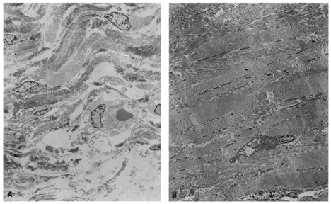
Ultrastructural studies show that developmental events of the sclera begin in the region anterior to the equator at approximately day 43.4,5 The late mesenchymal cells or very early fibroblasts of the anterior portion possess elongated nuclei and many glycogen granules and lipid vacuoles whereas those of the posterior portion possess round-to-oval nuclei and few glycogen granules and lipid vacuoles. The late mesenchymal cells or very early fibroblasts of the anterior and posterior portions contain many free ribosomes and polyribosomes, as well as immature rough-surfaced endoplasmic reticulum and Golgi complex. The early fibroblasts begin the synthesis of glycoproteins, glycosaminoglycans (especially hyaluronic acid), collagen, and elastin between day 43 and 50, thus filling the intercellular space. Developmental events directed from inside outward begin at week 7.2 with a marked increase in the inner portion in glycogen granules and lipid droplets of the cells, and in number and average diameter of collagen fibrils. Cytodevelopment of the sclera is characterized by decrease of ribosomes, polyribosomes, glycogen granules, and lipid vacuoles, and by increase of rough-surfaced endoplasmic reticulum and Golgi complex components. Development of intercellular substances is characterized by an increase in the number and average diameter of collagen fibrils (Fig. 1) and in the amount of elastic deposits with electron-translucent central cores. By week 10.9 there are no more differences between the inner and outer portions. By week 13 there are no more differences between the anterior and posterior portions. By week 24, fetal sclera has the same ultrastructural characteristics as adult sclera. Between week 6 and week 24 there is a threefold increase in thickness, possibly by progressive laying down of collagen fibrils on its inner aspect as more mature collagen fibrils are found in the outer part of the sclera and the younger smaller collagen fibrils on its inner aspect; thereafter the rate of increase diminishes rapidly.

Fig. 1. Transmission electron micrographs (x4000) of fetal sclera, week 16 of development (A), and adult sclera (B). Fetal sclera shows more fibroblasts and less packed and dense, intermingled arrangements of collagen bundless than adult sclera.

Defects in synthesis of extracellular matrix components during scleral development may account for conditions such as Marfan syndrome, osteogenesis imperfecta, pseudoxanthoma elasticum, Ehlers-Danlos syndrome, congenital myopia, and nanophthalmos.

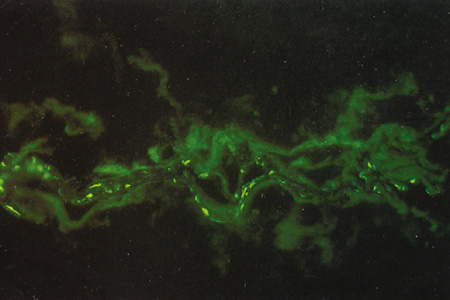
Immunohistochemical studies show that the extracellular matrix of fetal sclera has the collagens I, III, IV, V, and VI, the glycosaminoglycans dermatan sulfate, heparan sulfate, hyaluronic acid, and chondroitin sulfate, and the glycoproteins fibronectin, vitronectin, and laminin.9 Collagens II and VII are not present. Collagens play a major role in strength (collagen I), resiliency (collagen III) and structural integrity (collagens IV, V, VI); glycosaminoglycans in distensibility; and glycoproteins in cell adhesion, growth, differentiation and migration, as well as in other extracellular matrix component organization. Collagen VIII may also be present in human fetal sclera.10 Development of scleral extracellular matrix from fetus into the adult includes increase of collagens I, III, and VI, and decrease of collagen IV and VIII, hyaluronic acid (Fig. 2), fibronectin, vitronectin, and laminin.9,10 By the time the tissue is mature, collagen IV, although abundant in fetal tissue, has almost completely disappeared, except in blood vessels. Heparan sulfate is identified in fetal and adult sclera in small amounts. Increase of collagens I, III, and VI may account for the increase in strength, resiliency, and structural integrity of adult sclera. Decrease of hyaluronic acid may explain the decrease of distensibility of adult sclera. Decrease of fibronectin, vitronectin, and laminin may suggest they play a major role in directing developmental events during the younger gestational periods, including cell and extracellular matrix organization.

Fig. 2. Immunofluorescence microscopy (100×) using antihyaluronic acid antibodies. Fetal sclera, week 19 of development (A) and adult sclera (B). The moderate amount of hyaluronic acid seen in the 19-week fetal specimen steadily decreases through posterior gestational periods and is nearly absent in adult sclera.

POSTNATAL DEVELOPMENT AND AGE-RELATED CHARACTERISTICS

Scleral development is determined by the genetic signaling to the fibrocytes of the sclera, and by the concomitant development of the adjacent structures such as lens, retina, choroid, and the production of aqueous by the ciliary body. During this period of growth there is an increase in axial length in order to acquire a state of emmetropia. This increase in axial length occurs in two stages, an infantile phase which is up to 3 years and then a slower juvenile phase which is up to 13 years; after this age the eye is fully developed.

The postnatal sclera is relatively thin, allowing the pigment cells of the choroid to show through, giving a bluish color. It is also somewhat distensible, allowing the sclera to stretch as a result of increased intraocular pressure in infantile glaucoma (buphthalmic globe). The relatively thin, bluish, distensible, small, and translucent postnatal sclera gradually becomes thicker, whiter, less distensible, larger, and more opaque as the eye goes through childhood and puberty.

Although the adult sclera is poorly distensible, ectasias (localized protrusions of thin sclera) or staphylomas (localized protrusions of thin sclera lined by uveal tissue) can appear at any age after damage (trauma or inflammation). The water content of the adult sclera ranges from 65% to 75%.11 The sclera appears opaque if the water content is between 40% and 80% but becomes translucent if it falls below 40% or rises above 80%.12 This is especially evident in surgical procedures such as strabismus surgery or retinal detachment in which conjunctiva and extraocular muscles are temporally removed from the underlying sclera. The exposed sclera becomes dry and therefore appears translucent unless it is continuously moistened. Similar changes occur after removal of perilimbal conjunctiva in surgical procedures such as excision of pterygium or other limbal lesions. The exposed sclera, adjacent to small elevations of conjunctiva, becomes dry because of the interference in the lubricating effect of the tear film. The dry spots, called dellen, dissappear after rehydration of the area with artificial tears or eye patching.

The increase in transparency after inflammation is the result of rearrangement of the fibrils of the sclera and physiochemical changes; it is only rarely caused by true thinning of the sclera.

In the elderly, the sclera is even less distensible, has decreased water content, and contains fewer glycosaminoglycans.13 This is the result of a progressive cross-linking of the lysine residues of collagen and a decrease in the size of the interfibrillar spaces, possibly as the result of changes within the proteoglycans. Other age-related changes are the subconjunctival deposition of lipids such as cholesterol esters, free fatty acids, triglycerides, and sphingomyelin, which give the sclera a yellowish color.14,15 Calcium phosphate may also be deposited in small rectangular or ellipsoid areas with a vertical axis longer than the horizontal one (approximately 6 mm high and 1 mm wide) just anterior to the insertions of medial or lateral rectus muscles. These slate-gray areas are called senile scleral plaques and usually occur in individuals over 70 years of age.16,17 The cause is uncertain but some etiologic possibilities taken either individually or in combination include ischemia secondary to atherosclerosis of the anterior ciliary arteries,18 dehydration,19 constant stress by the rectus muscles,20 and actinic damage from solar irradiation.17 The collagen fibrils themselves become thicker and less uniform, especially in the region of the muscle insertions. Here the sclera becomes progressively thinned, increasing the color contrasts between one part of the sclera and the next.
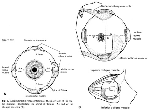
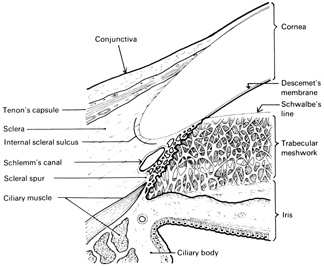
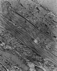
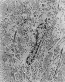
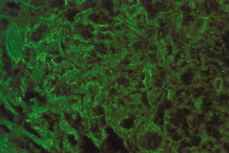
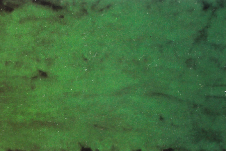
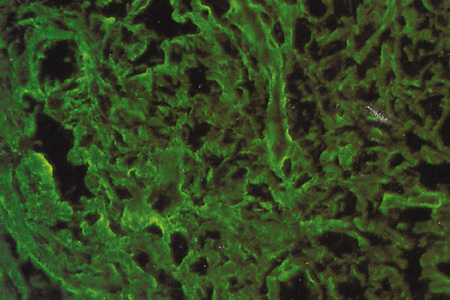
Back to Top
ANATOMY

GROSS AND MICROSCOPIC ANATOMY

The surface of the eyeball is formed by two spherical connective tissue segments of different sizes, one posterior and the other anterior.21 The posterior opaque one is the sclera, accounting for five-sixths of the eyeball with a radius of curvature of 12 mm. The anterior transparent one is the cornea, accounting for one-sixth of the eyeball, with a radius of curvature of 8 mm. Scleral thickness varies: the thickest part is near the optic nerve (1 mm); it decreases gradually at the equator (0.4 to 0.5 mm), reaching a minimum immediately behind the rectus muscle tendinous insertions (0.3 mm), and increases at the rectus muscles tendinous insertions (0.6 mm) and in the area adjacent to the limbus (0.8 mm). The collagenous fibrils of the rectus muscle tendons interweave with the collagenous fibrils of the sclera making the fusion inseparable. The traumatic scleral rupture usually occurs immediately behind the insertion of the recti, at the equator or in an area parallel to the limbus opposite from the site of the impact.13,22 It is important to remember how thin the sclera is behind the insertions of the recti when tendons must be sutured to the sclera in tendon recessions or advancements as part of strabismus surgery.

The rectus muscle insertions are progressively more posterior and follow a spiral pattern described by Tillaux and hence called the spiral of Tillaux (Fig. 3). The medial rectus inserts 5.5 mm posterior to the limbus; the inferior rectus, 6.5 mm; the lateral rectus, 6.9 mm; and the superior rectus, 7.7 mm. The insertions of the superior oblique and inferior oblique muscles are posterior to the equator. The long tendon for the superior oblique muscle inserts superiorly and slightly laterally, inferior to the superior rectus muscle; the line of insertion is convex posteriorly and laterally. The inferior oblique muscle inserts posterolaterally. Because this muscle has no tendon, the muscular fibers attach directly; the line of insertion is convex superiorly and laterally. The most posterior point lies 5 mm temporal to the optic nerve, external to the macula.

Fig. 3. Diagrammatic representation of the insertions of the rectus muscles, illustrating the spiral of Tillaux (A) and of the oblique muscles (B).

Tenon's capsule, the fascial sheath of the eyeball, supports the eyeball within the orbit separating it from the orbital fat and permits the eyeball movement produced by the extraocular muscles.21 Tenon's capsule is a membrane that extends anteriorly from the limbus backwards to ensheath the extraocular rectus muscles. Posteriorly, it envelopes the globe and fuses with the optic nerve dural sheath and with the sclera around the exit of the optic nerve. Close to the optic nerve, Tenon's capsule is penetrated by the long and short ciliary nerves and vessels, and by the vortex veins. It is closely connected to the underlying outer portion of the sclera or episclera by delicate lamellae, particularly at the limbus and at the muscle tendon insertions anteriorly and at the optic nerve dural sheath posteriorly. Tenon's capsule is thin at the limbus but becomes thicker about 3 mm backward until over the muscle tendon insertions. After being penetrated by the tendons of the six extraocular muscles, Tenon's capsule reflects on each tendon, forming a tubular sleeve. The tubular sleeves for the medial and lateral recti attach to the lacrimal and zygomatic bones, respectively; because they limit the action of these muscles on the eyeball they are called the medial and lateral check ligaments. Behind the muscle insertions Tenon's capsule becomes thinner except in the inferior part of the eyeball from one check ligament to the other where it thickens to form the suspensory ligament of Lockwood. Tenon's capsule is very thin posteriorly, especially at the optic nerve where it fuses with the dural sheath and with the sclera.

Scleral Foramina

Anatomically the sclera forms an incomplete sphere interrupted by two foramina, one anterior for the cornea and the other posterior for the optic nerve.

ANTERIOR SCLERAL FORAMEN.

The anterior scleral foramen is an anatomic concept of the sclera without the cornea. It is funnel shaped with an elliptical appearance externally (horizontal diameter of 11.6 mm and vertical diameter of 10.6 mm) and a circular appearance internally (diameter of 11.6 mm). The sclera merges with the cornea at the anterior scleral foramen forming the corneoscleral junction or limbus, an area measuring approximately 1.5 to 2 mm wide that has a convex corneal side and a concave scleral side; the scleral side is formed by the external scleral sulcus in its outer surface, and by the internal scleral sulcus in its inner surface (Fig. 4). The external layers of the internal scleral sulcus merge with the stroma of the cornea. The internal layers of the internal scleral sulcus contain the trabecular meshwork and Schlemm's canal anteriorly, and the scleral spur posteriorly; the trabecular meshwork merges with Descemet's membrane. Because the scleral spur attaches to the meridional ciliary muscle, tension on the scleral spur by the muscle opens the trabecular meshwork.23

Fig. 4. Diagrammatic representation of a longitudinal section through the region of the corneoscleral junction showing the peripheral cornea, the sclera, the conjunctiva, and Tenon's capsule, as well as the canal of Schlemm, the trabecular meshwork, and the iris.

POSTERIOR SCLERAL FORAMEN.

The sclera allows the exit of the optic nerve through the posterior scleral foramen. The posterior scleral foramen also is funnel shaped but in the sense opposite to the anterior scleral foramen: the external diameter is of 3 to 3.5 mm and the internal diameter is of 1.5 to 2 mm. The site of this perforation is located 3 mm medial to the midline and 1 mm below the horizontal meridian. In this region the outer two thirds of the sclera continue backward along the nerve to blend with the dural and arachnoid sheaths of the optic nerve. The inner third of the sclera provides small perforations covered by glial tissue that allow the passage of the axons of the optic nerve. This sievelike area, which is slightly concave and faces inward, is known as the lamina cribrosa.24 After piercing the lamina cribrosa, the axons of the optic nerve become myelinated. One of the small perforations is larger than the rest and permits the passage of the central retinal artery and vein. Because the lamina cribrosa is a relatively weak area, it tends to bulge outward with an increase of intraocular pressure, forming a cupped disc. Here also, since the axons of the optic nerve and the vessels are enclosed within tiny perforations covered by glial tissue, inflammatory swelling easily leads to axonal and vessel strangulation.

Layers of the Sclera

The sclera may be divided into three layers: the episclera, the scleral stroma, and the lamina fusca.

THE EPISCLERA.

The episclera is the superficial aspect of the sclera that is continuous superficially with Tenon's capsule and merges with the underlying scleral stroma. Unlike Tenon's capsule, it has the bundless of collagen circumferentially arranged; and unlike the scleral stroma, it has a rich blood supply anteriorly. The loosely and circumferentially arranged bundles of collagen intermingle with fibroblasts, melanocytes, elastic fibers, proteoglycans, and glycoproteins. The rich blood supply anteriorly is derived from the anterior ciliary arteries and has tight attachments to the subconjunctival tissue, the rectus muscle insertions, and Tenon's capsule. The scanty blood supply equatorially and posteriorly is derived from the posterior ciliary arteries and has loose attachments to the subconjunctival tissue and Tenon's capsule. The episclera is thickest anterior to the rectus muscle insertions and becomes progressively thinner toward the back of the eye.

THE SCLERAL STROMA.

The scleral stroma also consists of bundles of collagen intermingled with fibroblasts, melanocytes, elastic fibers, proteoglycans, and glycoproteins. However, the bundles of collagen are thicker, the fibroblasts are thinner, and the proteoglycans and glycoproteins are scantier than those in the episcleral collagen bundles. Unlike corneal collagen bundles, the thickness of the fibrils in scleral stroma varies in each bundle. The bundles of collagen interlace greatly with each other, giving to the sclera strength and resiliency. In the superficial layers of the scleral stroma the anterior circular arrangement and the posterior meridional arrangement are interrupted by whorls and loops, especially around the rectus and the oblique muscles. This arrangement resists the stresses imposed by the pull of the extraocular muscles while at the same time accommodating changes in the intraocular pressure. In the deep layers of the scleral stroma, where the influence of the stresses imposed by the extraocular muscles is absent, the bundles of collagen run meridionally from the corneoscleral junction to the optic nerve. A combination of variability in collagen fibril diameter, interlacing in bundles of collagen, and relative deficiency in water-binding substances accounts for the scleral dull white color.

THE LAMINA FUSCA.

The lamina fusca (the word fusca is derived from the Latin fuscus, which means dark) is the innermost portion of the sclera and receives its name from the large number of melanocytes that have migrated from the underlying choroid, giving to this portion a brown color. Because the lamina fusca has grooves for the passage of ciliary vessels and nerves (emissary canals), melanocytes may pass through, producing dark spots on the episclera. The spots are most commonly seen 3 to 4 mm from the limbus in the superior episclera. An exaggerated number of melanocytes may pass through the emissary canals and invade scleral stroma and episclera. This invasion may be either diffuse in individuals of races whose skin is darkly pigmented or patchy in cases of congenital melanosis oculi and oculodermal melanocytosis (nevus de Ota). Neoplasms, such as malignant melanomas of the uvea, also may emerge through the emissary canals. The lamina fusca is attached to the choroid by fine collagen fibrils, which are easily separated in choroidal detachments or effusions. The bundles of collagen in the lamina fusca become much thinner and are characterized by a great increase of elastic fibers. The ciliary muscle is very strongly attached to the deep surface of the sclera.

Blood Supply and Emissary Canals

The sclera has a low metabolic requirement because of the slow turnover of the collagen of which it is composed.

The episcleral blood supply is derived mainly from the anterior ciliary arteries anterior to the insertions of the rectus muscles and from the long and short posterior ciliary arteries posterior to these insertions. Except for the perforating vessels, scleral stroma is a relatively avascular structure that is supplied by the episcleral and, to a lesser degree, choroidal vascular networks.

The arteries, veins, and nerves traverse the sclera through emissary canals in varying degrees of obliquity.25 These canals are more commonly superiorly and inferiorly than nasally and temporally, and are separated from the sclera by a thin layer of loose connective tissue.

Anteriorly, the muscular arteries arising from the ophthalmic artery run forward as the anterior ciliary arteries. Approximately seven anterior ciliary arteries pass through the sclera just in front of the insertions of the rectus muscles in a slightly oblique direction from posterior to anterior (two anterior ciliary arteries to each rectus muscle, except the lateral rectus muscle, which has only one). The seven anterior ciliary arteries, after interconnecting through their lateral branches 1 to 5 mm behind the limbus, form a complete anastomotic ring within the episclera (i.e., the anterior episcleral arterial circle), which feeds the limbal, anterior conjunctival, and anterior episcleral tissues (Fig. 5). The anterior episcleral arterial circle broadly resolves into limbal arcades, an anterior conjunctival plexus, a superficial episcleral plexus, and a deep episcleral plexus (Fig. 6). Limbal arcades and anterior conjunctival plexus usually share their origins and form the most superficial layer of vessels. The superficial episcleral plexus lies within the parietal layer of the episclera and anastomoses at the limbus with the conjunctival plexus and with the deep episcleral plexus. The deep episcleral plexus lies within the visceral layer of the episclera and anastomoses with branches of the same plexus. In addition, extensions of the remaining anterior ciliary arterial branches (10 to 20 per eye) perforate the limbal sclera through emissary canals and meet the long posterior ciliary arteries in the ciliary muscle to form the major arterial circle of the iris. Anterior segment video angiography has shown the flow in these emissary canals to be from inside ouwards.26 Combination of low dose fluorescein and indocyanine green in anterior segment angiography may be helpful in detecting vascular changes that occur in scleral diseases.27 Although the anterior ciliary artery accompanying the lateral rectus muscle takes part of the anterior episcleral arterial circle, it directly gives only one or two and often no scleral perforating anterior ciliary arteries; the temporal scleral perforating arteries often arise from the superior and inferior anterior ciliary arteries. The anterior episcleral arterial circle and the major arterial circle of the iris communicate by scleral perforating anterior ciliary arterial branches which do not form a capillary bed in the sclera but rather provide nutrients to the uveal tract (Figs. 7 and 8).

Fig. 5. Scanning electron micrograph (20×) of a primate ocular vascular cast showing the interconnection of the anterior ciliary arteries (ACA) by means of their lateral branches to form the anterior episcleral circle (EC). (Courtesy of Dr. EM Van Buskirk.)

Fig. 6. Scanning electron micrograph (24×) of a human vascular cast of the anterior plexuses of the eye. The anterior ciliary arteries (ACA) form the anterior episcleral arterial circle which gives rise to limbal arcades, anterior conjunctival plexus, superficial episcleral plexus (SE) and deep episcleral plexus (DE). Note the interconnections between superficial episcleral plexus and deep episcleral plexus (arrows). (Courtesy of Dr. AW Fryczkowski.)

Fig. 7. Scanning electron micrograph (50×) of a primate vascular ocular vascular cast showing a scleral perforating anterior ciliary arterial branch connecting the anterior episcleral arterial circle and the major arterial circle of the iris. (Courtesy of Dr. EM Van Buskirk.)

Fig. 8. Diagrammatic representation of a transverse section of the globe showing the relationships between the anterior ciliary, short posterior ciliary, and long posterior ciliary arteries, and the anterior ciliary, short posterior ciliary, and vortex veins. Note the connection between the anterior episcleral arterial circle and the major arterial circle of the iris by scleral perforating anterior ciliary arterial branches.

The two long posterior ciliary arteries (medial and lateral), which also arise from the ophthalmic artery, enter the sclera 3.6 mm nasal to the optic nerve and 3.9 mm temporal to the optic nerve (Figs. 9 and 10). The arteries together with the nerves traverse the sclera through emissary canals in an oblique manner, from posterior to anterior, and enter the suprachoroidal space at the equator. They run forward to give arterial supply to the ciliary body and the iris. In addition, they meet the anterior ciliary arteries to form the major arterial circle of the iris. The major arterial circle of the iris is located in the stroma of the ciliary body and gives arterial supply to the iris. Surgery on the vertical, but not the horizontal rectus muscles may give rise to ischemic defects in iris.28–30 This finding seems to indicate that the anterior ciliary arteries contribute to the iris supply and that this contribution is critical in sectors of the globe that receive inadequate long posterior ciliary artery perfusion (vertical meridian). The superior and inferior anterior uvea are, therefore, at greater risk of ischemia after superior and inferior anterior ciliary occlusion after ligation of the respective muscles. The greater prevalence of emissary canals containing perforating anterior ciliary arteries in the vertical meridia may compensate for this deficit.25

Fig. 9. Diagrammatic representation of the vascular supply to the globe showing the relationships between the internal carotid, ophthalmic central retinal, long and short posterior ciliary, supraorbital, lacrimal, and anterior ciliary arteries.

Fig. 10. Diagrammatic representation of the sites of perforation of the sclera by the long and short posterior ciliary arteries, the short posterior ciliary veins, and the inferior and superior vortex veins.

The short posterior ciliary arteries arise from the ophthalmic artery as it crosses the optic nerve (see Fig. 9). After dividing into 10 to 20 branches, they perforate the sclera around the entrance of the optic nerve and supply the choroid as far as the equator of the eye. The vessels pass directly to the choroid without forming a capillary bed in the sclera. Some of the branches in the sclera run toward the equator and anastomose with the branches of the long posterior ciliary arteries to supply the posterior episclera. However, this posterior episcleral plexus is so thin that it gives a poor supply to the underlying sclera in the equator. Most of the nutrient requirements of the sclera in this area are supplied by the choroidal circulation. Some of the branches of the short posterior ciliary arteries around the optic disc form the incomplete vascular circle of Zinn-Haller near the inner scleral rim. The emissary canals for the short ciliary arteries and veins may be perpendicular, oblique, or spiral.

The limbal venous circle collects blood from the anterior conjunctival veins and limbal arcades, and drains into radial episcleral collecting veins. The episcleral collecting veins also receive blood from anterior episcleral veins and perforating scleral veins. Perforating scleral veins, which may also receive aqueous from Schlemm's canal, penetrate the sclera through different emissary canals than do the arteries. These canals, over the ciliary body, often also carry the ciliary nerves. As the episcleral collecting veins run across the sclera backward, they form the anterior ciliary veins which leave the anterior surface of the globe over the rectus muscles.

Posterior to the equator, the sclera is perforated obliquely by the emissary canals for the vortex veins (Fig. 11). Each eye usually contains from four to seven veins. Vortex veins drain the venous system of the choroid, ciliary body, and iris. One or more veins are in each quadrant. The superior vortex veins exit 8 mm posterior to the equator, close to the most posterior edge of the insertion of the superior oblique muscle. The inferior vortex veins exit 6 mm posterior to the equator (see Fig. 10).

Fig. 11. Diagrammatic representation of the relationships between the lacrimal, vortex, short posterior ciliary, supraorbital, superior and inferior ophthalmic, and central retinal veins.

Nerve Supply

The nerve supply of the sclera is surprisingly rich for a structure whose main function is protective. All the ciliary nerves are mixed nerves carrying motor, sensory, and autonomic nerve fibrils to and from cornea, sclera, ciliary body, iris, and choroid. The posterior ciliary nerves perforate the sclera around the optic nerve. The many short posterior ciliary nerves supply the posterior region of the sclera while the two long posterior ciliary nerves supply the anterior portion. The branches of the long posterior ciliary nerves end in the cornea, episclera, trabecular meshwork, and canal of Schlemm. As a result of this profuse sensory innervation, inflammation of the sclera may cause severe pain due in part for direct stimulation of the nerve endings by the inflammatory process and in part for indirect stimulation of the nerve endings by stretching secondary to edema. Since the extraocular muscles have their insertions in the sclera, the pain may increase with ocular movement.

A branch of the long posterior ciliary nerve (intrascleral nerve loops of Axenfeld) enters the ciliary body and then passes outwards through the whole thickness of the sclera and back into the ciliary body through the same canal. These nerve loops, found in 12% of eyes as a normal anatomic variation, form a clinically visible nodular elevation less than 1 mm of diameter, 4 to 7 mm posterior to the limbus, often between the rectus muscle insertions superiorly or inferiorly. They are usually associated with blood vessels and with melanocytes. Because the nerve loops may produce a brown spot on the sclera, they must be included in the differential diagnosis of primary or metastatic malignant melanoma.31 Their function is unknown and, even if they are slightly painful, they should not be removed.

In the episclera anterior to the vascular circle there are numerous free nerve endings staining for substance P and calcitonin gene-related peptide that are adjacent to the vessels and aqueous veins. These nerve endings may have a role in the regulation of the blood supply of the anterior segment and in the rate of aqueous outflow.32,33

Lymphatic Drainage

While lymphatic channels are present in conjunctiva, they are absent in episclera and sclera. Lymph from the superficial episcleral tissue drains into the subconjunctival space and then to the parotid node nasally and to the submandibular nodes temporally. Lymph from the scleral and deep episcleral tissues drain into the deep cervical nodes through the orbit and the jugular lymph trunks.

ULTRAMICROSCOPIC ANATOMY

Sclera

Ultramicroscopically, the sclera is composed of a matrix consisting of collagen, proteoglycans, glycoproteins, and elastin (Fig. 12). These are produced and degraded by scleral fibroblasts.

Fig. 12. Transmission electron micrograph (11,000×) of adult human sclera. Note the bundles of collagen, both longitudinal and transverse, with variable thickness of the fibrils in each bundle, and a spindle-shaped fibroblast along the bundles of collagen.

Collagen bundles consist of long branch fibrils with a macroperiodicity of about 64 nm (range, 35 to 75 nm) and a microperiodicity of 11 nm. Unlike the uniform corneal collagen, the collagen fibrils in sclera vary in diameter ranging from 28 to 300 nm, and their arrangement in the individual bundles is more random than that of the cornea.34 Collagen bundles in sclera vary in diameter ranging from 0.5 to 6.0 μm and form complex and irregular branching patterns, curving around the muscular insertions and the optic nerve. Collagen bundles in the outer region are thinner (0.5 to 2 μm) than those in the inner region; they usually run in a lamellar fashion whereas those in the inner region are interwoven randomly forming irregular and intermingled arrangements. These arrangements may account for the rigidity and flexibility against changes in intraocular pressure and for the opacity of the sclera. The turnover rate of scleral collagen is unknown but the healing of the wounds in scleral injuries is a slow process that takes months or years.

Many ultrastructural studies of scleral collagen fibrils have been carried out using transmission electron microscopy (TEM) and scanning electron microscopy (SEM), and more recently by atomic force microscopy (AFM), generally showing good agreement on collagen fibril dimensions. Different periodicities of scleral fibril of 67 nm have been identified by AFM and attributed to different inclination angle (5 degrees) of microfibrillar components.35,36

The fibrils at the emissary canals run parallel to the direction of the canal. Few of these fibrils attach to the wall of the vessel or nerve in the canal. A small amount of fine elastic fibers (10 to 12 nm in diameter) lie parallel to the collagen fibrils.37

The interdigitations are especially dense and the sclera thickest around the posterior pole, presumably to hold that area rigid; they thin out again near the optic disc but some fibrils pass across the disc at the lamina cribrosa.

Flat stellate or spindle-shaped cells, the fibroblasts, are few in number along the bundles of collagen (Fig. 13). The long axis of the cell and the large elliptical nucleus is parallel to the surface. The nucleus has one or two nucleoli and the chromatin is sparse. Quiescent fibroblasts contain a relative scanty cytoplasm with long mitochondria, a small Golgi complex, a few cisternal profiles of granular endoplasmic reticulum and occasional small fat droplets; however, in growing or repairing processes, the Golgi complex and the granular endoplasmic reticulum become prominent. The fibroblast elaborates the precursors of the amorphous ground substance components as well as the fibrillar proteins such as collagen and elastin. After secretion, the fibrils lie on the cell surface while full maturation occurs.

Fig. 13. Transmission electron micrograph (11,000×) of adult human sclera. Note the bundles of collagen, both longitudinal and transverse, interlacing greatly with each other, giving to the sclera the strength and resiliency. Fibroblasts are few in number along the bundless of collagen.

The amorphous ground substance composed of proteoglycans and glycoproteins fills intercellular and interfibrillar spaces. Proteoglycans, demonstrated by cuprolinic blue stain, are fine filaments of approximately 54 nm in length and 5 nm in diameter that are localized around, along, and radiating from the collagen fibrils.38

Vessels

Unlike elastic arteries (large-sized vessels), muscular arteries (medium-sized vessels), and arterioles (small-sized vessels), episcleral and scleral vessels appear to be capillaries and postcapillary venules which do not possess tunica media, which is a layer consisting chiefly of smooth muscle cells. Episcleral and perforating scleral vessels appear to possess a simple wall composed of continuous endothelial cells attached to an underlying basement membrane secreted by them, and a discontinuous layer of pericytes without smooth muscle cells (Fig. 14).9

Fig. 14. Transmission electron micrograph (7,500×) of an adult human episcleral vessel. Note the endothelial cells, the basement membrane, and the pericytes.

The irregularly shaped endothelial cells have a cytoplasm with mitochondria, rough-surfaced endoplasmic reticulum, smooth-surfaced endoplasmic reticulum, Golgi apparatus, and pinocytotic vesicles. Endothelial cells interconnect through thin areas of more or less tortuous interendothelial clefts composed of adjacent cell membranes. The basement membrane, which is almost parallel to the outer contour of the endothelial cells, consists of one to several dense layers with a less dense zone filling the space between them. Pericytes are attached to the basement membrane, often with one or more dense zones between them and the endothelial cell; their cytoplasm contains endoplasmic reticulum, mitochondria, Golgi apparatus, pinocytotic vesicles, and fine filaments. No smooth muscle cells are found. Bundles of collagen fibrils adjacent to the vessel walls merge into the surrounding connective tissue.

Episcleral and conjunctival vessels are permeable to tracers such as fluorescein-labeled dextrans of different molecular weight or horseradish peroxidase injected into the blood stream or into the anterior chamber39,40; tracers escape from the vessel lumen by crossing the thin interendothelial clefts. It can equally be expected that the aqueous humor that reaches the episcleral and conjunctival vessels through the canal of Schlemm and collector channels can freely diffuse into the episcleral loose connective tissues and the subconjunctival spaces across the walls of these permeable vessels. Episcleral and conjunctival vessels are not permeable to indocyanin green because this molecule binds 98% of the plasma proteins.27

Back to Top
IMMUNOHISTOCHEMISTRY
The structural and functional properties of the sclera are to a large extent determined by the composition and macromolecular organization of its extracellular matrix. The collagen and proteoglycan constituents of this matrix give the sclera a tensile strength to protect the eye from perforating injury.

Immunohistochemical studies on human adult sclera show that the adult extravascular sclera contains the collagens type I, III, V, VI, and VIII (Fig. 15), the glycosaminoglycans dermatan sufate and chondroitin sulfate with small amounts of hyaluronic acid and heparan sulfate, and the glycoproteins fibronectin and vitronectin.9 Scleral blood vessels have the collagens type IV, V, and VI, the glycosaminoglycans heparan sulfate and chondroitin sulfate (Fig. 16), and the glycoproteins fibronectin and laminin.

Fig. 15. Immunofluorescence microscopy of human adult sclera using anticollagen type I (A), anticollagen type II (B), and anticollagen type III (C), antibodies (100×). Collagens type I and III stain intensely in sclera whereas collagen type II is absent.

Fig. 16. Immunofluorescence microscopy of human adult sclera using antiglycosaminoglycan chondroitin sulfate antibody (100×). Chondroitin sulfate stains intensely in scleral blood vessels.

Back to Top
MOLECULAR STRUCTURE
The sclera consists of cells such as fibroblasts and of extracellular matrix components such as collagens, proteoglycans, glycoproteins, and elastin. Extracellular matrix components are synthesized by fibroblasts and are degraded by enzymes such as collagenases, proteoglycanases, glycoproteinases, and elastases.

COLLAGENS

Each collagen molecule is composed of three polypeptide α chains containing triple-helical and globular domains.41,42 The triple helical regions have a repeating triplet amino acid sequence, summarized as (Gly-X-Y)n, where X and Y are often proline and hydroxyproline, respectively. The triple-helical regions are stabilized by interchain hydrogen bonds. Collagen biosynthesis by scleral fibroblasts is a complex process that consists of several specific intracellular steps.9 At least five types of collagens have been detected in scleral tissue: types I, III, V, VI, and VIII.

Many new collagen species have been detected in recent years, but their specific significance in relation to the sclera has yet to be determined. Of these, type XII collagen is thought to be associated with type I fibrils in human sclera but is expressed as different isoforms with only the long form expressed in the sclera.43 Types XII and XIV may play a role in collagen fibrillogenesis of ocular connective tissues,44 and type XVIII has been implicated in the development of high myopia.45

PROTEOGLYCANS

Proteoglycans are composed of a protein core of varying length with covalently attached glycosaminoglycan chains.46,47 Glycosaminoglycan chains are linear polymers of disaccharide units, usually consisting of a hexuronic acid and a hexosamine residue. Glycosaminoglycan chains of one class can have different kinds of sugar residues, can have different lengths, and can be linked to different core proteins. Some glycosaminoglycans have a mechanical or structural function and others play a role in cell adhesion, migration, and proliferation.

The glycosaminoglycans dermatan sulfate, chondroitin sulfate, heparan sulfate, and hyaluronic acid have been detected in scleral tissue.9 Dermatan sulfate consists of disaccharide units with glucuronic acid and iduronic acid linked to sulfated N-acetylgalactosamine by β (1–3) and β (1–4) linkages; if only glucuronic acid is present, the glycosaminoglycan chain is called chondroitin sulfate. Heparan sulfate consists of disaccharide units with glucuronic acid and iduronic acid linked to sulfated N-acetylglucosamine by β (1–3) and β (1–4) linkages. Hyaluronic acid consists of disaccharide units of glucuronic acid linked to N-acetylglucosamine by β (1–3) and β (1–4) linkages; hyaluronic acid differs from the other glycosaminoglycans in that its molecular weight is much greater, the chains are not sulfated, and it is not linked to a protein core (it is not a proteoglycan).

Dermatan sulfate and chondroitin sulfate are the most abundant glycosaminoglycans in sclera while heparan sulfate and hyaluronic acid are present only in small amounts.9 Keratan sulfate has recently been detected although in small amounts.48 The proportion of glycosaminoglycans varies markedly in the sclera. Chondroitin sulfate increases from the equator to the posterior pole, the maximum concentration, 2 mm from the fovea. The peripapillary area contains a high concentration of dermatan sulfate. Heparan sulfate is in low concentration throughout the sclera but is highest anterior to the equator, particularly nasally and maximal at the limbus.49

The two small proteoglycans found in sclera, known as decorin and biglycan, have a central protein core of 37 kd carrying one (decorin) or two (biglycan) glycosaminoglycan chains of chondroitin sulfate-dermatan sulfate.48,50,51 The large proteoglycan found in sclera, known as aggrecan, have a central protein core of 220 kd carrying glycosaminoglycan chains of chondroitin sulfate-keratan sulfate and is typically found in cartilage.48 Decorin and biglycan are the most abundant small proteoglycans in sclera with only small amounts of aggrecan. Decorin, biglycan, and aggrecan proteoglycans are present throughout the full thickness of the tissue, although aggrecan is most abundant in the posterior sclera.

Proteoglycans from sclera share many features, including immunological cross reactivity, with proteoglycans from articular cartilage and from cornea. Because an immune response to proteoglycans from articular cartilage may lead to an immunological cross reaction with the proteoglycans from sclera and/or cornea, inflammatory joint diseases such as rheumatoid arthritis may be associated with scleritis and/or peripheral ulcerative keratitis.

Decorin binds at specific sites along collagen fibrils and may play a role in the organization of collagen fibrils. Because decorin may deccelerate fibril growth and increase fibril diameter, it may be important in scleral development and repair.52 Decorin also may serve as modulator of cell growth factors such as transforming growth factor-β (TGF-β), which is important in connective tissue remodeling in inflammation and fibrosis.53–55 Decorin may be an effector molecule in a negative feedback loop that regulates TGF-β activity: decorin inhibits TGF-β activity, and TGF-β stimulates the synthesis of decorin.

Another proteoglycan, lumican, has recently been identified in human sclera.56 Lumican is the classical proteoglycan of corneal stroma and is able to regulate collagen fibril formation and organization.57 Lumican-aggrecan interactions may play a role in age-related scleral extracellular matrix changes.56

GLYCOPROTEINS

Glycoproteins are composed of oligosaccharide with a mannose core linked to asparagine by an N-glycosidic linkage.58 At least three types of glycoproteins have been detected in scleral tissue: fibronectin, vitronectin, and laminin.9 Fibronectin is important in the organization of the pericellular and intercellular matrix by its ability to bind to collagen, fibroblasts, and glycosaminoglycans.59 Fibronectin participates in host defense by its ability to interact with C1q component of complement, fibrin, bacteria, and DNA.60 Fibronectin also plays a role in embryogenesis, wound healing, and homeostatic cell maintenance.61 Laminin is important in cell interaction; cells such as endothelial cells interact with extracellular matrix components such as glycosaminoglycans, nonintegrin proteins, and integrins through laminin by means of specific surface receptors.62 Laminin is also important in promotion of cell adhesion, growth, migration, and differentiation, as well as in assembly of basement membranes.63

ELASTIN

Elastin is composed primarily of nonpolar hydrophobic amino acids such as alanine, valine, isoleucine, and leucine, and contains little hydroxyproline and no hydroxylysine.64 Two unique amino acids, desmosine and isodesmosine, serve to cross-link the polypeptide chains. Small amounts of elastin have been detected in scleral tissue.9 Elastin fibrils are secreted by the scleral fibroblasts in response to stretching. They are mostly located in the innermost layers of the sclera and along the tension lines of the intraocular muscles.65

MATRIX-DEGRADING ENZYMES

Matrix metalloproteinases (MMPs) are a group of closely related enzymes formerly known as the interstitial collagenases. These enzymes are involved in the normal turnover of the matrix of both cartilage and sclera allowing balanced remodelling to be undertaken. Some of these enzymes are synthesized by scleral fibroblasts, whereas others are secreted by inflammatory cells such as neutrophils and macrophages.64,66–68 MMPs are zinc-containing Ca2+-dependent proteolytic enzymes, secreted in a latent form, and subsequently activated by proteolytic cleavage of an inhibitory domain. They are capable of degrading most extracellular matrix components. The MMPs include two types of interstitial collagenases that degrade fibrillar collagens; three types of stromelysin that degrade casein, proteoglycans and extracellular proteins; a small proteinase; and two gelatinases that degrade gelatin (denatured collagen), native collagen, elastin, and fibronectin. The activity of the MMPs is tightly controlled by a family of specific inhibitors-the tissue inhibitors of metalloproteinases (TIMPs).69 The balance between the enzymes and their inhibitors will determine the effects on matrix metabolism. In scleritis there is an imbalanced production of MMPs by scleral fibroblasts and B cells in relation to their inhibitors.70,71

The MMPs are triggered intracellularly by TGF-β, tumor necrosis factor-α (TNF-α), interleukin-1β (IL-1β), and platelet derived growth factor (PDGF).72,73 They are downregulated by IL-4,IL-10, glucocorticosteroids, and retinoid acid. These downregulators of MMPs upregulate TIMPs.

Back to Top
REFERENCES

1. Cook CS, Ozanics V, Jakobiec FA: Prenatal development of the eye and its anexa. In Tasman W, Jaeger EA, eds. Duane's Foundations of Clinical Ophthalmology. Philadelphia: Lippincott, 1991:1–93

2. Johnston MC, Noden DM, Hazelton RD, et al: Origins of avian ocular and periocular tissues. Exp Eye Res 29:27, 1979

3. Noden DM: Periocular mesenchyme: Neural crest and mesodermal interactions. In Tasman W, Jaeger EA, eds. Duane's Foundations of Clinical Ophthalmology. Philadelphia: Lippincott, 1982:1–23

4. Sellheyer K, Spitznas M: Development of the human sclera: A morphological study. Graefe's Arch Clin Exp Ophthalmol 226:89, 1988

5. Brusilovsky AI, Shapovalova EY: Morphological criteria of the human early embryonal histogenesis: Development of the ocular auxillary apparatus. Arkh Anat Gistol Embriol 100:34, 1991

6. Ozanics F, Rayborn M, Sagun D: Some aspects of corneal and scleral differentiation of the primate. Exp Eye Res 22:305, 1976

7. Newsome DA: Cartilage induction by retinal pigmented epithelium of the chick embryo. Dev Biol 27:575, 1972

8. Stewart PA, McCallion DJ: Establishment of the scleral cartilage in the chick. Dev Biol 46:383, 1975

9. Foster CS, Sainz de la Maza M: The Sclera. New York: Springer-Verlag, 1994:1–32

10. Tamura Y, Konomi H, Sawada H, et al: Tissue distribution of type VIII collagen in human adult and fetal eyes. Invest Ophthalmol Vis Sci 32:2636, 1991

11. Dische J: Biochemistry of connective tissues of the vertebrate eye. Int Rev Connect Tissue Res 5:209, 1970

12. Pau H: Double refraction of sclera and cornea. Graefes Arch Clin Exp Ophthalmol 156:415, 1955

13. Spencer WH: Sclera. In Spencer WH, ed. Ophthalmic Pathology, 3rd ed. Philadelphia: WB Saunders, 1985:389–422

14. Broekhuyse RM, Kuhlmann ED: Lipids in tissues of the eye: VI. Sphingomyelins and cholesterol esters in human sclera. Exp Eye Res 14:111, 1972

15. Broekhyse RM: The lipid composition of aging sclera and cornea. Ophthalmologica 171:82, 1975

16. Cogan DG, Kuwabara T: Focal senile translucency of the sclera. Arch Ophthalmol 62:604, 1959

17. Scroggs MW, Klintworth GK: Senile scleral plaques: A histopathologic study using energy-dispersive x-ray microanalysis. Hum Pathol 22:557, 1991

18. Boshoff PH: Hyaline scleral plaques. Arch Ophthalmol 28:503, 1942

19. Gasteiger H: Ueber senile Entartung der Lederhaut an den Ansatzstellen der Augenmuskeln. Klin Monatsbl Augenheilkd 98:767, 1937

20. Manschot WA: Senile scleral plaques and senile scleromalacia. Br J Ophthalmol 62:376, 1978

21. Snell RS, Lemp MA: Clinical Anatomy of the Eye. Boston: Blackwell Scientific, 1989

22. Schwartz A, Rathbun E: Scleral strength impairment and recovery after diathermy. Arch Ophthalmol 52:539, 1961

23. Moses RA, Grodzki WJ Jr: The scleral spur and scleral roll. Invest Ophthalmol Vis Sci 16:925, 1977

24. Anderson DR: Ultrastructure of human and monkey lamina cribrosa and optic nerve head. Arch Ophthalmol 82:800, 1969

25. Norn MS: Topography of scleral emissaries and sclera-perforating blood vessels. Acta Ophthalmol 63:320, 1985

26. Meyer PAR: Patterns of blood flow in episcleral vessels studies by low dose fluorescein videoangiography. Eye 2:533, 1988

27. Nieuwenhuizen J, Watson PG, Jager MJ, et al: The value of combining anterior segment fluorescein angiography with indocyanine green angiography in scleral inflammation. Ophthalmology 110:1653, 2003

28. Morrison JC, Van Buskirk EM: Anterior collateral circulation in the primate eye. Ophthalmology 90:707, 1983

29. Hayreh SS, Scott WE: Fluorescein iris angiography: II. Disturbances in iris circulation following strabismus operation on the various recti. Arch Ophthalmol 96:1390, 1978

30. Virdi PS, Hayreh SS: Anterior segment ischemia after recession of various recti: An experimental study. Ophthalmology 94:1258, 1987

31. Crandall AS, Yanoff M, Schaffer DB: Intrascleral nerve loop mistakenly identified as a foreign body. Arch Ophthalmol 95:497, 1977

32. Selbach JM, Schonfelder U, Funk RH: Arteriovenous anastomoses of the episcleral vasculature in the rabbit and rat eye. J Glaucoma 7:50, 1998

33. Selbach JM, Gottanka J, Wittnan M, et al: Efferent and afferent innervation of primate trabecular meshwork and scleral spur. Invest Ophthalmol Vis Sci 418:2184, 2000

34. Komai Y, Ushiki T: The three-dimensional organization of collagen fibrils in the human cornea and sclera. Invest Ophthalmol Vis Sci 32:2244, 1991

35. Meek KM, Fullwood NJ: Corneal and scleral collagens—A microscopist's perspective. Micron 32:261, 2001

36. Yamamoto S, Hashizume H, Hitomi J, et al: The subfibrillar arrangement of corneal and scleral collagen fibrils as revealed by scanning electron and atomic force microscopy. Arch Histol Cytol 63:127, 2000

37. Kanai A, Kaufman HE: Electron microscopic studies of the elastic fiber in human sclera. Invest Ophthalmol Vis Sci 11:816, 1972

38. Young RD: The ultrastructural organization of proteoglycans and collagen in human and rabbit scleral matrix. J Cell Sci 74:95, 1985

39. Raviola G: Conjunctival and episcleral blood vessels are permeable to blood-borne horseradish peroxidase. Invest Ophthalmol Vis Sci 24:725, 1983

40. Cole DF, Monro PAG: The use of fluorescein-labelled dextrans in investigation of aqueous humor outflow in the rabbit. Exp Eye Res 23:571, 1976

41. Kivirikko KI, Myllyla R: Biosynthesis of collagens. In Piez KA, Reddi AH, eds. Extracellular Matrix Biochemistry. New York, Elsevier, 1984:83–112

42. Postlethwaite AE, Kang AH: Fibroblasts. In Gallin JI, Goldstein IM, Snyderman R, eds. Inflammation: Basic Principles and Clinical Correlates. New York: Raven Press, 1988:747–777

43. Anderson S, SundarRaj S, Fite D: Developmentally regulated appearance of spliced variants of Type XII collagen in the cornea. Invest Ophthalmol Vis Sci 41:55, 2000

44. Young BB, Zhang G, Koch M, et al: The roles of type XII and XIV collagen in fibrillogenesis and matrix assembly in the developing cornea. J Cell Biochem 87:208, 2002

45. Suzuki OT, Sertie AL, der Kaloustian VM, et al: Molecular analysis of collagen XVIII reveals novel mutations, presence of a third isoform and possible genetic heterogeneity in Knobloch syndrome. Am J Human Genet 71:1320, 2002

46. Brandt KD: Glycosaminoglycans. In Kelley WN, Harris E Jr, Ruddy S, Sledge CB, eds. Textbook of Rheumatology. Philadelphia: WB Saunders, 1985:237–250

47. Heinegard D, Paulson M: Structure and metabolism of proteoglycans. In Piez KA, Reddi AH, eds. Extracellular Matrix Biochemistry. New York: Elsevier, 1984:277–322

48. Rada JA, Achen VR, Penugonda S, et al: Proteoglycan composition in the human sclera during growth and aging. Invest Ophthalmol Vis Sci 41;1639, 2000

49. Borcherding MS, Blacik LJ, Sitting RA, et al: Proteoglycans and collagen fiber organization in human corneoscleral tissue. Exp Eye Res 21:59, 1975

50. Cöster L: Structure and properties of dermatan sulphate proteoglycans. Biochem Soc Trans 19:866, 1991

51. Cöster L, Rosenberg LC, van der Rest M, et al: The dermatan sulfate proteoglycans of bovine sclera and their relationship to those of articular cartilage. J Biol Chem 262:3809, 1987

52. Neame PJ, Kay CJ, McQuillan DJ, et al: Independent modulation of collagen fibrillogenesis by decorin and lumican. Cell Mol Life Sci 57:859, 2000

53. Ruoslahti E, Yamaguchi Y: Proteoglycans as modulators of growth factor activities. Cell 64:867, 1991

54. Yamaguchi Y, Mann DM, Ruoslahti E: Negative regulation of transforming growth factor-β by the proteoglycan decorin. Nature 346:281, 1990

55. Takeuchi Y, Kodama Y, Matsumoto T: Bone matrix decorin binds transforming growth factor-β and enhances its bioactivity. J Biol Chem 269:32634, 1994

56. Dunlevy JR, Summers Rada JA: Interaction of lumican with aggrecan in the aging human sclera. Invest Ophthalmol Vis Sci 45:3849, 2004

57. Chakravarti S, Petroll WM, Hassell JR, et al: Corneal opacity in lumican-null mice: Defects in collagen fibril structure and packing in the posterior stroma. 41:3365, 2000

58. Hynes R: Molecular biology of fibronectin. Annu Rev Cell Biol 1:67, 1985

59. Yamada KM, Kennedy DW, Kimata K, et al: Characteristics of fibronectin interactions with glycosaminoglycans and identification of active proteolytic fragments. J Biol Chem 255:6055, 1980

60. Menzel EJ, Smolen JS, Liotta L, et al: Interaction of fibronectin with C1q and its collagen-like fragment. FEBS Lett 129:188, 1981

61. Kornblihtt AR, Vibe-Pedersen K, Baralle FE: Isolation and characterization of cDNA clones for human and bovine fibronectins. Proc Natl Acad Sci USA 80:3218, 1983

62. Albelda SM, Buck CA: Integrins and other cell adhesion molecules. FASEB J 4:2868, 1990

63. Kleinman HK, Cannon FB, Laurie GW, et al: Biological activities of laminin. J Cell Biochem 27:317, 1987

64. Gosline JM, Rosenbloom J: Elastin. In Piez KA, Reddi AH, eds. Extracellular Matrix Biochemistry. New York: Elsevier, 1984:191–226

65. Marshall GE: Human scleral elastic system: An immunoelectron microscopic study. Br J Ophthalmol 79:57, 1995

66. Wooley DE: Mammalian collagenases. In Piez KA, Reddi AH, eds. Extracellular Matrix Biochemistry. New York: Elsevier, 1984:119–151

67. Sandy JD, Brown HLG, Lowther DA: Degradation of proteoglycan in articular cartilage. Biochim Biophys Acta 543:536, 1978

68. Hakomori S, Fukuda M, Sekiguchi K, et al: Fibronectin, laminin, and other extracellular glycoproteins. In Piez KA, Reddi AH, eds. Extracellular Matrix Biochemistry. New York: Elsevier, 1984:229–264

69. Reynolds JJ: Collagenases and tissue inhibitors of metalloproteinases: A functional balance in tissue degradation. Oral Dis 2:70, 1996

70. Di Girolamo N, Visvanathan K, Lloyd A, et al. Expression of TNF-α by human plasma cells in chronic inflammation. J Leukoc Biol 61:667, 1997

71. Di Girolamo N, Lloyd A, McCluskey P, et al: Increased expression of matrix metalloproteinases in vivo in scleritis tissue and in vitro in cultured human scleral fibroblasts. Am J Pathol 150:653, 1997

72. Edwards DR, Leco KJ, Beaudry PP, el al: Differential effects of transforming growth factor-β1 on the expression of matrix metalloproteinases and tissue inhibitors of metalloproteinases in young and old human fibroblasts. Exp Gerontol 31:207, 1996

73. Wong WR, Kossodo S, Kochevar IE: Influence of cytokines on matrix metalloproteinases produced by fibroblasts cultured in monolayer and collagen gels. J Formos Med Assoc 100:377, 2001

Back to Top