|
|
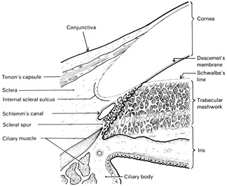
| Fig. 4. Diagrammatic representation of a longitudinal section through the region of the corneoscleral junction showing the peripheral cornea, the sclera, the conjunctiva, and Tenon's capsule, as well as the canal of Schlemm, the trabecular meshwork, and the iris. |