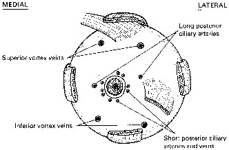
Fig. 10.
Diagrammatic representation of the sites of perforation of the sclera by the long and short posterior ciliary arteries, the short posterior ciliary veins, and the inferior and superior vortex veins.