Chapter 3
Periocular Mesenchyme: Neural Crest and Mesodermal Interactions
CHRISTINA WAHL and DREW M. NODEN
Main Menu   Table Of Contents

Search

DEFINING THE ENVIRONMENT AROUND THE EYE
THE BLUEPRINT: HEAD MESODERM, SONIC HEDGEHOG, AND PAX-6
FORMATION OF THE NEURAL CREST AND PERIOCULAR CONNECTIVE TISSUES
TERMINAL DIFFERENTIATION AND MORPHOGENESIS
REFERENCES

DEFINING THE ENVIRONMENT AROUND THE EYE
Periocular tissues consist of oculomotor muscles, connective tissues, glands, blood vessels, and nerves of the orbit within which the eye is supported and suspended. Although the progenitors of these tissues and their embryonic histories are distinct from those of the eye, they are nevertheless intimately integrated with ocular development. Most periocular tissues arise from three basic embryonic cell types—neurepithelium, neural crest, and mesoderm. In addition, ectoderm contributes the epidermis of the eyelids.

The objective of this chapter is to discuss what is known about early development and morphogenesis of the periocular area, and especially to consider various proposed mechanisms of spatial assembly and patterning of its principal contributors, the neural crest and mesoderm. Prepatterning of head tissues prior to gastrulation will be discussed, but in less detail. Readers interested in this subject should refer to a recent review of early development, currently a very exciting field. Advances in molecular techniques as well as improvements in older experimental approaches have made it possible to investigate this topic as never before. Much of the information in this chapter was gathered from avian, amphibian, fly, frog, mouse, and zebra fish species. Comparisons of growth patterns among them show enough similarity that we can, with confidence, extrapolate to the human embryo, for which only descriptive data exist.

Differentiation and spatial patterning of neural crest and mesoderm must occur before the two tissues can be successfully integrated. The major organizational problems that precede assembly of the periocular mesenchyme will be defined first. To construct the head of the embryo (in this instance, specifically the area around the eye), zygotic cells must overcome a series of developmental problems. The first major problem is establishment of the body axis; the second is formation of the mesoderm; the third is differentiation of the neural crest; and the fourth is integration and interaction of the mesoderm and neural crest.

Back to Top
THE BLUEPRINT: HEAD MESODERM, SONIC HEDGEHOG, AND PAX-6
The body axis is defined in the blastula, before the embryo exhibits any overt polarity, by molecular signaling that leads to establishment of a “node” or “head organizer.” Subsequent morphometric movements of cells occurring at the node during gastrulation lead to the definition of a shallow rostrocaudal depression known as the “primitive streak.” The issue facing the embryo at this early stage is twofold: First, mesoderm must be programmed to form at precise locations—typically, along the primitive streak in amniote embryos; second, it must be imbued with regionally specific properties such that, acting in concert with overlying ectoderm, it can effect the spatial organization of axial structures. This issue has been extensively studied in Xenopus. On the surface of the blastula, molecular signals govern the later appearance of mesoderm, creating regional “mesoderm maps.” The signaling molecule activin plays a dominant role in this early process.1,2 The blastula does not synthesize enough activin to promote mesoderm by itself, but it is influenced by the presence of maternally derived activin.3 Activin exerts a direct effect on the ectoderm by upregulation of the organizer signals: noggin,4goosecoid,5 and nodal.6 Goosecoid, in turn, upregulates chordin, which is known to directly promote formation of Spemann's “head organizer.”7 Goosecoid null mutants lack the organizer phenotype, and resultant embryos exhibit several craniofacial defects.8 Other maternal signaling molecules also contribute significantly to the spatial organization of mesoderm induction. For instance, upregulation of goosecoid depends on synergism with maternal fibroblast growth factor (bFGF). Acting as a competence factor, bFGF facilitates the action of activin to form mesoderm at the equator of the blastula, where vegetal and animal poles are in contact.5,9 Activin passively diffuses over at least 10 cell diameters; this suggests that it may form a “morphogen gradient.”10 Thus, the response generated depends on the developmental stage at activin signaling, as well as the distance from the activin source. As embryogenesis proceeds and different receptor populations appear, activin plays different roles.11 Formed from two β-subunit peptides of inhibin, activin has two binding proteins: follistatin and α2M.12 Although maternal follistatin will block the effect of activin, it does not inhibit formation of mesoderm.13 This suggests that multiple factors may be involved in mesoderm fate-mapping, although activin is clearly a major player.

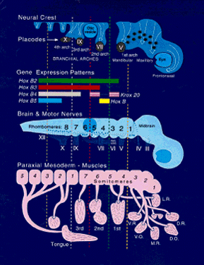
The body axis appears with the genesis of the node and primitive streak. HNF3 β, originally identified as a liver-specific transcription factor, is first found in the node, where it is required for proper morphogenesis of the primitive streak and notochord. In HNF3 β null mutants, anteroposterior patterning persists, but there is no node or notochord formation, and dorsoventral patterning is disturbed. Null embryos have abnormal streaks and affected foregut formation.14 The region just anterior to the node is called the “prechordal plate.” Mesoderm, largely responsible for dorsoventral and anteroposterior patterning of the body axis,15,16 also underlies the future eye-forming part of the prechordal plate, or “eye field,” before neural tube closure. This formation of mesoderm at the prechordal plate and its equivalents, which form the prosencephalon and diencephalon and related head structures will be discussed. Patterning of the prechordal plate differs substantially from that of the rest of the head and trunk. Caudal to the node there is clear rostrocaudal organization that is maintained by several genes. The Hox family of genes is the one best characterized.17 Development of most left-right body features follows this segmental plan, but the prosencephalon is not a part of this organization. It forms as a single, uninterrupted crescent-shaped area extending forward of the node, with no primitive streak. However, interruption of expression in the most anterior of the segmentally organized genes can have a direct impact on the anterior head segment. For example, the homeobox genes XLim1 (and its mammalian homologue Lim1) and KROX 20 are essential for establishment of anterior head structures. Both Lim-/- and KROX 20-/- mutants are truncated just rostral to rhombomere 3, in the metencephalon.18

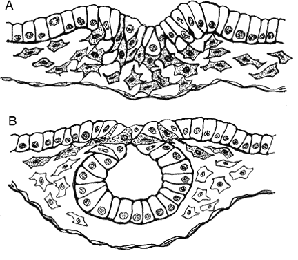
Mesoderm is generated in the prechordal region, as elsewhere, by way of a process known as epithelial-mesenchymal transformation (Fig. 1A), through which epithelial cells lose their tight junctions and move between openings in the underlying basement membrane. Mesenchyme is a uniquely embryonic connective tissue best characterized by the absence of tight junctions between its highly mobile cells and the heterogeneous nature of its extracellular matrix (ECM). Mesenchymal cells advance by “pulling” themselves through the filaments of the ECM, a process that depends on the presence of certain cell adhesion-specific molecules in the ECM and complementary matrix adhesion-specific molecules on the cell surfaces. This has been demonstrated both in vitro and in vivo. For instance, when ectodermal explants are treated with heparinase to remove glycosaminoglycans, morphogenetic movements are inhibited and no mesoderm is formed.19

Fig. 1. Epithelial-mesenchymal transformation. A. Mesodermal mesenchymal cells (stippled) are de-laminating from the region of the primitive streak. B. Neural crest cells (stippled) are de-laminating from the neural folds.

Mesoderm may be restricted, as opposed to induced, through the action of molecules such as bone morphogenetic proteins (BMPs), which are members of the transforming growth factor- β superfamily of signaling molecules. These molecules are important during the early fate-mapping process,20 although subsequently they may assume different signaling roles. BMP 3 inhibits noggin and goosecoid, thus restricting a subset of mesoderm contributing to the head and anterior trunk (“dorsal mesoderm”).21 Maternally derived BMP 2 promotes generalized mesoderm formation,22 whereas BMP 4 is more specific, inhibiting production of “dorsal” mesoderm and promoting differentiation of “ventral” structures, including the blood, pronephros, and visceral tissues.4

Ventral mesoderm induction can be regulated in part by suppression of the BMP proteins. An example is the recently characterized gene XIPOU2,23 which suppresses activation of goosecoid and XLim1 by activin, thus in turn suppressing BMP 4. If XIPOU2 is injected into the anterior pole of Xenopus blastulas, it reduces the eye field (as measured by Pax-6 gene expression), and the resultant tadpoles exhibit microphthalmia. The normal phenotype can be rescued by chordin. However, injection of XIPOU2 into the posterior vegetal pole produces axis duplication, two heads, and four eyes. This demonstrates the site-specificity of action of a given gene product, another feature that continues to change with development.

The formation of mesoderm from transformed ectodermal cells completes the three primary germ layers; ectoderm, mesenchymal mesoderm, and endoderm. Next, many important lineage commitments relative to the eye and periocular tissues become established, the body axis becomes more fully defined, and dorsoventral, mediolateral spatial coordinates are becoming established through molecular signaling. One of the most important signals involved in axis definition is sonic hedgehog (SHH), which is the mammalian homologue of the Drosophila hedgehog gene. Until recently, it was known that the SHH protein was expressed along the body axis and the posterior margins of limb buds in ways that suggested that the role of this protein involved axial patterning. The SHH knockout mouse demonstrates a role in defining the midline of the body, reinforcing bilaterality, and promoting limb morphogenesis.24 The null phenotype is lethal and includes incompletely fused neural tubes with poorly defined ventral midlines, and severe cyclopia, expressed as one eye in a single frontal socket, with an overlying proboscis and no olfactory placode.

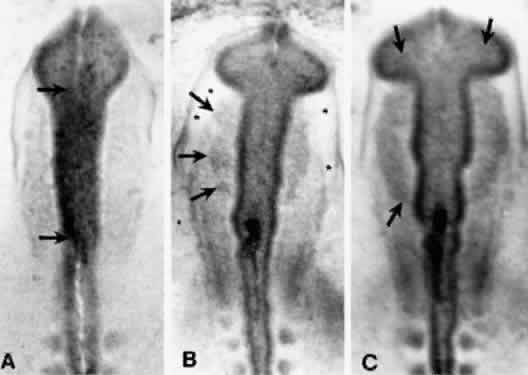
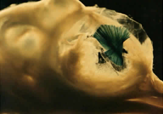
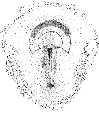
Cyclopia results from incomplete bilateralization of the eye field, which is initially located in the prechordal plate (Fig. 2; also, see elsewhere in these volumes). When the prechordal mesoderm underlying the eye field of Xenopus embryos is removed, only one retina is formed.25 Part of the future prosencephalon, which later forms both the telencephalon and diencephalon, is set aside to form the eyes. This single crescent, or eye field, arises rostral to the primitive streak. The eye field expresses a gene, known as Pax-6, which is essential for eye formation. Transplantation experiments show that pieces of the prechordal plate are able to suppress Pax-6 expression in the retina, but expression of Pax-6 in the prechordal ectoderm is not affected by the presence or location of early optic vesicles.26 This suggests that Pax-6 expression in the prechordal field is spatially segregated into two eye fields through the action of the prechordal mesoderm. Axial mesendoderm positively regulates the medially and ventrally expressed gene Nkx2.1 in the anterior head fold and suppresses laterally and dorsally expressed genes, such as Pax-6. Expression of Nkx2.1 is induced by the prechordal plate.27

Fig. 2. Pax-6 expression in the prechordal plate.25,26 The borders of the presumptive basal and alar lamina (inner solid line) and presumptive neurosomatic junction (outer solid line) are shown. The stippled crescent in the anterior prechordal plate represents the eye field, as recognized by Pax-6 expression.

Human anophthalmia, aniridia, Peters' anomaly, and autosomal dominant keratitis are known to be linked to Pax-6 deficiencies.28–31 In the Pax-6-deficient mouse Sey-sey, eyeless homozygote dysmorphologies include abnormal midline connective tissues,32 perhaps reflecting the default outcome of prechordal ectoderm in the absence of Pax-6. Pax-6 is progressively downregulated until it is restricted to the lens and olfactory placodes. In Sey-sey homozygote mice that fail to form placodes, optic vesicles are formed, but involute.33 Perhaps the ultimate role of Pax-6 in eye formation is most dramatically demonstrated in Drosophila, in which artificial upregulation of the Pax-6 homologue eyeless results in ectopic eye formation all over the body, including the legs, antennae, and wings!34 This startling result, coupled with the prevalence of Pax-6 homologues among species, led the authors to suggest that Pax-6 may be a “master control gene” for eye morphogenesis. It also demonstrates that the general functions of many key regulatory genes have been conserved through thousands of milleniums of animal evolution.

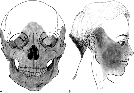
Usually, teratogenic agents are blamed for failure of the eye field to separate. For some years, veterinarians have been aware that pregnant ewes grazing on fields containing specimens of the weed Veratrum give birth to cyclopic lambs if they eat the plant during the first weeks of gestation.35 Earlier in this century, studies on eye formation led to the discovery that fish embryos exposed to magnesium in sufficient concentrations developed cyclopia.36 The percentage of larvae with cyclopia was dependent on the amount of magnesium in the water, whereas the severity of the phenotype (ranging from close-set eyes, to two eyes in one frontal orbit, to a true cyclops with one eye in a frontal socket) was determined by the length of the exposure. The most common teratogen affecting human births is ethanol, which disrupts the prosencephalon by limiting its size; the affected child may show microphthalmia, short palpebral fissures, deficiencies of the philtral region, and a long upper lip.37 The otherwise low incidence of cyclopia in nature suggests that chemical agents with the potential to cause major prosencephalic defects are not generally encountered, and that the window of development during which the embryo is vulnerable is short.

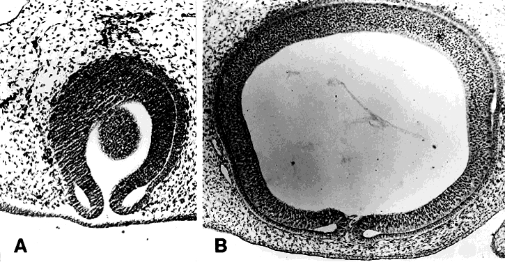
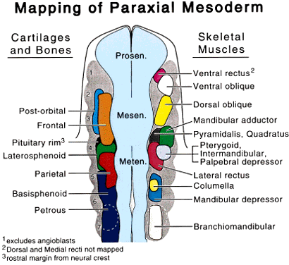
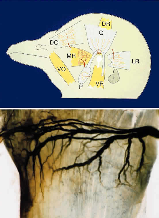
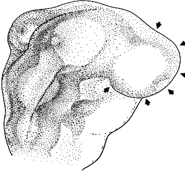
The periocular area undergoes marked changes in tissue relations as the brain grows. As the flat neural tube expands mediolaterally, its walls form paired lateral outpockets called optic vesicles, which later become the optic cups (Fig. 3). The prechordal and paraxial mesoderm originally underlying the rostral neural plate expands laterally and rostrocaudally in congruence with the neural tissue until the evagination of the optic vesicles pushes adjacent mesoderm caudally and ventrally. This leaves the optic vesicle devoid of an enveloping mesenchymal layer except on its caudal (future temporal) surface. The remaining mesenchymal space posterior to these rudimentary optic vesicles is characterized simply as “paraxial mesoderm.” This mesenchyme is not displaced or migratory but consists of a uniform population of mesodermal, stellate cells located almost exclusively in the dorsal and ventrolateral (12 to 6 o'clock) positions. The remaining periocular area is acellular, consisting solely of ECM in the space between the optic vesicle and overlying ectoderm.

Fig. 3. Formation of the optic vesicles as lateral bulges of the prosencephalon. This drawing is a dorsal view of the right side of an avian embryo at about 2.5 days development. The arrowheads indicate the superficial extent of the bulging right optic vesicle.

Thus, the neural epithelium of the optic vesicle is in direct apposition to the surface ectoderm along its lateral and anterior surfaces. Normal development of the eye depends on the absence of mesenchyme in these areas, as has been demonstrated by the mouse mutant extra-toes, in which invasion of mesenchymal cells along the lateral margin prevents normal formation of the lens.38 In normal animals, mesenchyme that may become trapped between the invaginating optic vesicle and thickening lens undergoes necrosis and is resorbed. In a congenitally anophthalmic mouse strain, this trapped mesenchymal population failed to undergo apoptosis, and the normal sequence of necrotic loci in the developing eye was lacking.39

It is the failure of the lens placode to contact the optic vesicle as a result of inappropriately placed mesenchyme that results in anophthalmia among these genetically defective animals.33,40 The lens placode also plays a key role in ocular expansion, perhaps related to generation of the vitreous. Growth of the retina is unaffected by eye size, because the retina will continue to proliferate beyond the confines of a miniaturized eye. Mesenchymal processes that lead to expansion of the globe do not control or otherwise affect the mitotic rate within the retina. Retinal convolutions may fill the eyecups of microphthalmic or “anophthalmic” animals, a phenomenon that can be duplicated experimentally by the use of teratogens.41

Back to Top
FORMATION OF THE NEURAL CREST AND PERIOCULAR CONNECTIVE TISSUES
If the optic vesicle were to remain devoid of surrounding mesenchymal cells, both its development and the potential to form the array of connective tissues necessary for orbital and corneal development would be absent. In the early delineation of the vertebrates, it is not known why the existing mesodermal cells did not simply migrate rostrally and circumscribe the globe. Instead, a new population of mesenchymal cells, the neural crest, was generated to serve this and many other functions.

The neural crest is formed around the time of closure of the dorsal lips of the neural tube, during a second period of epithelial-mesenchymal transformation limited to the angle in the epithelium between dorsal ectoderm and neural tube (see Fig. 1B). The neural crest is a highly migratory cell population; it carries great differentiation potential; it exerts inductive influences on almost every tissue that it invests; and it is essential in midfacial and orbital development. Students of comparative neuroanatomy hear the neural crest defined as “the single greatest innovation separating vertebrates from invertebrates.” The importance of this embryonic tissue extends beyond the fact that it forms all autonomic postganglionic neurons, the enteric nervous system, the outflow tract of the heart, and melanophores. In the formation of the peripheral glia, or Schwann cells, the neural crest furnishes a way to speed conduction velocities along axons. Saltatory conduction, arguably more than any other feature, explains why the world's largest animals are vertebrates. Much of the vertebrate head is neuroectodermal in origin, because most of its bones and all of its fibroblast-derived connective tissues, dermis, and perivascular smooth muscles arise from neural crest. Only the endothelium, glands, and muscles of the orbit are not crest derivatives. Thus, the formation and migration of the neural crest into the periorbital region signals the beginning of final tissue integration and assembly as required for scleral formation, development of the accommodative mechanism of the eye, and spatial patterning of the eye muscles.

The epithelio-mesenchymal transformation of neural crest cells is initiated during presomite stages.42 It begins at the mesencephalic level and is followed within a few hours by similar formations in the diencephalic and then metencephalic and more caudal regions. Cephalic crest cells start their migration at the 4-somite stage in humans43 and at the 5-somite stage in mouse44 and avian embryos.45–47 These cells do not arrive in the periocular region until the optic vesicle has formed and the periocular mesoderm has been displaced. Figure 4 shows the initial formation of the cephalic neural crest population in a series of living avian embryos. The crest population expands while the optic stalks begin to constrict. The enlarging cavity between the dorsal surface of each stalk and the overlying surface ectoderm is soon occupied by neural crest cells; some of these are diencephalic in origin, but most have moved rostrally from a mesencephalic origin.

Fig. 4. Three stages in the early migration of avian neural crest cells. These photographs were taken of living embryos; they were stained with neutral red to enhance contrast. In each case, the arrows indicate an edge of the crest population. A. Crest cells are just forming (see Fig. 1B). B. The leading edge of the crest population is beyond the dorsolateral margin of the mesencephalon. Note the appearance of the cell-free space (asterisks) between the mesoderm and the surface ectoderm. C. Crest cells are present dorsal to the site of optic stalk constricture. (Noden D: The migration and cytodifferentiation of cranial neural crest cells. In Pratt R (ed): Current Research Trends in Prenatal Cranio-Facial Development. New York, Elsevier North Holland, 1980)

Like mesodermal mesenchyme, movements of neural crest cells depend on molecular interactions with the ECM. In the chick embryo, neural crest cell movements are disrupted if HNK-1, an antibody to a carbohydrate moiety involved in cell adhesion, is injected into the ECM along migration pathways.48 In albino axolotls, it has been shown that fibronectin, tenascin, collagens I and VI, and a chick aggrecan stimulate migration of crest cells, although in vitro the aggrecan molecule inhibits cell migration.49 This latter result is a dramatic reminder that cell responses to specific molecules within three-dimensional, intact organisms may differ substantially from behaviors in culture! The molecular structure of the ECM and epithelial tissue boundaries thus delineate migration routes of the neural crest cell.50–53 Continual modification of the ECM by the mesenchyme provides temporal control over crest cell migration as substrate qualities are altered.54–56 Therefore, correct morphogenesis at this time depends first on the initial spatial guidance of crest cells by the mesenchyme; second, on temporal control of crest cell migration by the ECM; and third, on the behavior of the crest cells themselves en route to their final destinations. One mode of action of Pax-6 involves controlling neural crest migration by facilitating movement of anterior midbrain crest cells forward into periocular and nasal areas.57

In mammals, crest cells first appear in the mesencephalic region prior to neural fold apposition and neural tube closure. This apparently precocious emigration occurs because neural tube closure begins in the future occipital or cranial cervical region (at the level of somites 2 through 7 in humans and 4 through 6 in rodents) and then proceeds cranially and caudally. In humans, those parts of the mesencephalic crest population that contribute to the periocular area emerge during the 4- to 12-somite period.43 A second group of crest cells is reported to emerge from the optic vesicle and sulcus (or diencephalic neural folds) during the 13- to 20somite period,43 although this population has not been experimentally traced in other species (Fig. 5).

Fig. 5. Drawing of an embryo with prominent mesencephalic cranial flexure but before formation of the pontine flexure. The optic vesicles have formed eyecups, and the lens is present. The stippled area indicates neural crest. Dotted lines drawn from the neural folds show regions either previously traversed or currently occupied by crest cells that originated from the prosencephalic, mesencephalic, metencephalic, and myelencephalic regions of the neural tube, respectively. Neural crest vacates the dorsal mesenchymal regions around the neural tube and extensively populates the periocular region, branchial arches, and outflow tract regions of the heart.

Although cranial flexure precedes neural crest migration in many mammals, including the human,43,58 in the avian and some reptiles the head of the embryo is still in a flat plane as the crest cells leave the neuroectoderm. Crest cells begin to migrate en masse laterally from atop the middle of the mesencephalon. Immediately ahead of the moving front of crest cells, the ECM expands, creating an acellular space filled with hydrated glycosaminoglycans, laminin, and fibronectin. The neural crest population moves laterally through this acellular matrix, then proceeds ventrally, following the body's contours. The first cranial (mesencephalic) flexure is a ventral folding at the midbrain level that brings the forebrain and paired eyes ventrally and caudally more than 90° with respect to the axis of the trunk, to a position just rostral to the floor of the mesencephalon. Later, the second (pontine) flexure, which is a dorsal folding at the hindbrain level, elevates the eyes and forebrain, bringing them forward about 30° with respect to the axis of the trunk. In the human, the midbrain cephalic flexure occurs early, beginning at the 1- to 3-somite stage, forming a right angle between the long axis of the forebrain and that of the hindbrain.59 Embryos at this stage are approximately 20 days old. By the 29th day, the pontine flexure is evident. During this interval, the eyes have changed considerably. The vesicles have become optic cups, the lens placode has formed a lens vesicle, the pigment epithelium has begun to produce melanin, eye muscle precursor populations are evident in the periocular area, and the eye has more than doubled in size (the actual proportion of growth varies among species). Expansion of the optic vesicle coincident with cranial flexure also brings about changes in the relations of mesenchymal cells to each other, including new contributions of mesenchymal cells from caudal sites of origin along the hindbrain. The neural crest cells that form the remainder of the dorsally situated population continue to migrate along surfaces of the optic cups (Fig. 6). It is important to recognize that the genesis of the neural crest, whether at open or closed neural folds, and whether early or later relative to cranial flexures, does not affect the fundamental relations between the sites of neural crest origin and the terminal location of neural crest or mesodermal populations.47,50,60–62 Crest cells arriving at the limbic region are prevented from further movement into the future corneal site by the presence of a dense meshwork of extracellular fibers that coalesce between the lip of the optic cup and the surface ectoderm.

Fig. 6. Parasagittal sections through the optic cup of 2-day (A) and 3-day (B) chick embyros. A. The ventral lips of the optic cup have separated from the underlying surface ectoderm, but crest-derived mesenchyme has not penetrated this region. B. This space is now occupied by neural crest mesenchyme.

Many mesencephalic crest cells remain dorsal to the brain and migrate rostrally to join with those formed from the diencephalic neural folds. These populations appear to intermingle and then accumulate dorsal to the constricting optic stalks. Subsequently, some of these crest cells migrate ventrally along the caudal and cranial surfaces of each stalk (Fig. 7). Their movement around the optic stalk ceases when contact is made with the choroid fissure, which is tightly apposed to the ventral surface ectoderm. Asymmetric patterns of neural crest cell proliferation in the crest-derived mesenchyme immediately caudal to the choroid fissure cause cranial expansion of this tissue beneath the fissure, resulting in the formation of the maxillary and frontonasal processes.

Fig. 7. Transverse section showing the close apposition of the optic stalk (o.s.) and lateral margin of the optic vesicle (o.v.) to the ventral and lateral surface ectoderm. Arrows indicate appositions that prevent movement of crest cells beneath the eye until later stages. The thickening of the lateral surface ectoderm is the lens placode. (Johnston M, Noden D, Hazelton R et al: Origins of avian ocular and periocular tissues. Exp Eye Res 29:27, 1979)

Detailed analyses of the migratory patterns of cranial neural crest cells indicate that periocular neural crest includes cells originating at several axial levels (diencephalic through mid-mesencephalic) that display different migratory pathways. (Differences in crest migration patterns have been studied in chick45,50,61,63–65 and mouse66,67). The presence of paraxial mesoderm adjacent to the original temporal surface of the optic vesicle temporarily precludes the movements of postorbital crest cells in this region. In the adult, this is reflected in a small patch of temporal sclera derived from mesoderm but otherwise indistinguishable from the rest of the sclera.

In some areas, the crest cells proliferate rapidly; in others, they apparently fail to establish themselves. These behaviors are triggered by unique ECM environments, which are in part self-generated.68,69 Migrating crest cells will orient toward surfaces with abundant ECM,60 especially environments rich in highly hydrated compounds such as hyaluronate, which are thought to contribute to the swelling of intertissue spaces that precedes migratory events.70 Hyaluronate does not actually promote migration since enzymatic digestion in vivo causes collapse of the subectodermal space without disruption of the early crest cell movements.71 Antibodies to receptors for fibronectin and laminin, which are located along crest cell pathways,72 will inhibit the crest's ability to migrate normally from the neural fold.73 This suggests that these components of the ECM play a significant role in cell movement. Interestingly, however, crest cells are able to move out of a 2-day-old neural tube cultured in type 1 collagen containing neither laminin nor fibronectin.74,75 The appearance of type 1 collagen has been correlated more specifically with the onset of migrating crest cells than with the occurrence of either laminin or tenascin, which are widespread in distribution by the time crest cell emigration begins.76 These and other data, when taken collectively, indicate that a variety of often heterogeneous ECM compounds may affect the proliferation or direction of crest cell migration.

The entire midbrain population of crest cells vacates its site of origin. Many of these laterally and ventrally moving cells populate the maxillary process and, together with metencephalic crest cells, form the mandibular prominence. Some of these cells will contact the optic vesicle on its most lateral, temporal surface, just lateral to the area contacted by paraxial mesoderm. These cells accumulate in the angle formed between the vesicle and surface ectoderm and will later participate in formation of the cornea.

To study the movements of cells within heterogeneous populations, Le Douarin introduced a method of transplanting cells from the Japanese quail, Coturnix japonica, into the tissues of chicken embryos.47 Within the nucleus of each quail cell is a condensation of nucleolus-associated heterochromatin, unlike anything found in interphase chick cells. Because the marker is self-replicating, it can be followed for extended periods after transplantation, and the terminal locations and fates of all cells derived from the quail implant can be identified. To study the distributions of neural crest cells, small sections of the dorsal neural folds were replaced by quail tissues in homotypic grafts. The orbital and sclerotic cartilages of the resulting chimeric embryos always contained a mixture of quail and chick cells whether the graft was at the mesencephalic or prosencephalic level, whereas quail cells from the prosencephalon dominated the anteroventral part of the orbit and the sclerotic plates, and mesodermal cells from the mesencephalic primordium were located mainly in the dorsal region of these cartilages.77

Direct grafting of neural crest cells into mammalian embryos in vitro67,78 and microinjection of neural crest cells into mutant mouse host embryos in vivo,79 contribute information regarding crest migration patterns in mammals. To date, the results have been strikingly similar.66,80–82 This technique has also been successful in the chick.83 Additional data on the mouse are available on a small population of dorsoventrally moving crest cells in the head, which have not been seen at these early stages in the chick,84,85 although both species have dorsoventral pathways in the trunk.72 Lipophilic dye tracking of small populations of cranial crest cells has been useful in resolving some ambiguities from transplant experiments.86

The observations of Veermeij-Keers and Poelman42 focus more specifically on the high degree of cell proliferation, degeneration, and differentiation associated with neural crest. These processes should certainly be taken into account in considering the overall phenomenon of crest cell migration, because facial malformations may result from errors in any one or more. A specific example of the role of cell death in crest cell migration is seen in the chick hindbrain at the level of rhombomeres 3 and 5, where no crest is observed to emerge but cell death at the dorsal lips of the neural folds is marked. This suggests that progenitor crest cells are selectively eliminated in these areas.82

The movement of the crest cells is an example of one of the “broad strokes” in early morphogenesis that define characteristics inclusive of whole body regions rather than specific subsystems. A dysfunction in the crest cells or in the matrix in which they must proliferate and move, or in the signaling epithelia around them (e.g., prosencephalon, optic vesicle), can lead to syndromic developmental defects involving the midfacial region (Fig. 8). Examples of this type of defect include Waardenburg's syndrome (Pax-3 abnormalities87), primary cleft palates or lips, the craniofacial abnormalities of fetal alcohol syndrome, and other variants of holoprosencephaly. The various movements and rates of growth of all cell populations within the head must be precisely integrated for normal development.

Fig. 8. A. Sketch of human skull, frontal view, showing skeletal elements of neural crest origin (shaded). B. Human profile with shaded area indicating neural crest-derived tissues.

Certain relationships between various assemblies of cells during early tissue morphogenesis have been observed. For example, crest cell and mesodermal cell populations do not mingle at any time during the early stages; the interface between them is distinct. Later, cranial flexure and growth of the eye and branchial arches will cause this interface to become ambiguous. Penetrations by muscle-forming and vascular endothelium-forming mesoderm will continue to obscure the boundaries, and some periorbital skeletal tissues will derive from a dual mesodermal-crest origin.65,77,88

Pluripotency of crest cells in the periocular region has been demonstrated by transplanting periocular mesenchyme into the vicinity of embryonic gut tubes. The periocular crest was shown to differentiate into enteric ganglion and autonomic neurons, phenotypes these cells do not normally display.89 In a hormonally defined culture medium, mouse neural crest cells can also be induced to express a neuronal phenotype.90 Thus, the environment around pluripotent neural crest populations influences the timing, location, and phenotypic character of subsequent differentiation.

Regulatory molecules such as analogues of retinoic acid, corticosteroids, and other components of serum selectively affect the development of crest-derived cells (see reference 51 for review). Morphogenetic events are also affected by basement membranes,69,91 and in the quail, cultures containing basement membrane matrix stimulate the differentiation of catecholamine-positive cells among pluripotent crest precursors while inhibiting the differentiation of other crest derivatives.92 Normally, migrating crest cells encounter basement membranes along the neural tube, somites (in the trunk), and ectoderm—locations in which peripheral neurogenesis is promoted.

In the eye, the optic vesicle exerts an inductive effect on the periocular crest, stimulating the formation of the sclera.88,93–95 Similarly, anterior corneal epithelium promotes first the ingrowth of adjacent crest cells to establish the stroma and later the progressive, layered assembly of these cells into the compact, transparent cornea.96

In turn, the neural crest precursors that aggregate around the eye provide significant spatial programming information to nearby mesodermal cells (e.g., myoblasts) that move into the periocular region. Experiments in which presumptive jaw neural crest cells are transplanted to caudal head regions have resulted in the development of jaw-type structures (e.g., quadrate mandibular cartilages and bones) in an ectopic location.97,98 Furthermore, the assembly of myoblasts that move into this ectopic connective tissue-forming population is modified in accordance with the imposed jaw spatial organization.98,99 All this occurs before the overt differentiation of cartilages or myotendinous tissue. Thus, crest cells, destined to form skeletal elements, tendons, and muscular septa of the periocular region, exert a significant organizational influence on spatial morphogenesis of the myogenic cells with which they come in contact. Whether the periocular crest population generates this ability intrinsically or is itself responding to cues emanating from the optic vesicle is not known.

EXTRAOCULAR MUSCLES

In the spinal cord and hindbrain caudal to the ear, paraxial mesenchymal cells form new epithelial blocks, known as somites, by way of mesenchymal-epithelial transformation (see reference 52 for review). Subsequently, somites separate into rostral and caudal, medial and lateral portions, which represent early steps toward determination and delineation of the muscular, skeletal, dermal, and secondarily peripheral nervous system portions of each body “segment.” After the internal organization of the somites into discrete populations of myogenic, precartilaginous, and connective tissue components has occurred, many of these cells move out into the body walls and limbs, where they undergo final assembly and differentiation.

In the head, however, the situation is different. Somites do not form. Scanning electron microscope studies reveal the presence of loose, serially arranged mesenchymal condensations within the paraxial mesoderm that are termed somitomeres. These do not become epithelia as do the somitic mesodermal cells. Although somitomeres are indistinct (and controversial) in avian and mammalian embryos, they are strongly evident in the shark.100,101 It has been suggested that discontinuities in the dorsal, outer surface of the paraxial mesoderm resulting from somitomere formation may have a profound effect on the pattern of morphogenesis of the neural crest, but experimental evidence for this is lacking.

The six muscles of the oculomotor system, as well as the retractor bulbi muscles, arise from distinct myogenic condensations within the paraxial mesoderm.102 Exact maps of the origins of each muscle are obtained by injecting neurulating embryos with replication-incompetent retroviruses carrying a β-galactosidase marker. After the eye muscles assemble in the orbit, the embryo is fixed and stained to determine the location of the retroviral marker (Fig. 9). The resultant data show that the anteriormost muscle precursors are initially located lateral to, and then immediately caudal to, the optic vesicle. These are the medial rectus, ventral rectus, and ventral oblique muscles, which arise from a common myoblast condensation. The origins of some of these myoblasts can be traced back to a few progenitors that arise within prechordal mesoderm located immediately beneath the future eye-forming field in the neural plate. Ventrolateral to the mesencephalon lie the precursors of the dorsal rectus, followed by those of the superior oblique. The lateral rectus muscle arises even further caudally, ventrolateral to the metencephalon at the level of rhombomeres 3 and 4 (Fig. 10).

Fig. 9. Right-sided view of avian embryo, near hatching (19 days). This embryo was injected with β-galactosidase-marked retrovirus at 2 days' incubation. The injected area corresponded to deep paraxial mesenchyme at the mesencephalic level (see Fig. 10). The embryo was fixed, stained for the presence of marker, and dissected. The deep blue muscle (which carries the retrovirus) is the dorsal oblique. (Fixman J, Wahl C, Noden D: Unpublished data.)

Fig. 10. This map of the paraxial mesoderm summarizes the results of lineage tracing using retroviruses as shown in Figure 9. (Wahl C, Fixman J, Concoran P, Vilenchik M, Evans D, and Noden D).

Much of the available experimental evidence indicates that myogenic cells have little role in establishing the gross spatial patterning of voluntary muscles. Nonbranchial somites grafted into prospective wing regions form normal wing muscles103; trunk somites grafted into the head make apparently normal extrinsic ocular or masticatory muscles104; and quail myotomes transplanted into chicks develop into typical chick muscles.105 These results complement neural crest grafting experiments in establishing the neural crest as the dominant pattern-generating peripheral tissue in the head. However, Wahl and Noden106 and Wahl and colleagues102 have found that some migrating myoblasts exhibit eye muscle-specific features, and that neural contacts are established with extraocular muscle myoblasts before their migration from the hindbrain paraxial mesoderm and before they contact neural crest cells. These results do not lessen the importance of crest cells in spatial organization; in fact, they show that this process is initiated earlier while eye muscle progenitors are still within paraxial mesoderm and close to the brain. Cues provided by early neural contacts may act together with local cues from the crest within the periocular mesenchyme, thereby integrating the spatial and functional organization of muscles within the orbit.

Definition of myogenic cells within the paraxial mesoderm involves subtle changes on cell surfaces that enable myoblasts to coalesce and form condensed muscle primordia. Within this aggregate population, primary myotubes take shape, as described in a wide range of vertebrate embryos and neonates. Myotubes interact differently with the matrix than with undifferentiated mesenchymal cells. Laminin stimulates myoblast proliferation and process outgrowth.107 Fibronectin, hyaluronic acid, and prostaglandins are all involved in spatial organization of whole muscles.108 The myotubes themselves produce different types of collagen109 and display different nerve cell adhesion molecule distributions.110–112

There is also growing evidence that each whole muscle may have a unique “identity.” In the limb, muscles are known to exhibit individual patterns of innervation,113 a phenomenon that is also true of avian extraocular muscles (Fig. 11). What part of this uniqueness, or self-identity, is an intrinsic property of the myoblasts, and what part relies on the contribution of the muscle's connective tissues, is not clear. Myoblasts do have distinct identities before they reach the periocular area, as illustrated by the avian lateral rectus-pyramidalis/quadratus (LR-P/Q) complex. The pyramidalis (P) and quadratus (Q) muscles of avian eyes are homologues of mammalian retractor muscles, arising from similar origins. They lie between the globe of the eye and the rectus muscles, where their function is retraction of the nictitating membrane. The lateral rectus and the P/Q muscles arise as a single condensation from the paraxial mesoderm during neurulation (Fig. 12). Although the lateral rectus subset of the mesodermal condensation differentiates early, forming myotubes and becoming oriented in the orbit within a matter of 2 days, the P/Q precursors do not develop or proliferate for more than a week. During this lengthy period of embryogenesis, the P/Q cells move around the developing optic nerve and begin to proliferate and further differentiate only after most of the surrounding orbital tissues are in place.102 This shows that the two myoblast populations are distinct from each other before overt structural variation becomes apparent.

Fig. 11. Unique nerve branching patterns are found on avian extraocular eye muscles. These patterns are consistent in a recognizable way among embryos. A. Diagrammatic representation of major branching patterns for each eye muscle (dorsal rectus not shown). B. Photograph of oculomotor nerve traversing and branching across the medial rectus muscle (silver stain).

Fig. 12. Structural characteristics of muscle-nerve relations at stages 20 through 22 (chick). A. Section of stage-22 LR-P/Q anlage stained with antineurofilament antibodies to reveal two separate nerves entering the muscle. B and C. Sections from a stage-20 Bodian-stained embryo showing the spatial relations between the LR-P/Q anlage and sixth nerve axons. The sixth nerve bifurcates as it reaches the muscle rudiment. One bundle (red arrows in B, C) continues rostrally, then curves laterally to contact the less dense rostral portion of the muscle condensation, which will form the P and Q muscles. The other axonal bundle (black arrow on left in B) directly enters the LR primordium. The inset in B illustrates the onset of accessory abducens motor neuron migration in rhombomere 5 of an age-matched, DiI-labeled specimen. The arrow points to a migrating perikaryon. (Wahl C, Noden D, Baker R: Developmental relations between sixth nerve motor neurons and their targets in the chick embryo. Dev Dyn 201:198, 1994)

VASCULAR ELEMENTS

Angiogenic precursors are mesenchymal “wild cards.” Within embryonic mesenchyme they are universally invasive and move great distances through the embryo. Their behavior in epithelial contexts is more constrained. For example, the sites at which blood vessels penetrate the brain are initially defined at boundaries between rhombomeres (Fig. 13). Factors that may influence or control movements of angioblasts are not well studied. In Sturge-Weber syndrome, which produces “port wine stains” over parts of the face and neck innervated by the trigeminal nerve, correlations exist between the occurrence of upper facial dermal abnormalities and the incidence of ocular and leptomeningeal angiomas, which have not been described in patients whose dermal involvement is restricted to the mandibular and neck areas (reviewed in reference 65). Such syndromes may arise from local perturbations of the ECM, both initially, when the angiogenic precursors populate paraxial mesenchyme, and later on, when the vascular elements continue to exhibit uncontrolled growth in a localized region of the body instead of being down-regulated by local cues in the integument. In culture, increasing densities of fibronectin on precoated dishes causes endothelial cells to become more extended and to proliferate more rapidly.114 The importance of this integrin-mediated cellular attachment to the ECM in vivo has been demonstrated by blocking αvβ3 integrin binding with an agonist. Neovascularization was impaired, with clustering of angioblasts lacking normal protrusions, disruption of lumen formation, and abnormal vessel patterning.115

Fig. 13. Antibodies to quail endothelial cells were used to obtain this fluorescent photograph of the hindbrain of a quail embryo (comparable in age to that shown in Figure 5). Rhombomeres are numbered. Note that the blood vessels form with respect to the rhombomere boundaries and show a pattern of organization that resembles the segmentation of the neural tube. This is a transient phenomenon, disappearing within a day as both blood vessels and neural tube continue to develop and proliferate.

Blood vessel growth follows two modes of assembly. Initially, individual angioblasts migrate freely through the mesenchyme, then stop and initiate formation of isolated segments of capillaries. A second process of vascular sprouting (angiogenesis) also occurs and is the dominant process during later stages of organogenesis and in the adult. Endothelial cells of extant blood vessels extend long filapodia into the surrounding mesenchyme, then move in that direction, adding to the ends of the sprouts by active cell division. They may connect with proliferating isolated segments of blood vessels forming simultaneously through migratory action of the angioblasts.116–118

Migration and differentiation of angioblasts involve several surface molecules, as seen in other embryonic populations. Surface-bound oligosaccharide molecules involved with signaling and/or binding to the ECM are important in vascular assembly. Oligosaccharides are important during early ontogeny in events related to cell differentiation. Mouse embryos that are null mutants for aspartine-linked oligosaccharides are initially able to absorb these molecules through their endoderm from maternal sources. However, they later exhibit a variety of defects, including poor vascularization and left-right cardiac asymmetries.119

PERIOCULAR SKELETON

Most of the periocular mesenchyme is of neural crest origin; therefore, the periocular skeletal and connective tissues are formed largely from crest cells. In a 4.5-day-old chick embryo, the incursions of mesoderm into neural crest mesenchyme have been completed, and the elongation of maxillary and frontonasal prominences is under way. However, overt differentiation of periocular skeletal, muscular, and connective tissues has not begun. The neural crest-mesoderm boundary crosses the caudal margin of the future orbit, and deflections of the interface between crest and mesoderm will continue to appear as a result of differential growth of cephalic tissues (e.g., brain, olfactory pits, oral cavity). Thus, the relationships of the two mesenchymal populations to each other and to adjacent epithelial tissues will change somewhat.

Many osseous elements that surround the orbit develop within or around embryonic cartilages that form in the periocular mesenchyme, usually adjacent to an epithelium (e.g., brain, optic vesicle, or nasal epithelium). By the beginning of the second week of incubation, the following cartilages are adjacent to the eye: the interorbital septum, medially; the nasal labyrinth, rostrally; the postorbital cartilage, caudally; and the sphenoid cartilages, ventrally. The squamous, temporal, and zygomatic bones are located caudoventral to the eye.

Most of these cartilages form cranial to the interface and are thus of neural crest origin. However, the postorbital cartilage arises in the region of overlap between crest and mesoderm and is thus of dual origin. The sphenoid region of the chondrocranium crosses the interface, extending from the interorbital septum to the basioccipital cartilage/bone. In birds, the crest-derived and mesodermal parts of this cartilage meet lateral to the adenohypophysis, where they fuse to form a single element. Later, the presphenoid and basisphenoid bones form on the crest and mesodermal sides of the interface, respectively.

Many periocular bones arise from ossification centers that form in mesenchyme without a cartilaginous precursor (intramembranous or “dermal” bone). Included in this category are the nasal, maxillary (including palatal), lacrimal (or prefrontal), palatine, ethmoid, and zygomatic (jugal) bones. The frontal bone of the chick is chimeric, with the most rostral, supraorbital pair of ossification centers located cranial to the interface (i.e., in neural crest mesenchyme), and the remaining centers situated caudal to the interface. Although there is no direct evidence that precisely identifies the position of the interface in mammals, the mapping data show nearly identical pathways of crest cell migration in chicks and mice, and for the most part homologous skeletal structures can be identified. Furthermore, the frequent occurrence of an isolated interfrontal bone in cases of frontoparietal cranioschisis is consistent with a dual origin for the mammalian frontal bone.

SCLERAL AND CHOROIDAL TISSUES

The optic cup is intimately surrounded by vascular and pigmented choroid layers and a fibrous (or in birds a cartilaginous) sclera. The sclera is of dual origin, reflecting the location of the crest-mesodermal interface. It is thought that type II collagen and chondroitin sulfate proteoglycan are fundamental to skeletal morphogenesis in specifying a chondrogenic fate for responsive mesenchyme cells.120 Type II collagen is expressed around the optic vesicles in the mouse, correlating with expression observed earlier in the quail.121 Thus, collagen type II may “prepattern” mesenchyme around the optic vesicles for scleral formation (although at this time it is not possible to say what “prepatterning” actually involves). Insulin-like growth factor-2 (IGF-2) mRNA is expressed by periorbital mesenchyme as it differentiates to form sclera, but it is not expressed in mature sclera. Thus, the expression of IGF-2 by these cells may also play a role in determining the ultimate size or shape of the eye.122

As described previously, at the time of neural crest formation the caudomedial surface of the optic vesicle is apposed by paraxial and prechordal mesoderm. This apposition is maintained throughout the period of crest migration. As a result, scleral chondrocytes covering the globe caudal to the interface are mesodermal in origin. Those located medially to the sclera, including the area that surrounds the optic stalk, are derived from the neural crest.

The choroid layer is also of dual origin, but not as a result of topographic differences. The lining of all endothelial blood vessels is mesodermal, whereas all connective tissues and pigment cells of the choroid, including those that underlie the mesodermal part of the sclera, are of neural crest origin.123,124

The ciliary region is histologically one of the most complex regions of the eye. However, embryonically it follows the same pattern as the other periocular tissues. Both the ciliary region and the iris incorporate mesenchymal cells located between the mesenchymal shelf and the lip of the optic cup. This mesenchyme is situated cranial to the interface and, except for angiogenic cells, is of neural crest origin. The ciliary muscles, which are striated in birds, and the cells that form the pectinate ligament are also of neural crest origin.

Data on the iris are incomplete. In the few quail-chick chimeras that were maintained to hatching stages, the iris was found to contain a core of host mesenchymal cells surrounded by quail neural crest cells. In embryos injected at stages 9 through 10 in medial paraxial mesoderm along the mesencephalic-metencephalic border using replicationincompetent retroviruses, virally labeled cells were found in the caudal iris and sclera in locations spanning the 8 o'clock to 10 o'clock positions (Wahl and Noden, unpublished data). Descriptive accounts of iris formation support the original suggestion of Lews that iris muscles are formed by budding from the retinal and pigmented layers of the optic cup. However, in the absence of definitive experimental evidence, the possibility that the mesenchymal core is of mesodermal origin or, alternatively, that it might have formed from an area of the neural crest adjacent to that where the quail cells were grafted cannot be excluded.

MORPHOGENESIS OF THE CORNEA

During the fourth day of incubation in the chick, crest-derived mesenchymal and mesodermal angiogenic cells accumulate along the perimeter of the presumptive cornea. However, centripetal movement into the space between the lens and surface ectoderm is prevented by an annular fibrous meshwork. The definitive cornea is formed as a result of two waves of migration of mesenchymal cells.94,122,123

The fibrous meshwork is penetrated first by macrophagelike cells. These are followed by contiguous mesenchymal cells, which move into this region as a loose monolayer. This movement continues until a confluent layer, the posterior epithelium (corneal endothelium), is established beneath the acellular components of the primary stroma, which are products of the overlying anterior epithelium.124

Because the mesenchyme adjacent to the lip of the optic cup contains cells of both mesodermal and neural crest origin, descriptive accounts of this process cannot resolve the precise origins of the posterior epithelium. However, by surgically excising premigratory crest cells from the cranial mesencephalic region of a quail embryo and grafting them in the place of homologous cells of a chick embryo, the neural crest origin of the posterior epithelium has been proved.46,123,125–127

Subsequent to formation of the corneal endothelium, the depth of the acellular primary stroma doubles, an event that is correlated with a dramatic increase in the amount of hyaluronate in this region.128 In response, additional periocular mesenchymal cells invade the stromal region. These cells, all of which are neural crest in origin, subsequently form the keratocytes of the cornea.

Back to Top
TERMINAL DIFFERENTIATION AND MORPHOGENESIS
Each cell of the embryo reaches a point in development beyond which it is irrevocably committed to a unique phenotype. This event is not necessarily coincident for all cells within a given population, but the temporal and spatial order of differentiation must be coordinated for proper morphogenesis. In and around the eye there is an early framework of committed cells that provides the necessary cues that help define the migratory patterns and spatial organization of successive populations. Evidence of this process has been shown in amphibian eyecups, which develop neuronal locus specificity of the permanent anteroposterior and dorsoventral reference axes such that rotated or transplanted eye primordia retain specified axes.

Difficulties in assessing the stages of commitment during migration and differentiation of neural crest cells are being overcome as new molecular techniques become available. Pluripotent neural crest cell populations, which appear morphologically identical before the onset of migration, begin to develop chemical identities that distinguish them as subpopulations before phenotypic spatial or structural changes take place. Early neurogenic cells of the neural crest can be distinguished from nonneurogenic precursors by use of the human autoantibody Anti-Hu, which is derived from patients suffering from small-cell lung carcinoma and subacute sensory neuropathy.129 These antibodies recognize proteins that are expressed in both tumor cells and neurons of the sensory and peripheral nervous system. Using these antibodies in the quail embryo, it is possible to distinguish crest cells biased toward, though not necessarily fully committed to, either neurogenic or non-neurogenic pathways. It has been shown that the premigratory neural crest contains populations competent to form both neurogenic and non-neurogenic lineages, both in vitro and in vivo.51,130 The first cells to migrate are biased toward neurogenesis, whereas those that migrate later do not have this bias. Myogenic precursors are also temporally regulated and do not differentiate at the same rates.102

The appearance of specific chemicals in the local ECM environment has been correlated with differential induction of pluripotent crest cells. Brain-derived neurotrophic factor, probably acting in concert with a BMP released by the dorsal neural tube, causes an increase in the number of migrating crest cells that commit to sensory neuronogenesis.131,132 Recent analyses of intestinal hypoganglionosis (Hirschsprung's disease) in humans and animals have linked a specific deficit to an identified growth factor. Functional loss of either endothelin 3 or its receptor will reduce the rapid population growth needed to form a complete complement of enteric neurons from only a few hundred precursors.133 The results of identical mutations are variable, showing that multiple growth-enhancing factors act upon the neural crest population.

A highly detailed level of organizational information is necessary in cases such as the alignment of ganglion cell axons, in which proper visual perception depends on an orderly pattern of connectivity. The mechanisms responsible for generating this order are largely unknown. One theory that gained wide acceptance, and had considerable experimental support, was the chemoaffinity theory.134 This theory proposed that neurons acquire positionally dependent chemical labels and that the pattern of connections is due to the selective affinity between the labels carried on the growing axons and the cells to which they connect. A problem with this idea is that it is cumbersome, that is, the genome seems too small to allow for unique chemical labels for each ganglion cell. An alternate theory was that two molecules distributed across the retina in concentration gradients perpendicular to one another could impart positional labels to these cells.135,136 Evidence supporting an expanded version of the latter view has been mounting, because a monoclonal antibody that distinguishes between temporal and nasal retinal axons has been described137; aldehyde dehydrogenase has been shown to be distributed in a gradient across the dorsoventral axis of the retina,138 as have two different antigens (TOP,139 JONES140) and a p-40 protein.141,142 The homeobox-containing transcript Pax-2 is restricted to the ventral embryonic retina.143 Thus, the retina is composed of distinct addresses generated by possibly a dozen or more spatial and chemical signals.

The neural component of eye movement coordination is completed postnatally and is remarkably plastic, although the oculomotor system is among the most highly conserved functional units in vertebrate evolution.144 Fewer cues may be necessary for spatial organization of the eye muscles because they are not precisely positioned on the globe of the eye. There is a substantial normal range of insertion points for eye muscles, especially the inferior oblique muscle. However, eye movements are highly precise. This flexibility has been exploited in correcting various forms of strabismus during early childhood.

This chapter has addressed the origins and patterns of development of the periocular mesenchyme. Some of the tissue interactions and relationships that have been discussed here are summarized in Figure 14. Since the last edition of this chapter, the importance of the ECM, especially, in regulating cell migrations and tissue assemblage has been widely recognized. We are also beginning to understand the genetic underpinning of ocular and periocular assembly, which provides us with exciting possibilities for future research in this area. Molecular techniques have demonstrated that a remarkable conservation of mechanisms inherent to signaling events exists throughout the body during embryogenesis. This realization will lead us to better explain and understand developmental syndromes, many of which affect the eye and periocular mesenchyme.

Fig. 14. Interactions and relationships between the neural crest, placodes, gene expression patterns, brain and motor nerves, and paraxial mesoderm muscles. (acc., accessory; abd., abducens; L.R., lateral rectus; D.R., dorsal rectus; D.O., dorsal oblique (superior oblique); M.R., medial rectus; V.O., ventral oblique (inferior oblique); V.R., ventral rectus)

Back to Top
REFERENCES

1. Johnston D, Nusslein-Vollhard C: The origin of patterns and polarity in the Drosophila embryo. Cell 68:201, 1992

2. Jessell T, Melton D: Diffusible factors in vertebrate embryo induction. Cell 68:257, 1992

3. Ariizumi T, Asashima M: Control of the embryonic body plan by activin during amphibian development. Zoological Science (Tokyo) 12(5):509, 1995

4. Re'em-Kalma Y, Lamb T, Frank D: Competition between noggin and BMP4 activities may regulate dorsalization during Xenopus development. Proc Natl Acad Sci USA 92(26):12141, 1995

5. Joore J, Fasciana C, Speksnijder E et al: Regulation of the zebrafish goosecoid promoter by mesoderm inducing factors and Xwnt 1. Mech Dev 55(1):3, 1996

6. Jones C, Kuehn M, Hogan B et al: Nodal related signals induce axial mesoderm and dorsalize mesoderm during gastrulation. Development 121(11):3651, 1995

7. Sasai Y, Lu B, Steinbeisser H et al: Xenopus chordin: a novel dorsalizing factor activated by organizer-specific homeobox genes. Cell 79(5):779, 1994

8. Yamada G, Mansouri A, Torres M et al: Targeted mutation of the murine goosecoid gene results in craniofacial defects and neonatal death. Development 121(9):2917, 1995

9. Cornell R, Musci T, Kimelman D: FGF is a prospective competence factor for early activin-type signals in Xenopus mesoderm induction. Development 121(8):2429, 1995

10. Gurdon J, Harger P, Mitchell A, Lemaire P: Activin signalling and response to a morphogen gradient. Nature 371(6497):487, 1994

11. Kinoshita K, Asashima M: Effect of activin and lithium on isolated Xenopus animal blastomeres and response alteration at the midblastula transition. Development 121(6): 1581, 1995

12. Nakano H, Uchiyama H, Fukui A et al: Comparison of mesoderm-inducing activity with monomeric and dimeric inhibin alpha and beta-A subunits on Xenopus ectoderm. Horm Res 44 (suppl 2):15, 1995

13. Kessler D, Melton D: Induction of dorsal mesoderm by soluble, mature Vg1 protein. Development 121(7):2155, 1995

14. Ang S, Wierda A, Wong D et al: The formation and maintenance of the definitive endoderm lineage in the mouse: involvement of HNF 3-forkhead proteins. Development 119(4):1301, 1993

15. Slack J, Tannahill D: Mechanisms of antero-posterior axis specification in vertebrates: lessons from the amphibians. Development 114:285, 1992

16. Sive H: The frog prince-ss: a molecular formula for antero-ventral patterning in Xenopus. Genes Dev 7:1, 1993

17. Krumlauf R: Molecular approaches to the segmentation of the hindbrain. Trends Neurosci 13(8):335, 1990

18. Shawlot W, Behringer R: Mosaic analysis of Lim1 function in the head organizer. Santa Cruz Conference on Developmental Biology, Santa Cruz, California, June 23-July 3, 1996

19. Itoh K, Sokol S: Heparan sulphate proteoglycans are required for mesoderm formation in Xenopus embryos. Development 120(9):2703, 1994

20. Hemmati-Brivanlo A, Melton A: A truncated activin receptor dominantly inhibits mesoderm induction and formation of axial structures in Xenopus embryos. Nature 359:609, 1992

21. Moos M Jr, Wang S, Krinks M: Anti-dorsalizing morphogenetic protein is a novel TGFβ homolog expressed in the Spemann organizer. Development 121(12):4293, 1995

22. Clement J, Fettes P, Knoechel S et al: BMP2 in the early development of Xenopus laevis. Mech Dev 52(2–3):357, 1995

23. Witta S, Agarwal V, Sato S: XIPOU 2, a noggin-inducible gene, has direct neuralizing activity. Development 121(3): 721, 1995

24. Chiang C, Litingtung Y, Lee E et al: Cyclopia and defective axial patterning in mice lacking sonic hedgehog gene function. Nature 383:407, 1996

25. Li H-S, Tierny C, Wen L et al: A single morphogenetic field gives rise to two retina primordia under the influence of the prechordal mesoderm. Santa Cruz Conference on Developmental Biology, Santa Cruz, California, June 23-July 3, 1996

26. Li HS, Yang JM, Jacobson RD et al: Pax-6 is first expressed in a region of ectoderm anterior to the early neural plate: implications for stepwise determination of the lens. Dev Biol 162(1):181, 1994

27. Shimamura K, Rubenstein J: Inductive interactions direct early regionalization in the mouse forebrain. Santa Cruz Conference on Developmental Biology, Santa Cruz, California, June 23-July 3, 1996

28. Hanson IM, Fletcher JM, Jordan T et al: Mutations at the PAX6 locus are found in heterogeneous anterior segment malformations including Peters' anomaly. Nat Genet 6(2): 168, 1994

29. Glaser T, Walton DS, Maas RL: Genomic structure, evolutionary conservation and aniridia mutations in the human PAX6 gene. Nat Genet 2(3):232, 1992

30. Jordan T, Hanson L, Zalemyev D et al: The human PAX6 gene is mutated in two patients with aniridia. Nat Genet 1(5):328, 1992

31. Mirzayan F, Pearce W-G, Macdonald I, Walter M: Mutation of the Pax-6 gene in patients with autosomal dominant keratitis. Am J Hum Genet 57(3):539, 1995

32. Grindley JC, Davidson DR, Hill RE: The role of Pax-6 in eye and nasal development. Development 121(5):1433, 1995

33. Kaufman M, Chang H, Shaw J: Craniofacial abnormalities in homozygous small eye (Sey-sey) embryos and newborn mice. J Anat 186 (3):607, 1995

34. Halder G, Callaerts P, Gehring WJ: Induction of ectopic eyes by targeted expression of the eyeless gene in Drosophila. Science 267(5205):1788, 1995

35. Evans H, Ingalls T, Binns W: Natural and experimental cephalic deformities of sheep. Arch Environ Health 113: 706, 1996

36. Stockard C: The development of artificially produced cyclopean fish—“The Magnesium Embryo.” J Exp Zool 6(2): 285, 1909

37. Sulik K, Johnston M: Sequence of developmental alterations following acute ethanol exposure in mice: craniofacial features of the fetal alcohol syndrome. Am J Anat 166:257, 1983

38. Franz T, Besecke A: The development of the eye in homozygotes of the mouse mutant Extra-toes. Anat Embryol (Berl) 184(4):355, 1991

39. Silver J, Hughes AFW: The relationship between morphogenetic cell death and the development of congenital anophthalmia. J Comp Neurol 157:281, 1974

40. Breitman ML, Bryce DM, Giddens E et al: Analysis of lens fate and eye morphogenesis in transgenic mice ablated for cells of the lens lineage. Development 106:457, 1989

41. Narbaitz R, Marino I: Experimental induction of microphthalmia in the chick embryo with single dose of cisplatin. Teratology 37:127, 1988

42. Veermeij-Keers, Poelman RE: The neural crest: a study on cell degeneration and the improbability of cell migration in mouse embryos. Neth J Zool 30:74, 1980

43. O'Rahilly R, Gardner E: The timing and sequence of events in the development of the human nervous system during embryonic period proper. Z Anat Entwickl-Gesch 134:1, 1971

44. Verwoerd CD, van Oostrom CG: Cephalic neural crest and placodes. Adv Anat Embryol Cell Biol 58:1, 1981

45. Johnston MC: A radioautographic study of the migration and fate of cranial neural crest cells in the chick embryo. Anat Rec 156:143, 1966

46. Johnston MC: Regional embryology: aspects relevant to the embryogenesis of craniofacial malformations. In Converse JM, Pruzanski S (eds): Proceedings of the International Conference on Craniofacial Malformations. St. Louis, CV Mosby, 1974

47. LeLievre CS, LeDouarin NM: Mesenchymal derivatives of the neural crest: analysis of chimeric quail and chick embryos. J Embryol Exp Morphol 34:125, 1975

48. Bronner-Fraser M: Perturbation of cranial neural crest migration by the HNK-1 antibody. Dev Biol 123(2):321, 1987

49. Olsson L, Svensson K, Perris R: Effects of extracellular matrix molecules on subepidermal neural crest cell migration in wild type and white mutant (dd) axolotl embryos. Pigment Cell Res 9(1):18, 1996

50. Noden DM: An analysis of the migratory behavior of avian cephalic neural crest cells. Dev Biol 42:106, 1975

51. Marusich MF, Weston JA: Development of the neural crest. Curr Opin Genet Dev 1:221, 1991

52. Hay ED: Role of cell-matrix contacts in cell migration and epithelio-mesenchymal transformation. Cell Differentiation and Development 32:367, 1990

53. Bronner-Fraser M: Environmental influences on neural crest migration. J Neurobiol 24(2):233, 1993

54. Weston JA, Butler SL: Temporal factors affecting localization of neural crest cells in the chicken embryo. Dev Biol 14:246, 1966

55. Erickson CA, Perris R: The role of cell-cell and cell-matrix interactions in the morphogenesis of the neural crest. Dev Biol 159:60, 1993

56. Perris R, Krotoski D, Lallier T et al: Spatial and temporal changes in the distribution of proteoglycans during avian neural crest development. Development 111:583, 1991

57. Matsuo T, Osumi-Yamashita N, Noji S et al: A mutation in the Pax-6 gene in rat small eye is associated with impaired migration of midbrain crest cells. Nat Genet 3(4):299, 1993

58. O'Rahilly R, Gardner E: The initial development of the human brain. Acta Anat 104:123, 1979

59. Gilbert P: The origin and development of the human extrinsic eye muscles. Contrib Embryol 246:61, 1957

60. Tosney KW: The segregation and early migration of cranial neural crest cells in the avian. Dev Biol 89:13, 1982

61. Meier S: The distribution of cranial neural crest cells during ocular morphogenesis. Prog Clin Biol Res 82:1, 1982

62. Morriss GM, Thorogood P: An approach to cranial neural crest cell migration and differentiation in mammalian embryos. In Johnson, MH (ed): Development in Mammals, Vol 3. Amsterdam, Elsevier North Holland, 1978

63. Meier S: Development of the chick embryo mesoblast: pro-nephros, lateral plate, and early vasculature. J Embryol Exp Morphol 55:291, 1980b

64. Couly GF, LeDouarin NM: Mapping of the early neural primordium in quail-chick chimeras: I. Developmental relationships between placodes, facial ectoderm, and prosencephalon. Dev Biol 110:422, 1985

65. Couly GF, LeDouarin NM: Mapping of the neural primordium in quail-chick chimeras. II. The prosencephalic neural plate and neural folds: implications for the genesis of cephalic human congenital abnormalities. Dev Biol 120: 198, 1987

66. Tan SS, Morriss-Kay GM: The development and distribution of the cranial neural crest in the rat embryo. Cell Tissue Res 240:403, 1985

67. Tan SS, Morriss-Kay GM: Analysis of cranial neural crest cell migration and early fates in postimplantation rat chimeras. J Embryol Exp Morphol 98:21, 1986

68. Pintar J: Distribution and synthesis of glycosaminoglycans during quail neural crest morphogenesis. Dev Biol 67: 444, 1978

69. Hay ED: Cell Biology of the Extracellular Matrix. New York, Plenum, 1981

70. Pratt RM, Larsen MA, Johnston MC: Migration of cranial neural crest cells in a cell-free, hyaluronate-rich matrix. Dev Biol 44:298, 1975

71. Anderson CB, Meier S: Effect of hyaluronidase treatment on the distribution of cranial neural crest cells in the chick embryo. J Exp Zool 221:329, 1981

72. Newgreen DF, Thiery JP: Fibronectin in early avian embryos: synthesis and distribution along the migration pathways of neural crest cells. Cell Tissue Res 211:269, 1980

73. Bronner-Fraser M: An antibody to a receptor for fibronectin and laminin perturbs cranial neural crest development in vivo. Dev Biol 117(2):528, 1986

74. Tucker R, Erickson E: Morphology and behavior of quail neural crest cells in artificial 3-D extracellular matrices. Dev Biol 104:390, 1984

75. Bilozur M, Hay ED: Neural crest cell migration in 3D extracellular matrix utilizes laminin, fibronectin, or collagen. Dev Biol 125:19, 1988

76. McCarthy RA, Hay ED: Collagen 1, laminin, and tenascin: ultrastructure and correlation with avian neural crest formation. Int J Dev Biol 35:437, 1991

77. LeLievre CS: Participation of neural crest-derived cells in the genesis of the skull in birds. J Embryol Exp Morphol 47:17, 1978

78. Chan W, Tam P: A morphological and experimental study of the mesencephalic neural crest cells in the mouse embryo using wheat-germ agglutinin-gold conjugates as the cell marker. Development 102:427, 1988

79. Huszar D, Sharpe A, Jaenisch R: Migration and proliferation of cultured neural crest cells in W mutant neural crest chimeras. Development 112:131, 1991

80. Bronner-Fraser M, Fraser S: Migrating neural crest cells in the trunk of the avian embryo are multipotent. Development 112:4913, 1991

81. Serbedzija GN, Bronner-Fraser M, Fraser S: Vital dye analysis of cranial neural crest cell migrations in the mouse embryo. Development 116(2):297, 1992

82. Lumsden A, Sprawson N, Graham A: Segmental origin and migration of neural crest cells in the hindbrain region of the chick embryo. Development 113:1281, 1991

83. Bronner ME, Cohen AM: Migratory patterns of cloned neural crest melanocytes injected into host chicken embryos. Proc Natl Acad Sci USA 76:1843, 1979

84. Bronner-Fraser M, Cohen AM: Analysis of the neural crest ventral pathway using injected tracer cells. Dev Biol 77: 130, 1980

85. Bronner-Fraser M, Stern C: Effects of mesodermal tissue on avian neural crest cell migration. Dev Biol 143:213, 1991

86. Bronner-Fraser M, Fraser S: Cell lineage analysis reveals multipotency of some avian neural crest cells. Nature 335:161, 1988

87. Mansouri A, Stoykova A, Gruss P: Pax genes in development. J Cell Sci suppl 18:35, 1994

88. Noden DM: The control of avian cephalic neural crest cytodifferentiation. I. Skeletal and connective tissues. Dev Biol 67:296, 1978

89. Smith-Thomas L, Davis J, Epstein M: The gut supports neurogenic differentiation of periocular mesenchyme, a chondrogenic neural-crest derived cell population. Dev Biol 115(2):293, 1986

90. Boisseau S, Simonneau M: Mammalian neuronal differentiation: early expression of a neuronal phenotype from mouse neural crest cells in a chemically defined culture medium. Development 106:665, 1989

91. Sanes JR: Roles of extracellular matrix in neuronal development. Annu Rev Physiol 45:581, 1983

92. Maxwell GD, Forbes ME: Exogenous basement-membrane-like matrix stimulates adrenergic development in avian neural crest cultures. Development 101:767, 1987

93. Stewart PA, McCallion DJ: Establishment of the scleral cartilage in the chick. Dev Biol 46:383, 1975

94. Newsome DA: Cartilage induction by retinal pigmented epithelium of the chick embryo. Dev Biol 27:575, 1972

95. Newsome DA: In vitro stimulation of cartilage in embryonic neural crest cells by production of retinal pigment epithelium. Dev Biol 49:496, 1976

96. Hay ED, Revel JP: Fine structure of the developing avian cornea. In Wolsky A, Chen PS (eds): Monographs in Developmental Biology, Vol 1. Basel, S Karger AG, 1969

97. Horstadius S, Sellman S: Experimentelle Untersuchungen uber die Determination des Knorpeligen Kopfskelettes bei Urodelen. Nova Acta Regiae Soc Sci Upsaliensis (Series IV) 13:1, 1946

98. Noden D: The role of the neural crest in patterning of avian cranial skeletal, connective, and muscle tissues. Dev Biol 96:257, 1983

99. Balfour FM: The development of elasmobranch fishes. J Anat Physiol 1876–1878

100. Muller G: Effects of skeletal change on muscle pattern formation. Bibl Anat 29:91, 1986

101. Gilland E: Morphology and development of head mesoderm in early embryos of Squalus acanthias. American Zoologist 25(4):93A, 1985

102. Wahl CM, Noden DM, Baker R: Developmental relations between VIth nerve motor neurons and their targets in the chick embryo. Dev Dyn 201:191, 1995

103. Chevallier A, Kieny M, Mauger A: Limb-somite relationships: origin of the limb musculature. J Embryol Exp Morphol 41:245, 1977

104. Noden D: Patterning of avian craniofacial muscles. Dev Biol 116:347, 1986

105. Jacob H, Christ B, Grim M: Problems of muscle pattern formation and of neuromuscular relations in avian limb development. In Kelley RO, Goetinck PF, MacCabe JA (eds): Limb Development and Regeneration, Part B, pp 333–341. New York, Alan R. Liss, 1982

106. Wahl CM, Noden DM: Temporal and spatial specification of developing chick eye muscles and nerves. Keystone Symposium on Muscle Morphogenesis, Snowbird, UT, 1994

107. Ocalan M, Goodman SL, Kuhl U et al: Laminin alters cell shape and stimulates motility and proliferation of murine skeletal myoblasts. Dev Biol 125:158, 1988

108. McClearn D, Noden DM: Ontogeny of architectural complexity in embryonic quail visceral arch muscles. Am J Anat 183:277, 1988

109. Bailey AJ, Shellswell GB, Duance VC: Identification and change of collagen types in differentiating myoblasts and developing chick muscle. Nature 278:67, 1979

110. Covault J, Sanes JR: Distribution of N-CAM in synaptic and extrasynaptic portions of developing and adult skeletal muscle. J Cell Biol 102:716, 1986

111. Covault J, Merlie JP, Goridis C, Sanes JR: Molecular forms of N-CAM and its RNA in developing and denervated skeletal muscle. J Cell Biol 102:716, 1986

112. Tosney KW, Watanabe M, Landmesser L, Rutishauser U: The distribution of N-CAM in the chick hindlimb during axon outgrowth and synaptogenesis. Dev Biol 114:437, 1986

113. Dahm L, Landmesser L: The regulation of intramuscular nerve branching during normal development and following activity blockade. Dev Biol 130:621, 1988

114. Ingber DE: Fibronectin controls capillary endothelial cell growth by modulating cell shape. Proc Natl Acad Sci USA 87:3579, 1990

115. Drake C, Cheresh D, Little C: An antagonist of integrin αvβ3 prevents maturation of blood vessels during embryonic neovascularization. J Cell Sci 108:2655, 1995

116. Noden D, Poelman R, Gittenberger-de Groot A: Cell origins and tissue boundaries during outflow tract development. Trends in Cardiovascular Medicine 5(2):69, 1995

117. Coffin J, Poole T: Embryonic vascular development: immunohistochemical identification of the origin and subsequent morphogenesis of the major vessel primordia in quail embryos. Development 102:735, 1988

118. Kurz H, Gaertner T, Eggli P, Christ B: First blood vessels in the avian neural tube are formed by a combination of dorsal angioblast immigration and ventral sprouting of endothelial cells. Dev Biol 173(1):133, 1996

119. Marth J: Synthesis and Diversity of Vertebrate Oligosaccharides. Society for Developmental Biology, Mid-Atlantic Regional Conference. NIH, Bethesda, MD, October 10–11, 1996

120. Thorogood P: The developmental specification of the vertebrate skull. Development 103 (suppl):141, 1988

121. Wood A, Ashhurst D, Corbett A, Thorogood P: The transient expression of type II collagen at tissue interfaces during mammalian craniofacial development. Development 111:955, 1991

122. Cuthbertson R, Beck F, Senior P et al: Insulin-like growth factor II may play a local role in the regulation of ocular size. Development 107:123, 1989

123. Noden DM: The control of avian cephalic neural crest cytodifferentiation. II. Neural tissues. Dev Biol 67:313, 1978

124. Johnston MC, Noden DM, Hazelton RD et al: Origins of avian ocular and periocular tissues. Exp Eye Res 29:27, 1979

125. Bard JBL, Hay ED, Meller SM: Formation of the endothelium of the avian cornea: a study of cell movement in vivo. Dev Biol 42:315, 1975

126. Nelson GA, Revel JP: Scanning electron microscopic study of cell movements in the corneal endothelium of the avian embryo. Dev Biol 42:315, 1975

127. Dodson TW, Hay ED: Secretion of collagenous stroma by isolated epithelium grown in vitro. Exp Cell Res 65:215, 1971

128. Toole BP, Trelstad RL: Hyaluronate production and removal during corneal development in the chick. Dev Biol 26:28, 1971

129. Hunt RK, Jacobson M: Specificity of positional information in retinal ganglion cells of Xenopus: stability of the specified state. Proc Natl Acad Sci USA 69:2860, 1972

130. Hunt RK, Jacobson M: Development of neuronal locus specificity in Xenopus retinal ganglion cells after surgical eye transection or after fusion of whole eyes. Dev Biol 40:1, 1974

131. Marusich MF, Weston JA: Identification of early neurogenic cells in the neural crest lineage. Dev Biol 149:295, 1992

132. Sieber-Blum M, Ito K, Richardson MK et al: Distribution of pluripotent neural crest cells in the embryo and the role of brain-derived neurotrophic factor in the commitment to primary sensory neuron lineage. J Neurobiol 24(2):173, 1993

133. Hofstra R, Osinga J, Tan-Sindhunata G et al: A homozygous mutation in the endothelin-3 gene associated with a combined Waardenburg type 2 and Hirschsprung phenotype (Shah-Waardenburg syndrome). Nat Genet 12(4): 445, 1996

134. Sperry RW: Chemoaffinity in the orderly growth of nerve fiber patterns and connections. Proc Natl Acad Sci USA 50:703, 1963

135. Fraser S: A differential adhesion approach to the patterning of nerve connections. Dev Biol 79:453, 1980

136. Gierer A: Model for the retino-tectal projection. Proc R Soc Lond (Biol) 218:77, 1983

137. McLoon S: A monoclonal antibody that distinguishes between temporal and nasal retinal axons. J Neurosci 11(5): 1470, 1991

138. McCaffery P, Tempst P, Lara G, Drager UC: Aldehyde dehydrogenase is a positional marker in the retina. Development 112:693, 1991

139. Trisler G, Schneider MD, Nirenberg M: A topographic gradient of molecules in retina can be used to identify neuron position. Proc Natl Acad Sci USA 78:2145, 1981

140. Constantine-Paton M, Blum AS, Mendez-Otero R, Barnstable CJ: A cell surface molecule distributed in a dorso-ventral gradient in the perinatal rat retina. Nature 324: 459, 1986

141. McCaffery P, Neve RL, Drager UC: A dorso-ventral asymmetry in the embryonic retina defined by protein conformation. Proc Natl Acad Sci USA 87:8570, 1990

142. Rabacchi SA, Neve RL, Drager UC: A positional marker for the dorsal embryonic retina is homologous to the high-affinity laminin receptor. Development 109:521, 1990

143. Nornes HO, Dressler GR, Knapik EW et al: Spatially and temporally restricted expression of Pax2 during murine neurogenesis. Development 109:797, 1990

144. Baker R: A contemporary view of the phylogenetic history of eye muscles and motoneurons. In Shimazu H, Shinoda Y (eds): Vestibular and Brainstem Control of the Eye, Head, and Body Movements. Japan Scientific Societies Press, pp 3–19. Tokyo, Karger Basel, 1992

Back to Top