|
|
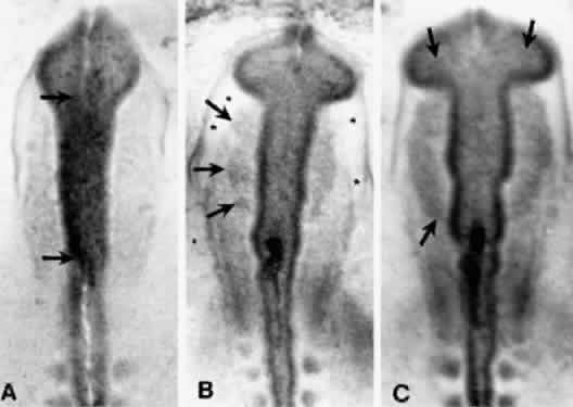
| Fig. 4. Three stages in the early migration of avian neural crest cells. These photographs were taken of living embryos; they were stained with neutral red to enhance contrast. In each case, the arrows indicate an edge of the crest population. A. Crest cells are just forming (see Fig. 1B). B. The leading edge of the crest population is beyond the dorsolateral margin of the mesencephalon. Note the appearance of the cell-free space (asterisks) between the mesoderm and the surface ectoderm. C. Crest cells are present dorsal to the site of optic stalk constricture. (Noden D: The migration and cytodifferentiation of cranial neural crest cells. In Pratt R (ed): Current Research Trends in Prenatal Cranio-Facial Development. New York, Elsevier North Holland, 1980) |