|
|
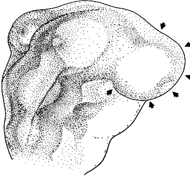
| Fig. 3. Formation of the optic vesicles as lateral bulges of the prosencephalon. This drawing is a dorsal view of the right side of an avian embryo at about 2.5 days development. The arrowheads indicate the superficial extent of the bulging right optic vesicle. |