Chapter 109
Surgical Management of Retinoblastoma
CAROL L. SHIELDS and JERRY A. SHIELDS
Main Menu   Table Of Contents

Search

GENERAL CONSIDERATIONS
ENUCLEATION
EPISCLERAL PLAQUE RADIOTHERAPY
CRYOTHERAPY
PHOTOCOAGULATION
THERMOTHERAPY
INTRAVENOUS CHEMOREDUCTION
SUBCONJUNCTIVAL CHEMOREDUCTION
ORBITAL EXENTERATION
COMBINED THERAPEUTIC MODALITIES
ASSESSMENT OF TREATMENT RESPONSE
FOLLOW-UP PROCEDURES
TRILATERAL RETINOBLASTOMA
SUMMARY
REFERENCES

GENERAL CONSIDERATIONS
The management of retinoblastoma can be remarkably complex. Each case should be individualized according to the entire clinical situation. Proper management requires the ability to use various instruments, familiarity with the disease and expected outcomes, and above all, experience in dealing with retinoblastoma-related problems.1–7 Several options are available for the treatment of retinoblastoma, and the method selected depends on the size and extent of the tumors, visual potential of the eye, laterality of involvement, and the patient's general systemic status. The methods that we currently advocate include enucleation, chemoreduction, external beam irradiation, scleral plaque irradiation, photocoagulation, cryotherapy, thermotherapy, and chemotherapy.1,8

Fine-needle aspiration biopsy has been advocated to assist in the diagnosis of certain intraocular tumors.9 Only 2% of 10,000 eyes with intraocular tumors have been managed with fine-needle aspiration biopsy, since the diagnosis of an intraocular tumor usually is based on clinical features without the need for cytologic confirmation.9 Furthermore, we do not recommend fine-needle aspiration biopsy in the diagnosis of retinoblastoma. Retinoblastoma is a loosely cohesive, friable tumor that could be disseminated locally. Therefore, we believe that needle biopsy should be avoided in cases of suspected retinoblastoma and reserved for eyes that strongly suggest a simulating disease such as Coats' disease or ocular toxocariasis, where the diagnosis of retinoblastoma is remote but should be ruled out before proceeding with therapy. In these cases, consultation with an ocular oncologist familiar with retinoblastoma and simulating conditions may avoid the need for fine-needle biopsy.

In many cases, it may be necessary to use various combinations of treatment to achieve a satisfactory result. This chapter covers the current management of retinoblastoma with emphasis on surgical techniques and laser methods. The nonsurgical aspects of retinoblastoma management, such as external beam radiotherapy, chemotherapy, and genetic counseling, are covered in recent textbooks on intraocular tumors.1,10

Back to Top
ENUCLEATION
Although several therapeutic approaches are available, enucleation is one of the most commonly used modalities in the management of retinoblastoma. However, there has been a trend recently toward using more conservative methods of treatment.11

INDICATIONS

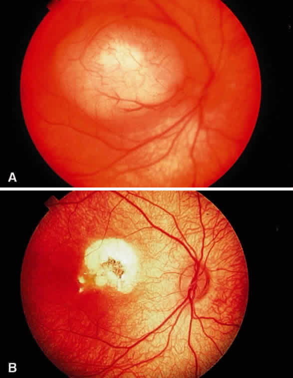
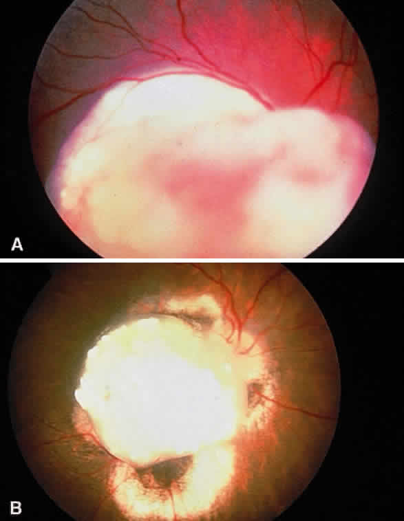
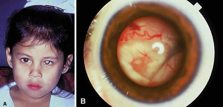
Enucleation probably is indicated for all unilateral cases in which the tumor fills most of the globe and in which there is little hope of salvaging any viable retina or useful vision (Fig. 1). If half of the retina is free from tumor, then other methods of treatment can be considered, as long as parents have been fully informed as to the possibilities of metastasis, the complications of treatment, and the risk for ultimate enucleation. Other indications for enucleation include the presence of neovascular glaucoma in an eye with retinoblastoma and the suspicion of optic nerve, choroidal, or orbital tumor extension. Seeding of retinoblastoma onto the pars plana or into the anterior chamber are important findings that often lead to enucleation.

Fig. 1. Advanced retinoblastoma requiring enucleation. A. Facial photograph demonstrating unilateral leukocoria. B. Photograph of the anterior segment revealing total retinal detachment from exophytic retinoblastoma.

In bilateral cases, the eye with the most advanced tumor traditionally has been enucleated and the less involved eye managed with irradiation or other methods. If the most advanced eye has sparing of more than half of the retina, an attempt can be made to salvage both eyes with treatment. If both eyes have far-advanced tumors and there is no hope of any vision, bilateral enucleation may be necessary. Trying chemoreduction, bilateral external beam irradiation, or both with close follow-up may be justified if the parents are fully informed and refuse bilateral enucleation.

TECHNIQUE

The technique of enucleation for retinoblastoma is slightly different from the standard enucleation performed by most ophthalmic surgeons.12,13 Since retinoblastoma is a loosely cohesive malignant tumor and the scleral wall in these young children can be thin, precautions should be taken to be extremely gentle during the procedure. A lateral canthotomy can be performed first if the child has a small, tight orbit. A peritomy is made for 360° at the limbus. Tenon's fascia is separated between the rectus muscles.

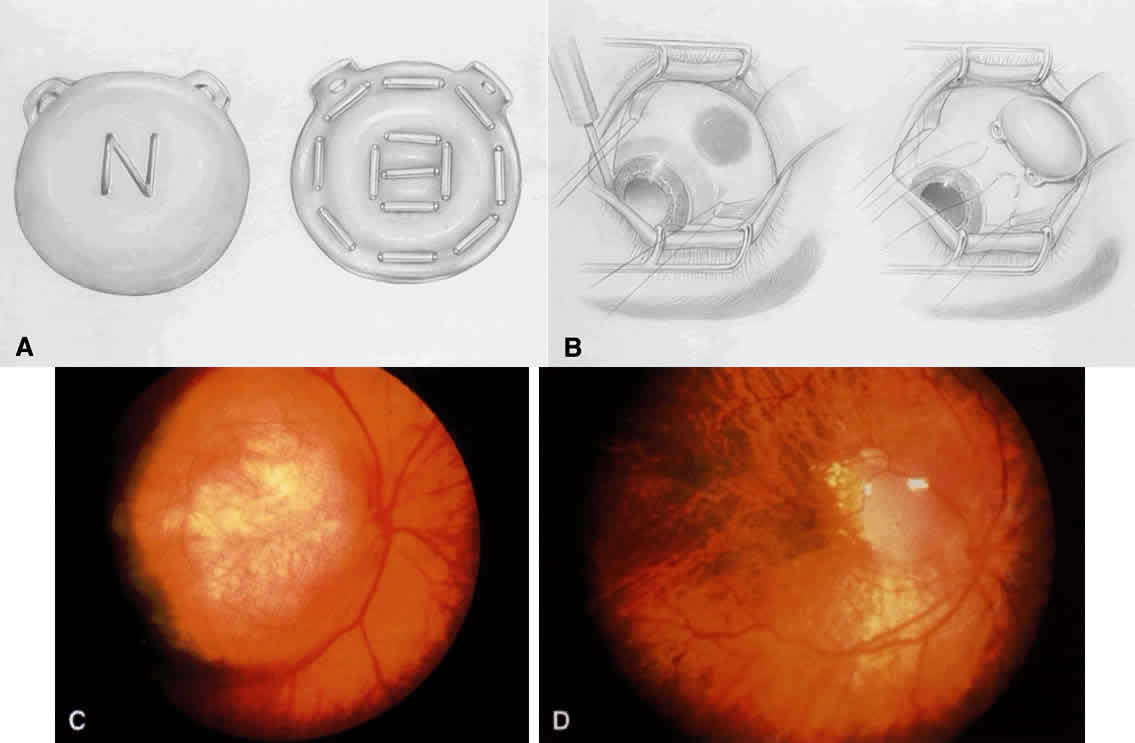
If the surgeon is not placing a motility implant, then the procedure is as follows (Fig. 2). The rectus muscles are individually gently hooked and cut with scissors near their insertion onto the sclera. The inferior and superior oblique muscle are sequentially hooked with two muscle hooks, clamped, and cut.

Fig. 2. Enucleation and placement of motility implant. A. After peritomy, the rectus muscles are tagged with vicryl suture. B. The medial rectus muscle stump is grasped, and the optic nerve is cut posteriorly in the orbit with enucleation scissors. C. The scleral wrapped motility implant is placed, and the four rectus muscles are sutured through cut windows into their anatomic positions. D. A cross-section of the orbit with a motility implant shows the attached rectus muscles. In the anterior portion of the implant is a sleeved peg system that fits into an indentation on the posterior surface of the prosthesis.

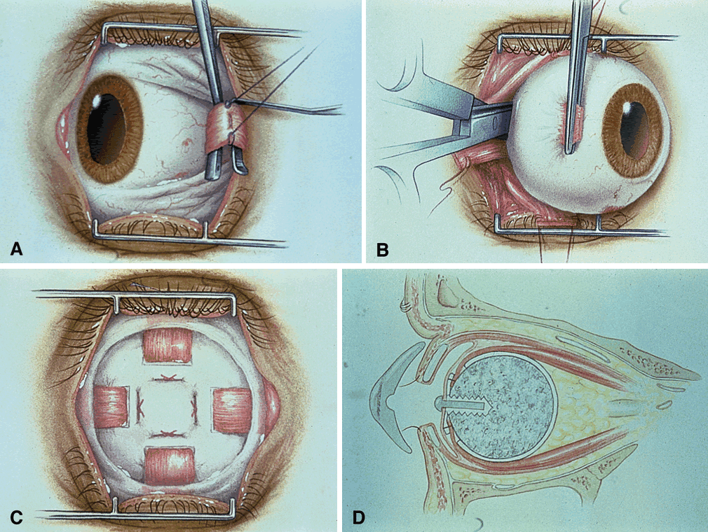
If the surgeon is placing a motility implant such as hydroxyapatite or13 polyethylene, then the technique is as follows. The rectus muscles are sequentially isolated on muscle hooks and tagged with double-armed 5-0 vicryl sutures near their insertion and cut at the insertion. The oblique muscles are isolated and cut without tagging. Orientation of the muscles and their respective sutures should be accurate.

The next step is cutting the optic nerve. Since the main route of extension of retinoblastoma is through the optic nerve and subarachnoid space, it is advisable to obtain as long a section of optic nerve as possible at the time of enucleation. This can be accomplished by placing a hemostat on the stump of the severed medial rectus muscle for traction. This permits the globe to be gently pulled forward, stretching the optic nerve when it is cut deep in the orbit. A firm grip with a hemostat provides better traction than silk sutures placed through the muscle insertions. By using this technique, the globe can be removed intact, and a long section of optic nerve generally can be obtained.

Enucleation scissors with long tips that have a slight curve are better than short scissors with a sharp curve for obtaining a sizable section of optic nerve. Snares or clamps on the optic nerve are not recommended, since they induce more trauma and can produce crush artifact in the optic nerve. This can lead to difficulty for the pathologist in differentiating meningothelial cells from crushed retinoblastoma cells.

The optic nerve is cut and the eye removed. The socket is compressed with gauze until adequate hemostasis is obtained. An appropriately sized implant measuring 18 to 20 mm in diameter is placed in posterior Tenon's capsule. If there is a standard ball implant, then the horizontal rectus muscles along with overlying Tenon's capsule are tied horizontally to each other, and the vertical rectus muscle and Tenon's capsule are then tied vertically to each other. If there is a motility implant, then the four rectus muscles are attached with anatomic orientation to the implant. The cut rectus muscles are pulled through windows that have been cut in the scleral wrap, covering the implant. Currently, we use the hydroxyapatite orbital motility implant, and it is wrapped with betadine-soaked sclera13 or prepared bovine pericardium.

Closure of the socket includes 5-0 vicryl sutures placed in an interrupted vertical fashion in the deep and superficial layer of Tenon's capsule. The conjunctiva is closed with running 5-0 vicryl sutures. A conformer is placed between the lids and the conjunctiva. Antibiotic ointment is instilled, and an absorbent pressure patch is applied for 24 hours. In 6 weeks, the patient visits an ocularist for permanent prosthesis fitting.

Motility implants can provide remarkable prosthesis movement. In some cases, a peg can be placed into the motility implant to improve coupling of the movement of orbital implant with the prosthesis. The peg placement generally is delayed for at least 6 months after enucleation to allow adequate fibrovascularization of the implant. In children with retinoblastoma, we generally postpone the peg placement until the teen-age years, when the patient is more cooperative and the procedure can be performed under local anesthesia.

Some authors recommend that frozen sections of the optic nerve be performed at the time of enucleation to detect tumor extension.14 Since posterior optic nerve extension is rare in our experience and since histopathologic interpretation can be difficult, we do not routinely perform frozen sections in such cases. However, it seems reasonable to consider frozen sections in cases where optic nerve extension is strongly suspected.

COMPLICATIONS

There are few complications of enucleation.15 Hemorrhage at the time of surgery may be controlled with orbital compression. Postoperative ecchymosis and edema of the eyelids usually subside with use of a pressure patch and ice compresses postoperatively. Chemosis of the conjunctiva may occur and cause the conformer to temporarily protrude or become displaced. This problem is remedied by pressure patching and topical ointment. Patients who are receiving chemotherapy may develop socket infection, which can be managed with appropriate antibiotic therapy. Long-term complications include ptosis, orbital fat atrophy, or superior sulcus fat atrophy. These problems can be managed by adjustment of the prosthesis by the ocularist or by reconstructive plastic surgery of the eyelid or orbit.

Motility implants pose a higher risk for postoperative problems such as conjunctival thinning, implant exposure, implant infection, and implant extrusion.15–17 The hydroxyapatite peg system can extrude and lead to granulation tissue.18 However, careful attention to the surgical placement of the hydroxyapatite-integrated implant minimizes complications.15

RESULTS

The cosmetic results of enucleation for retinoblastoma generally are excellent. If the child had external beam radiotherapy in addition to enucleation, then the cosmetic result often is less satisfactory, related to radiation-induced orbital fat atrophy and a sunken appearance to the prosthesis, as well as decreased tear production with chronic discharge mucous drying on the prosthesis. The use of the integrated hydroxyapatite implant with rectus muscles attached improves the motility of the prosthesis.

HANDLING OF THE GLOBE AFTER ENUCLEATION

Retinoblastoma research, including genetic DNA analysis, requires fresh tumor tissue. The ophthalmologist who performs enucleation for retinoblastoma should be familiar with a safe technique to harvest adequate tissue for research within minutes after enucleation and still provide good histopathologic sections.12 Fresh tumor tissue or other ocular tissue should be harvested immediately after enucleation before cytolysis occurs. This technique requires close communication and cooperation with the researchers and the ophthalmic pathologist.

Immediately after the eye is enucleated, a piece of optic nerve is transected, and the proximal portion (toward the brain) of the optic nerve is marked with an indelible marking pencil and placed in 10% buffered formalin. This should be done immediately, before the globe is manipulated or opened. The pathologist is instructed to study the first sections through the proximal end of the nerve looking for tumor extension.

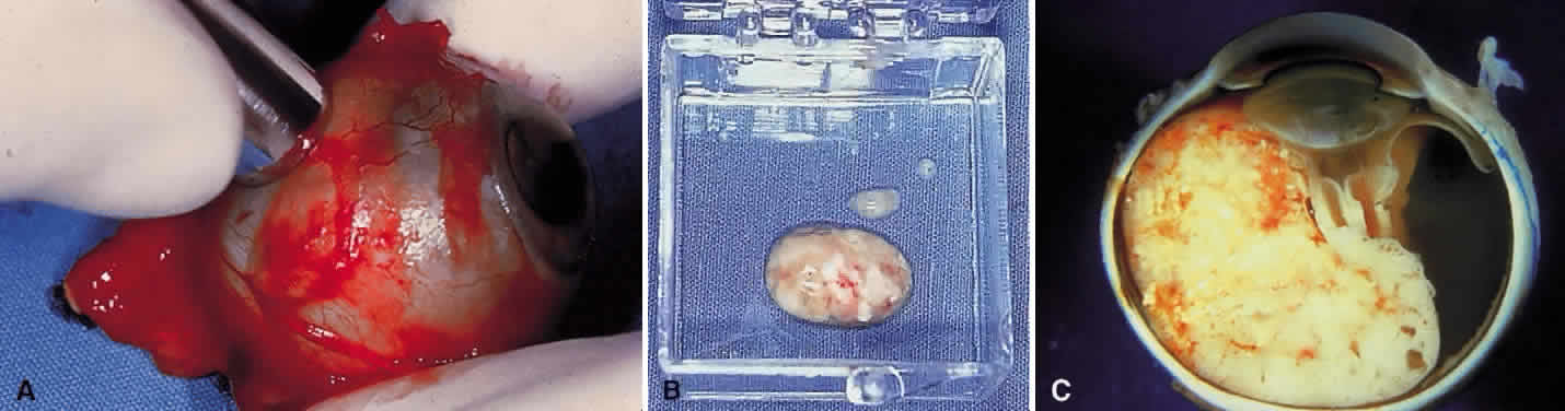
The area of the base of the tumor then is accurately marked on the sclera, and a scleral window is outlined to slightly overlap the tumor margin. A scleral window measuring 8 mm in diameter then is cut with a trephine (Fig. 3). The scleral opening should reveal typical soft white tumor tissue, which is partially removed with scissors or curretted without spillage and placed in tissue culture or submitted fresh. The tissue is immediately taken to the genetic research laboratory and prepared for DNA analysis or other studies. The sclera generally retains its spherical shape without collapse. The opened globe then is gently immersed in 10% formalin and submitted for routine histopathologic studies. It is important that the window be almost 90° from the center of the tumor base. This allows the pathologist to later make a pupillary-optic nerve section through the center of the tumor, avoiding the scleral window, which is left in the minor calotte. If done properly, the quality of the histopathologic sections will be excellent. The tray, instruments, and gloves used to harvest fresh tissue are contaminated with tumor and should be properly disposed.

Fig. 3. Fresh tissue harvesting. A. Fresh tissue on a separate tray for DNA analysis. B. Fresh tissue in a container to be immediately sent to the research laboratory. C. Photograph of the gross sectioned globe with large exophytic retinoblastoma.

Back to Top
EPISCLERAL PLAQUE RADIOTHERAPY
For years, various techniques of external beam radiotherapy represented the only available method of irradiation for retinoblastoma.19–21 It is still one of the favored methods for advanced tumors involving the entire eye, especially when there is extensive vitreous seeding. Because of the concern for the development of related second cancers after external radiotherapy and, less importantly, cosmetic problems,22,23 episcleral radioactive plaques have been used increasingly. These serious problems are much less common with plaque treatment. We believe that we achieve similar results with fewer complications with an iodine 125 plaque than with external beam irradiation in properly selected cases.

INDICATIONS

Relative indications for a radioactive plaque include a retinoblastoma that is less than 15 mm in diameter and 9 mm in thickness. Custom-designed plaques with proper shielding are essential for tumors near the optic disc. This treatment can be used for both unilateral and bilateral cases. Plaque treatment can be repeated on a single eye to retreat the same tumor or treat one at a new site. It can be used when mild to moderate vitreous seeding is present over the tumor. Recurrent or residual tumors that have been uncontrolled with external beam irradiation, photocoagulation, thermotherapy, chemothermotherapy, or cryotherapy may be treated by plaque radiotherapy.

TECHNIQUE

The ocular oncologist must first document the size and extent of the tumors and associated findings such as subretinal fluid, subretinal seeds, and vitreous seeds with clinical drawings and ultrasonography. Along with an experienced radiation oncologist who is familiar with retinoblastoma and plaque radiotherapy, the plaque isotope, size, configuration, seed distribution, dose, and dose rate are prescribed (Fig. 4). We have used several radioisotopes, including cobalt 60, ruthenium 106, iridium 192, and iodine 125. Currently, we generally use only iodine 125 radioactive seeds embedded into a gold-shielded plaque because of the facility for custom design and shielding with this low-dose gamma radiation device. The full radiation dose is delivered over an average of 2 days with the child kept in the hospital for radiation precautions.

Fig. 4. Plaque radiotherapy. A. Drawing of a plaque showing the gold shield on the outer surface (left) and the embedded radioactive iodine seeds on the inner surface (right). B. Localization of the intraocular tumor by transillumination (left) and placement of the episcleral sutures for the plaque (right). C. Macular retinoblastoma in a 3-month-old boy before plaque treatment. D. Regressed macular retinoblastoma 2 years after plaque radiotherapy. Notice the healthy optic disc.

The surgical technique of radioactive plaque application involves a conjunctival peritomy and exposure of the sclera in the area of the tumor. Transillumination and indirect ophthalmoscopy with gentle scleral indentation then are used to localize and mark the margins of the tumor on the sclera (see Fig. 4). A dummy plaque is placed on the sclera overlying the tumor, and nonabsorbable scleral sutures are placed through the superficial sclera in alignment with the holes in the arms of the plaque. The dummy plaque then is removed, and the active plaque is carefully is grasped with forceps and placed on the sclera in correct alignment with the tumor. The active plaque then is secured to the sclera in this position by tying the preplaced sutures through the holes in the arms of the plaque.

During the plaque application, standard irradiation precautions are followed. The plaque is surgically removed after the appropriate dose has been delivered. For retinoblastoma, the usual dose is 3500 to 4500 cGy to the tumor apex. Results of plaque therapy for selected cases of retinoblastoma are encouraging.24–29

COMPLICATIONS

The potential complications of plaque therapy, such as cataract and radiation retinopathy, are the same as those occurring with external beam irradiation. In our 25 years of experience with plaque radiotherapy, we have not witnessed a secondary cancer directly related to a solitary radioactive plaque. The calculated radiation dose to the orbit is far less with a shielded plaque (less than 200 cGy) than with external beam radiation (4000 cGy). As more follow-up data become available, such complications may become apparent.

RESULTS

Most tumors show a dramatic response to irradiation within the first 4 weeks after removal of the plaque. The regression patterns that are noted are similar to those seen with external beam irradiation. A successfully irradiated retinoblastoma usually appears as a shrunken white mass that resembles cottage cheese. There may be pigmentary alterations and scar tissue around the regressed tumor.

We have reported our preliminary results with episcleral plaque radiotherapy for retinoblastoma.24–28 In an evaluation of 103 consecutive patients with retinoblastoma treated by solitary plaque application, local tumor control rate was 87%.25 The average tumor had a 7-mm base and 4-mm thickness. The median dose to the tumor apex was 4000 cGy and to the tumor base was 15,000 cGy, delivered over a mean duration of 65 hours. In 30% of cases, plaque radiotherapy was the primary treatment, and in 70% of cases, it was used as a secondary treatment after failure of other methods, most often failure of external beam radiotherapy.25 In the cases where plaque was used as a secondary treatment, the eyes were otherwise destined for enucleation but salvaged with plaque treatment in nearly 90% of cases.26 The visual outcome varied with tumor size and location, as well as radiation problems of retinopathy and papillopathy. The visual outcome was good in 62% of cases, and the measured vision was 20/20 to 20/30 in over half of the cases.25 On the basis of these studies, we conclude that plaque radiotherapy can be used successfully as a primary treatment of selected cases of unilateral or bilateral retinoblastoma or as a supplemental treatment after other treatment methods fail.24–28

Back to Top
CRYOTHERAPY
Cryotherapy is an effective method of treating selected small retinoblastomas.30,31 Cryotherapy has the advantage of preserving the internal limiting membrane of the retina, which may be a natural barrier to the spread of tumor cells into the vitreous.

INDICATIONS

Cryotherapy may be used as the primary treatment for small peripheral retinoblastomas that are located between the equator and ora serrata. It also is used to treat residual or recurrent tumors in the peripheral fundus after incomplete eradication with external beam irradiation. Such recurrences usually occur near the ora serrata and probably occur secondary to incomplete irradiation of the most anterior portions of the retina. We believe that cryotherapy is not appropriate if there is seeding of tumor cells into the overlying vitreous cavity. With the advent of chemoreduction (discussed later), cryotherapy is used on normal peripheral retinal tissue to induce exudation of intravenous chemotherapy into the eye.

TECHNIQUE

Cryotherapy for retinoblastoma should be administered by the triple freeze-thaw technique. Using careful indirect ophthalmoscopy, with the cryoprobe used as a scleral depressor, the tumor is elevated on the tip, and freezing is applied until the surrounding retina turns white and ice crystals appear in the overlying vitreous (Fig. 5). After adequate freezing of the entire mass, the tumor is allowed to thaw. Then, it is refrozen immediately, and the sequence is repeated. Three successive freeze-thaw applications usually are adequate. It may be necessary to repeat the treatment in 3 to 4 weeks if there is still ophthalmoscopic evidence of viable tumor.

Fig. 5. Cryotherapy. A. Small retinoblastoma on the tip of a cryoprobe. B. Appearance of retinoblastoma during freezing phase. C. Appearance of retinoblastoma after cryotherapy, displaying flat atrophic scar.

COMPLICATIONS

There are few major complications of cryotherapy for retinoblastoma. All patients develop transient conjunctival chemosis and often mild eyelid edema. These findings resolve over a few days. Local vitreous hemorrhage or transient localized retinal detachment (ablatio fugax) may occur at the time of treatment, but this usually resolves within a few weeks to months. Pigment migration and proliferation secondary to cryotherapy usually do not cause visual problems. In rare cases, cryotherapy may stimulate vitreoretinal traction, leading to a direct or contracoup retinal tear.32 Excessive cryotherapy to the ciliary body region, however, could lead to possible ciliary body shutdown, ocular hypotension, or rarely, to phthisis bulbi.

RESULTS

We have reported our results of treatment of 67 retinoblastomas in 47 eyes of 45 patients that were treated with triple freeze-thaw cryotherapy.30 Tumor destruction was achieved with one or more cryotherapy applications in all cases in which the tumor was no greater than 2.5 mm in diameter and 1.0 mm in thickness, and in which the tumor was confined to the sensory retina without seeding into the adjacent vitreous. Cryotherapy appears to be generally successful for tumors up to 3.5 mm in diameter and 2.0 mm in thickness, but more than one treatment may be necessary.

Back to Top
PHOTOCOAGULATION
Photocoagulation can be used for selected small retinoblastomas.33–35 It may be used as primary treatment in some patients or as supplementary treatment in patients who were initially treated with irradiation or cryotherapy. Xenon arc photocoagulation was the first photocoagulator used, but its size and operation were cumbersome. Recently, we have found that the indirect ophthalmoscope laser delivery system using argon or diode is adequate to treat small retinoblastomas. When it is administered properly, photocoagulation has fewer complications than irradiation.

INDICATIONS

Photocoagulation is indicated for small tumors confined to the retina that do not involve the optic disc or the macula. It is probably contraindicated if there is ophthalmoscopic evidence of vitreous seeding, choroidal invasion, or involvement of the fovea, optic disc, or pars plana. This technique does not eliminate tumor cells in the vitreous. It probably would not destroy tumor cells in the choroid and possibly could promote dissemination of the tumor in such cases. If it is used on the optic disc or fovea, photocoagulation would result in marked visual loss.

TECHNIQUE

Two rows of confluent burns are placed around the tumor using enough power to obtain a whitening of the surrounding retina and closure of the retinal vessels that supply the tumor (Fig. 6). The treatment should be only heavy enough to obliterate the retinal vessels. Within a few weeks, the tumor should regress into a flat or excavated scar. It may be necessary to apply a second and third treatment in some cases.

Fig. 6. Laser photocoagulation. A. Small retinoblastoma near the fovea inferiorly. B. Immediately after delimiting argon laser photocoagulation, the treated retina appears white. C. Several months later, the tumor scar is flat.

COMPLICATIONS

There are few complications of properly administered photocoagulation for retinoblastoma. Vitreoretinal traction may lead to macular pucker or retinal detachment if treatment is heavy. A transient, localized retinal detachment (ablatio fugax) may occur, but it usually resolves in a few months. If Bruch's membrane is ruptured by heavy photocoagulation, choroidovitreal neovascularization may occur. Atrophic retinal tear at the site of laser photocoagulation can occur and may lead to rhegmatogenous retinal detachment.32 Minor complications such as small retinal hemorrhages at the time of treatment usually do not produce serious consequences.

RESULTS

We have reported our results of 45 retinoblastomas treated with xenon photocoagulation.33 Photocoagulation alone was successful in eradicating 76% of the tumors, whereas in 24% of the tumors, supplemental treatment with other modalities was necessary. In cases where the tumor was less than or equal to 3.0 mm in diameter and 2.0 mm in thickness and was confined to the sensory retina without vitreous seeding, tumor destruction usually was achieved with photocoagulation. We later studied our results of indirect argon laser photocoagulation of 30 retinoblastomas.34 The average tumor size was 2-mm base and 1-mm thickness. Using a mean of 350 mW of continuous laser, the tumors were controlled in 70% of cases. Complications were few, with foveal distortion in three eyes from treatment of tumors in the macula.

Back to Top
THERMOTHERAPY
Thermotherapy is a method of delivering heat to the eye using ultrasound, microwaves, or infrared radiation. The heat can be delivered to the whole eye,36 or it can be focused on one portion of the eye.37 We generally use focused heat delivered directly to intraocular retinoblastoma from an infrared radiation device. The goal is to deliver a temperature of 42° to 60°C, a temperature that is below the coagulative threshold. In its upper levels, heat can cause direct cell death, and in the lower levels, heat works synergistically with radiation and chemotherapy to induce cell death. The combination of heat and chemotherapy is termed chemothermotherapy and the combination of heat and radiation is termed thermoradiotherapy.

INDICATIONS

Thermotherapy generally is reserved for small retinoblastomas measuring up to 3 mm in thickness with no vitreous seeds and no subretinal fluid. Larger tumors or those with mild vitreous seeding or subretinal fluid usually are treated initially with chemoreduction and then followed with thermotherapy, a technique termed chemothermotherapy.

TECHNIQUE

The technique of thermotherapy involves the application of infrared radiation to heat tumor cells to 50° to 60°C. The energy is delivered through the pupil (transpupillary) or the sclera (transcleral), depending on the tumor size and location. The tumor is treated directly on its surface, and over a period of 5 to 30 minutes, the mass assumes a gray-white opaque appearance, indicating adequate treatment. The treatment is repeated on a monthly basis until complete regression of the tumor is obtained.

For slightly larger tumors, thermotherapy is coupled with chemotherapy for better control using special protocols.37–39 In an early protocol, there were two parts comprising the treatment: part A (chemothermotherapy) and part B (thermotherapy alone).39 Both parts A and B were delivered for the completion of one cycle within 1 week. Part A involved the administration of systemic intravenous carboplatin followed within 4 hours by thermotherapy to the retinoblastoma for 20 to 30 minutes delivered by an infrared radiation device through the operating microscope. Part B, occurring within 4 to 7 days of part A, provided delivery of thermotherapy to the tumor without adjuvant chemotherapy. The tumors best suited for this treatment were posterior pole tumors within 6 mm of the fovea and optic disc, 6 mm or less in base and 3 mm or less in thickness. Two or three complete cycles were necessary for tumor regression.

We use a different technique that is practical.37,40,41 For patients on a chemoreduction protocol, we allow one cycle of chemoreduction to shrink the tumors, and then we treat the regressed mass with thermotherapy coupled with each subsequent cycle up to six cycles. The thermotherapy is delivered within 4 hours of the same day of chemotherapy delivery, thereby achieving the benefit of chemotherapy for both tumor reduction and consolidation37 (Fig. 7). Under general anesthesia, thermotherapy is delivered for 5 to 20 minutes per tumor. Each tumor is individually heated until a sufficient color change is visualized, indicating adequate treatment. Steroid ointment is applied to the eye, and patching is not necessary.

Fig. 7. Thermotherapy. A. Macular retinoblastoma in an 18-day-old girl. B. After chemoreduction and coupled thermotherapy, the tumor regressed to a calcified scar.

COMPLICATIONS

In a study of 188 retinoblastomas treated with thermotherapy and chemothermotherapy, the complications included mild focal iris atrophy in 64%, peripheral focal lens opacity in 24%, retinal traction in 5%, retinal vascular obstruction in 2%, and ablatio fugax 2%.37 There were no cases of corneal scarring, central lens opacity, iris or retinal neovascularization, or rhegmatogenous retinal detachment. More treatment sessions and larger tumor base were factors predictive of iris atrophy.37

RESULTS

Thermotherapy, especially chemothermotherapy, has been found to be successful for treating small and medium retinoblastomas, achieving complete tumor control in 86% of tumors.37 It is important to be selective when identifying tumors suitable for this treatment. In our experience, small tumors having a 4-mm base and 2-mm thickness without subretinal fluid show the best response. Thermotherapy is especially suited for small tumors adjacent to the fovea and optic nerve, where radiation or laser photocoagulation possibly would induce more profound visual loss. It is a time-intensive, tedious process and requires many advanced facilities to work cooperatively.

Back to Top
INTRAVENOUS CHEMOREDUCTION
Chemoreduction is a method of reducing tumor volume using chemotherapy to allow for more focused, less damaging therapeutic measures. It has evolved to be an important measure in the initial management of retinoblastoma.42,43

INDICATIONS

Chemoreduction is reserved for eyes with advanced retinoblastoma that otherwise would require enucleation or external beam radiotherapy. In addition, it is used for eyes with medium and small retinoblastoma in which tumor shrinkage would provide much improved visual outcome. If a patient has retinoblastoma that can be adequately treated with laser photocoagulation, thermotherapy, cryotherapy, or plaque radiotherapy, then chemoreduction should not be used.

TECHNIQUE

The technique of chemoreduction involves intravenous route of delivery of chemotherapy agents. In special instances, the chemotherapy is delivered locally by the subconjunctival route. The chemotherapy agents vary, depending on the preference of the pediatric oncologist. For intravenous chemotherapy, we currently use carboplatin, etoposide, and vincristine for six cycles.44 Focal therapy to the individual regressed tumors is delivered at cycle 2 through cycle 6. The preferred focal therapy includes one or more of the following methods: laser photocoagulation, thermotherapy, cryotherapy, and plaque radiotherapy.44

COMPLICATIONS

The complications of chemoreduction include transient bone marrow suppression with a risk for infection, hair loss, hearing loss, and renal toxicity. There is concern for the risk of induction of second cancers, but this is predicted to be low because of the low-dose, short-term treatment.43

RESULTS

Overall, chemoreduction is an effective initial measure for treatment of retinoblastoma. Notice that definitive focal therapy is necessary once tumor reduction is achieved (Fig. 8). The ocular salvage rate has improved with chemoreduction.44,45 In the past, the ocular salvage rate in advanced retinoblastoma treated with external beam radiotherapy alone was 30%, whereas the ocular salvage rate in eyes with treated chemoreduction before external beam radiotherapy was nearly 70%.45

Fig. 8. Chemoreduction. A. Juxtapapillary retinoblastoma overhanging the optic disc and involving the foveola. B. After chemoreduction, the tumor regressed to a calcified scar, and thermotherapy was applied.

Data from our department report similar promising results with chemoreduction for retinoblastoma.43,44 Overall, we found a mean decrease of 35% in tumor base and nearly 50% in tumor thickness with chemoreduction.43 Subretinal fluid resolved in 76% of cases that had total retinal detachment, and both vitreous and subretinal seeds showed regression with the treatment.46

It remains uncertain which eye or patient should be submitted to chemoreduction. In Reese-Ellsworth groups 1, 2, 3, and 4, we have been successful in saving most eyes using chemoreduction combined with adjuvant methods.44 However, in Reese-Ellsworth group 5 eyes, the risk for enucleation increases.44 In eyes with Reese-Ellsworth group 5 retinoblastoma, we found that complete tumor control occurred in over 70% using chemoreduction and carefully applied adjuvant treatment.47 However, this treatment is tedious and requires careful follow-up.

Back to Top
SUBCONJUNCTIVAL CHEMOREDUCTION
There is increasing interest in local delivery of chemoreduction for intraocular retinoblastoma. It has been shown in animal models that carboplatin penetrates the sclera into the vitreous cavity, allowing for effective doses within the eye with minimal toxicity.48–50 We have used local subconjunctival injection of carboplatin under protocol in humans as both a secondary treatment and a primary treatment. Within 3 to 4 weeks, there usually is tumor response with regression, but the response may not be lasting, thereby requiring consolidation with other more lasting methods. Further investigation into the methods and results of local chemotherapy delivery for intraocular retinoblastoma are underway.
Back to Top
ORBITAL EXENTERATION
Orbital exenteration is an aggressive surgical technique reserved for certain primary orbital malignancies or malignant tumors that demonstrate orbital invasion and show insufficient response to chemotherapy and radiotherapy.51,52 Rarely is exenteration necessary for the management of retinoblastoma in the United States. However, in less developed countries, exenteration is used more frequently as patients present at a more advanced stage with orbital involvement.

INDICATIONS

Exenteration for retinoblastoma is indicated if there is macroscopic orbital invasion or orbital recurrence of retinoblastoma that is progressing despite irradiation and chemotherapy. Microscopic invasion of the orbit, found on histopathologic evaluation of the globe, should be treated by orbital radiotherapy and systemic chemotherapy. In addition, choroidal and optic nerve invasion of retinoblastoma generally is treated with systemic chemotherapy, and in certain instances, external beam radiotherapy.53–55 Exenteration is reserved for advanced tumor in the orbit, which is resistant to conservative treatment.

TECHNIQUE

The techniques of orbital exenteration are discussed in recent textbooks and are briefly considered here.51,52,56 The technique of orbital exenteration varies, but it usually involves the removal of all of the orbital content within the periosteum. If the eyelids are not involved by tumor, then they can be preserved with the “eyelid-sparing” technique. Under general anesthesia, a skin incision is made 2 mm above the upper eyelid margin and 2 mm below the lower eyelid margin to connect at the medial and lateral canthus. A skin-muscle flap is created within the eyelids up to the orbital rim. The periosteum is incised 2 mm outside of the orbital rim. The periosteum is elevated off of the orbital bone for 360° at the rim and back to the orbital apex. A clamp is placed at the eyelid margin for retraction, and the contents of the orbit are lifted out of the bony socket after the apex is cut. Thrombin-soaked gauze is used to temporarily pack the orbital socket. After orbital hemostasis, the eyelids are sewn together, and this skin-muscle complex retracts and lines the orbital bones, providing a concave, cutaneous bed for the prosthesis.

COMPLICATIONS

Complications of exenteration include orbital hematoma, serous fluid collection, infection from the adjacent sinus, dural wound leak at the apex, and wound dehiscence. These usually occur within the first 48 hours after surgery, therefore early follow-up is important.

RESULTS

Most patients who have the eyelid-sparing exenteration heal within the first 2 weeks from surgery and are fitted with an orbital prosthesis about 1 month from the time of surgery. The healing from the eyelid-sparing technique is much faster and more complete than the eyelid removal technique, which generally takes up to 2 or more years to granulate adequately.51

Back to Top
COMBINED THERAPEUTIC MODALITIES
The management of retinoblastoma can be challenging, and the physician must often make difficult decisions regarding the use of various treatment modalities. The clinician should be able to recognize residual tumor from recurrent tumor and anticipate the most appropriate supplemental treatment method. This generally requires considerable training and experience.

If the tumor is unilateral and large, enucleation or chemoreduction plus adjuvant focal treatments are options. If the histopathologic findings in the enucleated eye show no sign of optic nerve or choroidal invasion, then systemic chemotherapy is not provided. However, if the histopathologic study reveals choroidal or optic nerve invasion,53,54 suggesting a poor prognosis, then chemotherapy is advised. If there is extensive invasion of the optic nerve or orbital involvement, then supplementary chemotherapy and irradiation to the anophthalmic socket are indicated.55 Usually, 3500 to 4000 cGy in divided doses over a 4 to 6 weeks is adequate.

In bilateral cases in which attempts are being made to save the remaining eye, combined treatment may be more complex. After chemoreduction, external beam radiotherapy, or scleral plaque irradiation, a tumor may remain viable, as suggested by persistent dilated retinal blood vessels feeding the tumor or enlarging mass. Supplementary photocoagulation, thermotherapy, or repeat plaque radiotherapy may be necessary in such cases to eradicate the residual viable tumor.

If the tumor recurs as a small growth near the ora serrata, supplementary transcleral thermotherapy or freeze-thaw cryotherapy may be used. If such a peripheral recurrence is greater than 3 mm in elevation and has vitreous seeding, a supplementary radioactive plaque may be applied.

Back to Top
ASSESSMENT OF TREATMENT RESPONSE
It is important to be able to determine the response to treatment using the methods previously outlined. For eyes that have been enucleated, the prosthesis should be removed on each subsequent visit and the anophthalmic socket inspected and palpated for recurrent orbital tumor. For eyes not enucleated, careful indirect ophthalmoscopic examination is the best method of determining the status of the remaining or residual tumors and for recognizing radiation retinopathy or other complications. Ultrasonography can be used to document the decrease in tumor size after treatment.
Back to Top
FOLLOW-UP PROCEDURES
After the initial treatment, a follow-up plan should be devised for each patient, depending on the clinical and pathologic findings. In general, patients with unilateral sporadic retinoblastoma who are treated by enucleation should be evaluated periodically as follows:
  1. Examination under anesthesia every 3 months until 1 year of age
  2. Examination under anesthesia every 4 months between the ages of 1 and 3 years
  3. Examination under anesthesia every 6 months between the ages of 3 and 5 years
  4. Examination yearly in the office without anesthesia after 5 years of age

Children being treated by methods other than enucleation, whether they have unilateral or bilateral disease, should be followed more closely. The child should be examined under anesthesia 1 month after photocoagulation, cryotherapy, or thermotherapy or after the termination of a course of irradiation. Children on chemotherapy protocols should be examined under anesthesia monthly until control is achieved and the chemotherapy is discontinued. The frequency of subsequent examinations depends on the response to previous treatment and varies from patient to patient. Each examination should concentrate on inspection and palpation of the anophthalmic socket and indirect ophthalmoscopic examination of the remaining eyes, both for evidence of new tumors and to evaluate the effect of treatment on known tumors.

Back to Top
TRILATERAL RETINOBLASTOMA
Trilateral retinoblastoma is a term used to describe the association of bilateral retinoblastoma and neuroblastic tumor in the pineal gland or other midline structures. A report from our department reveals that this tumor occurs in children 4 years of age or younger, often with a fatal outcome.57 Overall, trilateral retinoblastoma occurs in 3% of all children with retinoblastoma and in up to 8% of those with bilateral disease.57 Magnetic resonance imaging or computed tomography are essential in making the diagnosis. The disease is highly fatal despite aggressive treatment with chemotherapy, radiation therapy, or gamma knife therapy.58 Longer survival has been correlated with earlier tumor diagnosis in asymptomatic patients. We advise that brain magnetic resonance imaging or computed tomography be performed twice a year in all children with retinoblastoma, most importantly, in those with bilateral disease until 5 years of age. Trilateral retinoblastoma is a major cause of mortality in children within the first 5 years after diagnosis of bilateral retinoblastoma.59 Of the nearly 20 patients we have managed with pinealoblastoma over the last decades, 2 have survived for up to 4 years with aggressive chemotherapy. On the other hand, we have recently observed no cases of pinealoblastoma in 147 children treated with initial chemoreduction followed for 1 to 4 years. Although follow-up is limited, it is tempting to speculate that chemoreduction may reduce the risk for the development of pinealoblastoma.
Back to Top
SUMMARY
The management of retinoblastoma has evolved tremendously over the last century, and there is a recent trend toward focal conservative treatments. This results primarily from earlier detection of the disease, when the tumors are in a smaller stage of development, as well as advanced, more focused treatment modalities. Enucleation still is used for advanced retinoblastoma, especially when there may be invasion of the optic nerve, choroid, or orbit. The hydroxyapatite implant has provided improved cosmetic rehabilitation of the socket after enucleation. External beam radiotherapy continues to be an important method of treating advanced retinoblastoma, especially when there is diffuse vitreous seeding. Plaque radiotherapy is a useful tool for controlling medium or small retinoblastomas, especially tumors with focal vitreous seeding or those that recur after failure of other methods. Cryotherapy and photocoagulation provide excellent control of small tumors, and advanced laser delivery systems have improved the visualization and ease of treatment of retinoblastoma. Thermotherapy and chemothermotherapy are the newest focal methods that are showing promising results for small to medium-sized retinoblastoma. Chemoreduction, a technique using chemotherapy to a reduce tumor volume, allowing for focal consolidation treatment, has allowed many children with advanced retinoblastoma to avoid external beam radiotherapy and enucleation. Chemoreduction combined with cryotherapy, thermotherapy, and plaque radiotherapy plays an important role in the management of many children with retinoblastoma.
Back to Top
REFERENCES

1. Shields JA, Shields CL: Management and prognosis of retinoblastoma. In Shields JA, Shields CL (eds): Intraocular Tumors: A Text and Atlas. Philadelphia, WB Saunders, pp 377–391, 1992

2. Shields JA, Shields CL, Donoso LA, Lieb WE: Changing concepts in the management of retinoblastoma. Ophthalmic Surg 21:72, 1990

3. Ellsworth RM: The practical management of retinoblastoma. Trans Am Ophthalmol Soc 67:462, 1969

4. Shields JA, Shields CL: Current management of retinoblastoma. Mayo Clin Proc 69:50, 1994

5. Abramson DH, Niksarli K, Ellsworth, Servodidio CA: Changing trends in the management of retinoblastoma: 1951-1965 vs 1966-1980. J Pediatr Ophthalmol Strabismus 31:32, 1994

6. Dudgeon J: Retinoblastoma: Trends in conservative management. Br J Ophthalmol 79:104, 1995

7. Shields JA: The 1992 Paul Henkind Memorial Lecture: Misconceptions and techniques in the management of retinoblastoma. Retina 12:320, 1992

8. Shields CL, Shields JA, De Potter P: Newer treatment modalities for retinoblastoma. Curr Opin Ophthalmol 7:20, 1996

9. Shields JA, Shields CL, Ehya H et al: The 1992 Urwick Lecture: Fine needle aspiration biopsy of suspected intraocular tumors. Ophthalmology 100:1677, 1993

10. Shields JA, Shields CL: Atlas of Intraocular Tumors. Philadelphia, Lippincott Williams & Wilkins, 1999:207–242

11. Shields JA, Shields CL, Sivalingam V: Decreasing frequency of enucleation in patients with retinoblastoma. Am J Ophthalmol 108:185, 1989

12. Shields JA, Shields CL, De Potter P: Enucleation technique for children with retinoblastoma. J Ped Ophthalmol Strabismus 29:213, 1992

13. Shields CL, Shields JA, DePotter P: Hydroxyapatite orbital implant after enucleation: Experience with initial 100 consecutive cases. Arch Ophthalmol 110:333, 1992

14. Karcioglu ZA, Haik BG, Gordon RA: Frozen section of the optic nerve in retinoblastoma surgery. Ophthalmology 95:674, 1988

15. Shields CL, Shields JA, Singh AD, et al: Lack of complications of the hydroxyapatite orbital implant in 250 consecutive cases. Trans Am Ophthalmol Soc 91:177, 1993

16. Kim YD, Golberg RA, Shorr N, Steinsapir KD: Management of exposed hydroxyapatite orbital implants. Opthalomology 101:1709, 1994

17. Goldberg RA, Holds JB, Ebrahimpour J: Exposed hydroxyapatite orbital implants: Report of six cases. Opthalomology 99:831, 1992

18. Edelstein C, Shields CL, De Potter P, Shields JA: Complications of motility peg placement for the hydroxyapatite orbital implant. Ophthalmology 104:1616, 1997

19. Hungerford JL, Toma NMG, Plowman PN, Kingston JE: External beam radiotherapy for retinoblastoma. I. Whole eye technique. Br J Ophthalmol 79:109, 1995

20. Toma NMG, Hungerford JL, Plowman PN et al: External beam radiotherapy for retinoblastoma. II. Lens sparing technique. Br J Ophthalmol 79:112, 1995

21. Singh AD, Garway-Heath D, Love S et al: Relationship of regression pattern to recurrence in retinoblastoma. Br J Ophthalmol 77:12, 1993

22. Roarty JD, McLean IW, Zimmerman LE: Incidence of second neoplasms in patients with retinoblastoma. Ophthalmology 95:1583, 1988

23. Eng C, Li FP, Abramson DH et al: Mortality from second tumors among long-term survivors of retinoblastoma. J Natl Cancer Inst 85:1121, 1993

24. Shields JA, Giblin ME, Shields CL et al: Episcleral plaque radiotherapy for retinoblastoma. Ophthalmology 96:530, 1989

25. Shields CL, Shields JA, Minelli S et al: Plaque radiotherapy in the management of retinoblastoma: Use as a primary and secondary treatment. Ophthalmology 100:216, 1993

26. Shields JA, Shields CL, Hernandez JC et al: Plaque radiotherapy for residual or recurrent retinoblastoma in 91 cases. J Pediatr Ophthalmol Strabismus 31:242, 1994

27. Shields CL, Shields JA, Minelli S et al: Regression of retinoblastoma after plaque radiotherapy. Am J Ophthalmol 115:181, 1993

28. Hernandez JC, Brady LW, Shields CL, Shields JA: Conservative treatment of retinoblastoma: The use of plaque brachytherapy. Am J Clin Oncol 16:397, 1993

29. Desjardins L, Levy C, Labib A et al: An experience of the use of radioactive plaques after failure of external beam radiation in the treatment of retinoblastoma. Ophthalmic Paediatr Genet 14:39, 1993

30. Shields JA, Parsons H, Shields CL, Giblin ME: The role of cryotherapy in the management of retinoblastoma. Am J Ophthalmol 108:260, 1989

31. Tolentino FI, Tablante RT: Cryotherapy of retinoblastoma. Arch Ophthalmol 87:52, 1972

32. Baumal CR, Shields CL, Shields JA, Tasman WS: Surgical repair of rhegmatogenous retinal detachment after treatment for retinoblastoma. Ophthalmology 1998;105:2134

33. Shields JA, Shields CL, Parsons H, Giblin ME: The role of photocoagulation in the management of retinoblastoma. Arch Ophthalmol 108:205, 1990

34. Shields CL, Shields JA, Kiratli H, Depotter P: Treatment of retinoblastoma with indirect ophthalmoscope laser photocoagulation. J Pediatr Ophthalmol Strabismus 32:317, 1995

35. Shields JA: The 1993 H. Christian Zweng Memorial Lecture: The expanding role of laser photocoagulation for intraocular tumors. Retina 14:310, 1994

36. Legendijk JJW: A microwave heating technique for the hyperthermic treatment of tumors in the eye, especially retinoblastoma. Phys Med Biol 27:1313, 1995

37. Shields CL, Shields JA, Santos C et al: Thermotherapy for retinoblastoma. Arch Ophthalmol 117:885, 1999

38. Kaneko A: Conservative therapy of retinoblastoma using hyperthermia. CRC 2:178, 1993

39. Murphree AL, Munier FL: Retinoblastoma. In Ryan SJ (ed): Retina, 2nd ed, pp 605–606. St Louis, CV Mosby, 1994

40. Shields JA, Shields CL, DePotter P, Needle M: Bilateral macular retinoblastoma managed by chemoreduction and chemothermotherapy. Arch Ophthalmol 114:1426, 1996

41. Shields CL: Turning up the heat on retinoblastoma. Rev Ophthalmol 4:116, 1997

42. Ferris FL, Chew EY: A new era for the treatment of retinoblastoma. Arch Ophthalmol 114:1412, 1996

43. Shields CL, DePotter P, Himmelstein B et al: Chemoreduction in the initial management of intraocular retinoblastoma. Arch Ophthalmol 114:1330, 1996

44. Shields CL, Shields JA, Needle M et al: Combined chemoreduction and adjuvant treatment for intraocular retinoblastoma. Ophthalmology 104:2101, 1997

45. Kingston JE, Hungerford JL, Madreperla SA, Plowman PN: Results of combined chemotherapy and radiotherapy for advanced intraocular retinoblastoma. Arch Ophthalmol 114: 1339, 1996

46. Shields CL, Shields JA, Himmelstein B et al: The effect of chemoreduction on retinoblastoma-induced retinal detachment. J Pediatr Ophthalmol Strabismus 34:165, 1997

47. Gunduz K, Shields CL, Shields JA et al: The outcome of chemoreduction treatment in patients with Reese-Ellsworth Group V retinoblastoma. Arch Ophthalmol 116:1613, 1998

48. Mendelsohn ME, Abramson DH, Madden T et al: Intraocular concentrations of chemotherapeutic agents after systemic or local administration. Arch Ophthalmol 116:1209, 1998

49. Murray TG, Cicciarelli N, O'Brien JM et al: Subconjunctival carboplatin therapy and cryotherapy in the treatment of transgenic murine retinoblastoma. Arch Ophthalmol 115: 1286, 1997

50. Harbour JW, Murray TG, Hamasaki D et al: Local carboplatin therapy in transgenic murine retinoblastoma. Invest Ophthalmol Vis Sci 37:1892, 1996

51. Shields JA, Shields CL, Suvarnamani C et al: Orbital exenteration with eyelid sparing: Indications, technique and results. Ophthalmic Surg 22:292, 1991

52. Shields JA: Basic principles of management: Orbital exenteration. In Shields JA (ed): Diagnosis and Management of Orbital Tumors, pp 63–64. Philadelphia, WB Saunders, 1989

53. Shields CL, Shields JA, Baez KA et al: Optic nerve invasion of retinoblastoma: Metastatic potential and clinical risk factors. Cancer 73:692, 1994

54. Shields CL, Shields JA, Baez KA et al: Choroidal invasion of retinoblastoma: Metastatic potential and clinical risk factors. Br J Ophthalmol 77:544, 1993

55. Kiratli H, Bilgic S, Ozerdem U: Management of massive orbital involvement of intraocular retinoblastoma. Ophthalmology 105:322, 1998

56. Shields JA, Shields CL: Atlas of Orbital Tumors. Philadelphia, Lippincott Williams & Wilkins, 1999:231

57. DePotter P, Shields CL, Shields JA: Clinical variations of trilateral retinoblastoma: A report of 13 cases. J Pediatri Ophthalmol Strabismus 31:26, 1994

58. Marcus DM, Brooks SE, Leff G et al: Trilateral retinoblastoma: Insights into histogenesis and management. Survey Ophthalmol 43:59, 1998

59. Blach LE, McCormick B, Abramson DH, Ellsworth RM: Trilateral retinoblastoma: Incidence and outcome. A decade of experience. Int J Radiat Oncol Biol Phys 29:729, 1994

Back to Top