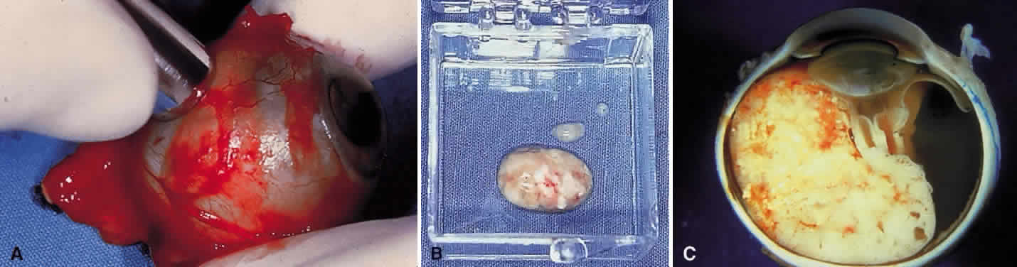
Fig. 3.
Fresh tissue harvesting.
A.
Fresh tissue on a separate tray for DNA analysis.
B.
Fresh tissue in a container to be immediately sent to the research laboratory.
C.
Photograph of the gross sectioned globe with large exophytic retinoblastoma.