|
|
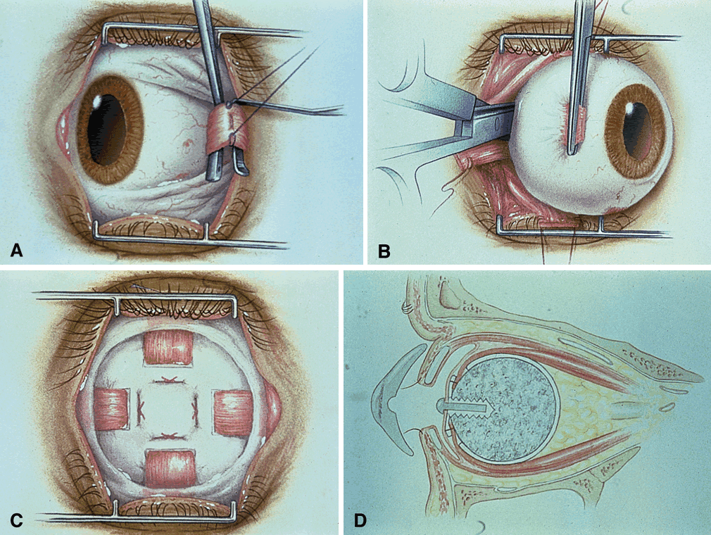
| Fig. 2. Enucleation and placement of motility implant. A. After peritomy, the rectus muscles are tagged with vicryl suture. B. The medial rectus muscle stump is grasped, and the optic nerve is cut posteriorly in the orbit with enucleation scissors. C. The scleral wrapped motility implant is placed, and the four rectus muscles are sutured through cut windows into their anatomic positions. D. A cross-section of the orbit with a motility implant shows the attached rectus muscles. In the anterior portion of the implant is a sleeved peg system that fits into an indentation on the posterior surface of the prosthesis. |