Chapter 86
Superior Oblique Muscle Surgery
RICHARD A. SAUNDERS and AMY K. HUTCHINSON
Main Menu   Table Of Contents

Search

SUPERIOR OBLIQUE STRENGTHENING PROCEDURES
SUPERIOR OBLIQUE WEAKENING PROCEDURES
REFERENCES

Defects in superior oblique muscle function can be either congenital or acquired. Both are manifested clinically as incomitant hypertropia, A-pattern strabismus, torticollis, and vertical or torsional diplopia. Patients undergo surgery to correct the superior oblique muscle function most often to weaken an “overacting” muscle or strengthen an “underacting” muscle. Superior oblique muscle surgery is occasionally performed to compensate for trochlear dysfunction (e.g., Brown's syndrome) or as part of the management of strabismus problems not directly involving the superior oblique muscle. In most cases, superior oblique muscle dysfunction presents as an isolated finding, associated only with other strabismus disorders. A few may be part of recognized syndromes (Apert's, Crouzon's), craniocervical junction deformities (Chiari's malformation), or neural tube defects (meningomyelocele).1,2 Familial transmission has been reported.3–5 Occasionally, myasthenia gravis may present as superior oblique muscle weakness.6

Surgery to strengthen or weaken the action of the superior oblique muscle is almost always accomplished by operating on the reflected tendon. Unlike the rectus muscles, which insert near the corneoscleral limbus, the oblique muscles insert behind the equator of the globe. Superior oblique muscle contraction produces a vector of force directed anteriorly and medially with respect to the orbit. The relative proportion of vertical, torsional, and horizontal forces exerted on the eye depends on direction of gaze. In adduction, the primary action of the superior oblique muscle is depression, but this becomes intorsion when the eye is abducted. Any procedure that strengthens or weakens the action of the superior oblique muscle therefore has multiple, gaze-dependent effects on ocular alignment. These complex and sometimes confusing actions can be intimidating to the ophthalmic surgeon. It is this unusual anatomy, difficult exposure, and unpredictable surgical outcomes that undoubtedly led to von Graefe's frequently quoted admonition (“Noli me tangere”) that surgery on the superior oblique muscle should not be attempted.7

It was not until 1935 that Wheeler8 showed that direct surgery on the superior oblique tendon could be performed to strengthen the action of an underacting superior oblique muscle. In 1942, Hughes and Bogart9 described a procedure to weaken the action of the superior oblique muscle by recessing the trochlea. Later that decade, Berke10 described and carefully illustrated superior oblique tendon tenotomy. His operation, with subsequent modifications, became the standard method of correcting superior oblique muscle overaction.11 Nevertheless, the reluctance of most ophthalmic surgeons to operate on the superior oblique muscle was reiterated by Fink12 as recently as 1947.

As a result of better understanding of the anatomy and physiology of the superior oblique tendon and trochlea, improved surgical instruments and sutures, and innovative surgical techniques, superior oblique muscle surgery is now more commonly performed. Appropriate surgical indications include alignment defects manifesting as clinical overaction or underaction of the superior oblique muscle(s), such as A-pattern esotropia and exotropia, superior oblique muscle palsy, and primary anatomic anomalies such as Brown's syndrome. Nevertheless, surgical procedures on the superior oblique tendon require clear indications, understanding of the superior oblique muscle anatomy and function, surgical expertise, and experience in the management of cyclovertical muscle disorders. When properly performed, superior oblique muscle surgery is safe, effective, and yields results that cannot readily be achieved by other methods.

Back to Top
SUPERIOR OBLIQUE STRENGTHENING PROCEDURES

INDICATIONS: SUPERIOR OBLIQUE MUSCLE PALSY

The superior oblique muscle may need to be strengthened to correct vertical strabismus in the setting of a clinically underacting muscle. The main indication for superior oblique strengthening is congenital or acquired superior oblique muscle palsy. Less frequently, strengthening may be helpful to treat residual inferior oblique muscle overaction and V-pattern after inferior oblique muscle weakening in patients with persistent underaction of one or both superior oblique muscles.

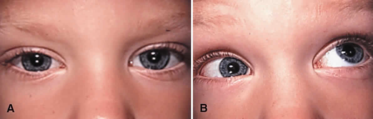
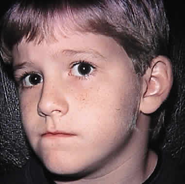
Surgery to correct unilateral, congenital superior oblique muscle palsy is the single most common indication for superior oblique muscle strengthening. It has traditionally been thought to be caused by dysfunction of the fourth (trochlear) cranial nerve. Patients typically present with an incomitant hypertropia characterized by elevation of the affected eye in adduction and overaction of the ipsilateral inferior oblique muscle. Diagnosis is normally supported (but not “diagnosed”) using Parks' three-step test.13 Compensatory torticollis is present in approximately 75% of cases, with the face turn or head tilt toward the opposite shoulder.14,15 This is often first noted when the child begins to walk. However, the presence of a vertical deviation in primary gaze position may be overlooked if the physician fails to evaluate ocular alignment with the child's head held erect. Chronic torticollis can result in asymmetric facial development and postural plagiocephaly (Fig. 1).16–18 Some authors believe that proper head positioning during sleep is more important than performing early surgery in preventing the facial asymmetry.19 It is not known whether early strabismus surgery can prevent or reverse this deformity.

Fig. 1. An 8-year-old boy with postural plagiocephaly associated with congenital left superior oblique muscle palsy. Chronic torticollis has produced atrophic facial development on the right side.

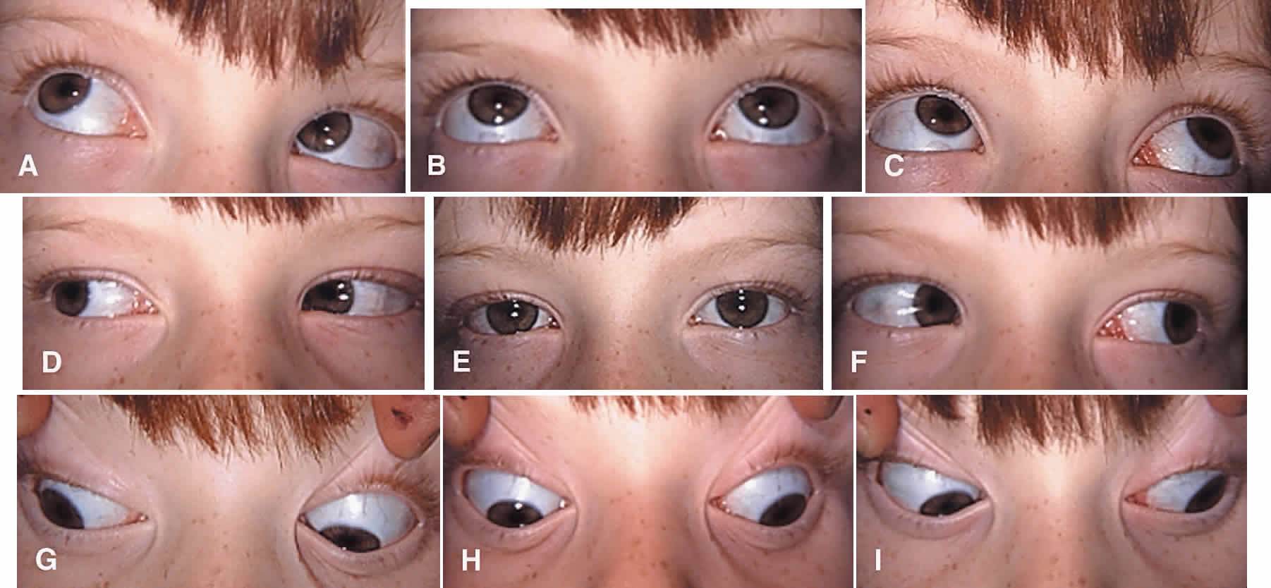
Bilateral superior oblique muscle palsy occurs most commonly after head trauma and affects all ages. In children, bilateral superior muscle palsy is much more common than unilateral palsy and may be occult.20–22 However, primary overaction of the inferior oblique muscles is common in strabismic children and may be a source of diagnostic confusion. This latter entity can usually be distinguished from bilateral superior oblique muscle palsy by its association with horizontal strabismus (usually esotropia) in primary gaze position, a large V-pattern (which is most pronounced between primary position and up gaze), and a negative result to Bielschowsky's head tilt test. Although bilateral superior oblique muscle palsy may also present with a V-pattern, it is usually smaller in magnitude and characterized by an esotropia in down gaze rather than an exotropia in up gaze. These patients often adopt a compensatory chin-down position to facilitate fusion.

The preoperative diagnosis of bilateral superior oblique muscle palsy can be confirmed with intraoperative traction testing or direct assessment of superior oblique tendon laxity after surgical retrieval.23,24 Clinically subtle, bilateral superior oblique muscle palsy can usually be distinguished from a strictly unilateral palsy by a smaller than expected hyperdeviation in primary gaze position accompanied by reversal of the hypertropia in the field of action of the apparently normal superior oblique muscle or on Bielschowsky's head tilt test. Assessing the oblique fields of gaze for a reversal of the hypertropia is essential to detecting bilaterality in some cases.22 If closed head trauma can be excluded by history, the diagnosis of congenitally absent or hypoplastic superior oblique muscles or tendons (alone or combined) should be considered, especially in patients with other craniofacial anomalies.1 Imaging studies may demonstrate attenuation or absence of the superior oblique muscle.25,26

Treatment for congenital superior oblique muscle palsy can generally be delayed until after infancy. This is fortunate, because reliable prism and cover measurements are difficult to obtain in young children. Fusion is usually preserved in children who exhibit torticollis, and the risk of developing suppression and amblyopia is low.14 Patients with trochlear nerve injury from closed head trauma or other forms of reversible superior oblique muscle palsy should not be surgically treated for at least 6 months after onset or until spontaneous improvement has ceased.

Three main indications for corrective surgery exist: asthenopia, a manifest vertical strabismus in primary gaze or reading positions, and compensatory torticollis. Large vertical vergence amplitudes to maintain fusion are typically present in patients with long-standing deviations of several years or more. However, motor fusional effort may cause asthenopic symptoms. Alternatively, patients may complain of loss of binocular vision, a cosmetically objectionable hypertropia, or vertical diplopia when their deviation is manifest. Compensatory torticollis is common and often the primary reason for ophthalmologic consultation in children. Older children and adults with acquired, bilateral superior oblique muscle palsy may complain of torsional diplopia in the absence of vertical strabismus when forced to look down or use bifocal glasses. Cyclodeviation may also be noted in acquired vertical strabismus caused by restrictive ophthalmopathy or myasthenia gravis.27 Adaptations to tilting of the visual environment can occur in patients with acquired cyclotropia but are more deeply rooted in patients with congenital cyclotropia.28

To choose an appropriate operation, it is usually necessary to quantitate the vertical and horizontal alignment defects using prism and cover measurements or Maddox rod testing. When possible, these measurements should be obtained in the diagnostic gaze positions at distance and the near reading position. This task is time consuming but worthwhile, given that these measurements will determine the type and amount of surgery performed. In addition, important ancillary information can be obtained, such as the strength of vertical vergence amplitudes, a clinically significant horizontal deviation, or the presence of occult bilateral involvement. Assessment of subjective ocular torsion using the double Maddox rod or objective excyclotropia by fundus examination is often helpful in determining the surgical approach to be taken.

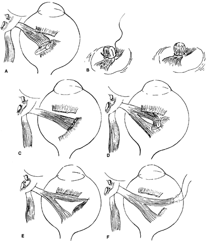
Successful results can be achieved with various surgical approaches (Fig. 2).29–36 Superior oblique muscle strengthening operations are usually performed in conjunction with surgery on other cyclovertical muscles. Their most common uses are (1) to augment ipsilateral inferior oblique muscle weakening in patients with at least 30 diopters (D) of hypertropia in lateral gaze or 20 D of hypertropia in primary gaze; (2) to augment ipsilateral superior rectus or contralateral inferior rectus muscle recession or posterior fixation in patients with increased hypertropia in down gaze; (3) to correct excyclotropia in the presence of a small hypertropia in primary gaze position; and (4) to improve the function of a congenitally lax or redundant tendon.37 In fact, torticollis may be difficult or impossible to eliminate in some children without a superior oblique tendon tuck or similar strengthening procedure.38 The Harada-Ito procedure and its modifications enhance intorsion and are used to correct excyclotropia when little or no vertical misalignment in primary gaze position exists.39–41 Its use, therefore, is limited primarily to correcting torsional diplopia in patients with bilateral superior oblique muscle palsy. Small horizontal deviations associated with superior oblique muscle palsy tend to resolve spontaneously after vertical realignment of the eyes. In our experience, concurrent horizontal rectus muscle surgery often (and for unknown reasons) leads to overcorrection and is usually unnecessary.

Fig. 2. Some useful superior oblique strengthening procedures. A and B. Superior oblique tendon tuck. C. Superior oblique tendon resection. D. Anterior fiber tuck. E. Harada-Ito procedure. F. Adjustable Harada-Ito procedure.

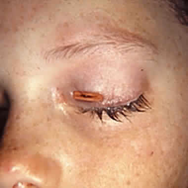
Superior oblique muscle strengthening is occasionally performed as an isolated procedure, often after surgical undercorrection following inferior oblique muscle weakening. Indications for this approach are persistent torticollis associated with a small residual hypertropia, excyclotropia, and clinical underaction of the paretic superior oblique muscle. A superior oblique tendon tuck is an appropriate primary procedure in patients with less than 10 D of hypertropia in primary gaze position, at least 5 degrees of subjective extorsion, and little or no overaction of the ipsilateral inferior oblique muscle. This situation is normally found in unilateral or bilateral posttraumatic superior oblique muscle palsy. Finally, patients with direct trauma to the superior oblique tendon after penetrating injuries of the upper eyelid and orbit may have a repairable injury and should have the superior portion of the globe carefully explored and severed portions of the tendon reapproximated if technically feasible.42

CONTRAINDICATIONS

Contraindications to superior oblique muscle strengthening are few, but important. Patients with small-angle, vertical deviations of less than 10 D in the fields of action of both the ipsilateral superior oblique and inferior oblique muscles and no torsional symptoms are not good candidates for superior oblique muscle strengthening procedures but can sometimes be managed successfully with prism-fitted glasses. However, prisms are rarely appropriate in children. Patients with preoperative underaction of the ipsilateral inferior oblique muscle or concurrent Brown's syndrome following trochlear injury should not undergo any procedure that could further compromise elevation of the eye. Similarly, the presence of a taught superior oblique tendon identified at the time of surgery, either by traction testing or direct inspection, should be a cause for concern. Such patients develop symptomatic postoperative Brown's syndrome following after minimal shortening of the superior oblique tendon.23,37,43–45 (Our only tuck takedown over 2 decades was required in a patient with posttraumatic superior oblique muscle palsy who had only 4 mm of tendon tuck performed!) Finally, the absence of excyclotropia or vertical incomitance in lateral gaze should prompt the surgeon to consider operating on alternative muscles. This is because superior oblique muscle strengthening typically produces between 5 and 10 degrees of intorsion in primary gaze position and has its greatest vertical effect in ad-duction. Although clinically important postoperative Brown's syndrome is uncommon, many patients have a mild, long-term elevation deficiency in the operated eye, particularly if the ipsilateral inferior oblique muscle has also been weakened. They often report vertical diplopia in extreme up gaze, which might pose problems in certain occupations (e.g., pilot, auto mechanic) or athletic activities requiring a full visual field of single binocular vision. Surgeons who lack full understanding of the patient's motility problem or who are insecure about their approach to the superior oblique tendon should not undertake superior oblique muscle surgery.

SURGICAL PROCEDURES

The superior oblique tendon tuck is the most frequently performed superior oblique muscle strengthening procedure. It is almost always performed unilaterally in patients with congenital superior oblique muscle palsy or acquired paresis in which marked tendon laxity is demonstrated by intraoperative traction testing.23,24 Although technically more difficult than tendon tucking, superior oblique tendon plication, advancement, or resection may be used in lieu of a tuck.46 In cases of acquired superior oblique muscle palsy in which tendon laxity is not present, the preferred surgical approach generally involves weakening the antagonist inferior oblique muscle or other vertical rectus muscles. The Harada-Ito procedure is usually performed bilaterally and is generally reserved for patients with acquired, bilateral superior oblique palsy to correct torsional diplopia.44

Tendon Tuck

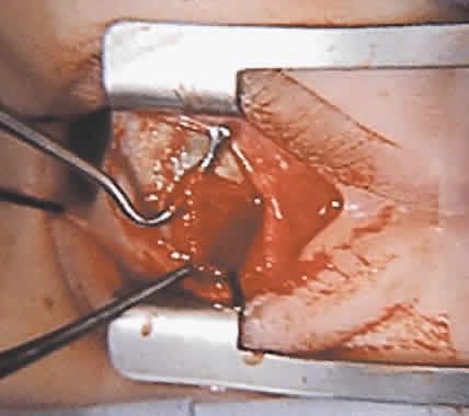
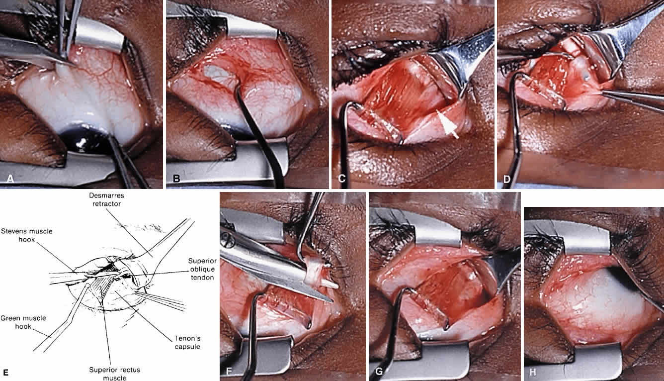
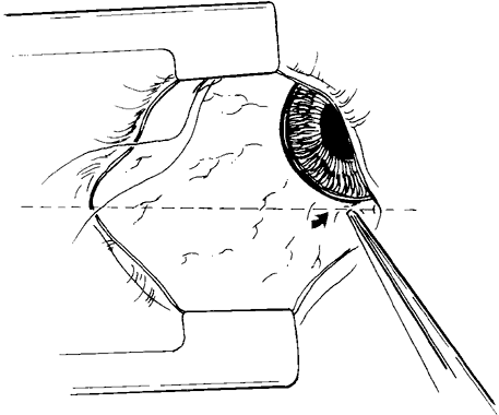
To perform the procedure, either general anesthesia or local anesthesia with intravenous sedation may be selected. Following anesthesia, it is often helpful to assess tendon laxity by performing an exaggerated traction test.47 A normal superior oblique tendon can be differentiated from a lax tendon when the globe is rotated upward, inward, and retropulsed into the orbit (Fig. 3).23

Fig. 3. A to C. Exaggerated torsional traction test performed in a patient with congenital right superior oblique muscle palsy. D. The right superior oblique tendon could not be felt as the right eye was elevated, adducted, and retropulsed into the orbit.

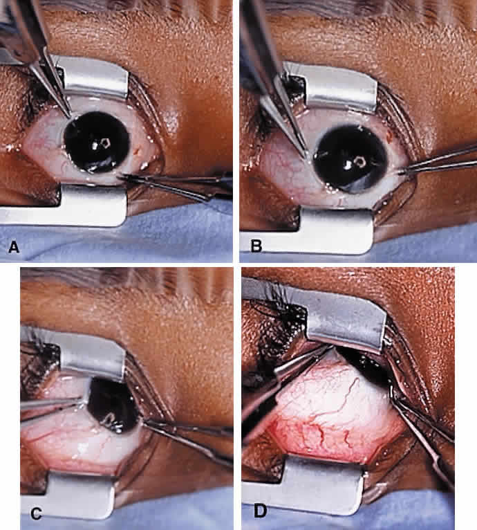
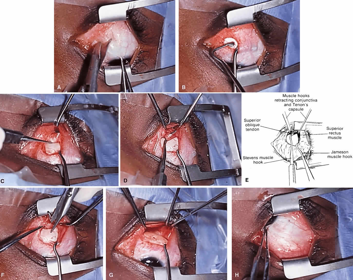
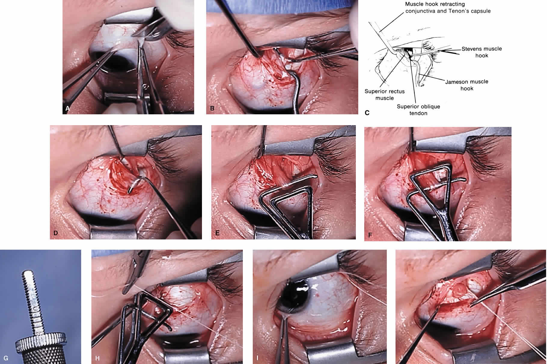
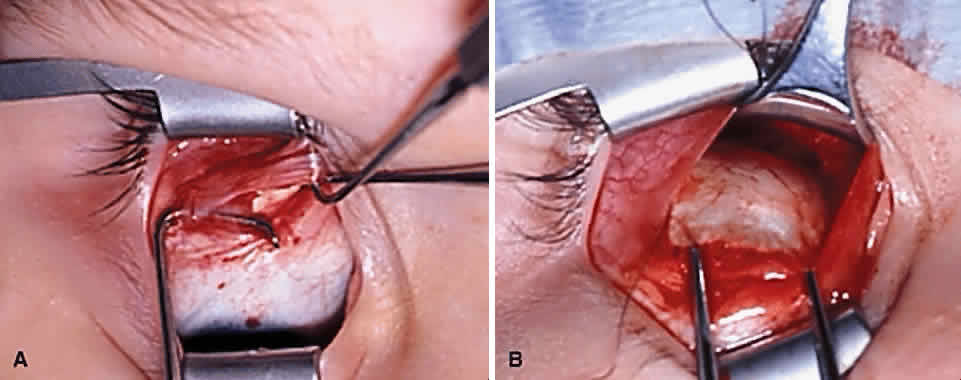
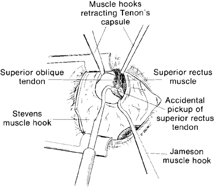
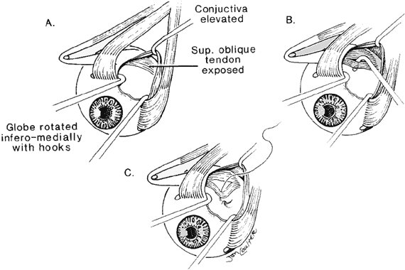
The superior oblique tendon tuck is best performed near the tendon's insertion as described by McLean.48 The surgical procedure is performed through a superior temporal cul-de-sac approach (Fig. 4). With the globe depressed, an incision is made through the conjunctiva and Tenon's fascia just temporal to the lateral border of the superior rectus muscle and parallel to the corneoscleral limbus. The superior rectus muscle is engaged on a muscle hook and the globe adducted and positioned in maximal depression. The lateral rectus muscle may also be used to assist positioning the globe. Using two small muscle hooks, the conjunctival incision is stretched open posteriorly in a triangular fashion. The lateral border of the superior rectus muscle is elevated and displaced medially. The globe is then examined for the glistening, white fibers of the superior oblique tendon running flush against the sclera and directed anteromedially. The anterior portion of the reflected tendon typically inserts under the lateral border of the superior rectus muscle, 5 to 7 mm posterior to its insertion. However, this location may vary, and anatomic anomalies are common (Fig. 5).12,37,47,48 The superior oblique tendon looks very different near its scleral insertion than 10 mm more proximally, where it becomes cordlike and enters Tenon's fascia after passing under the belly of the superior rectus muscle. Inexperienced surgeons have mistaken the lateral portion of the tendinous insertion of the superior rectus muscle for the superior oblique tendon (Fig. 6).49 However, the superior rectus muscle travels toward the orbital apex and contains prominent anterior ciliary blood vessels, whereas the superior oblique tendon is directed toward the trochlea and is usually avascular. In addition, direct traction on the superior oblique tendon can easily be palpated at the trochlea by pressing a finger over the eyelid in the superior nasal quadrant of the orbit.10 This is a reliable sign that the tendon (and not some other structure) has been engaged on the muscle hook.

Fig. 4. Superior oblique tendon tuck procedure in the left eye. A. A conjunctival incision is made temporal to the insertion of the superior rectus muscle. B. and C. With the superior rectus muscle engaged on a Jameson muscle hook, the globe is held in maximal depression and a Stevens tenotomy hook is passed under the superior oblique tendon near its scleral insertion. D. The superior oblique tendon is retrieved from the orbit and tendon laxity is assessed. E. The tendon is removed from the muscle hook and placed on a Bishop tendon tucker. F. The tucker is tightened until snug. G. The amount of tendon shortening (in mm) can be read directly above the screw knob of the tucker. H. Redundant tendon is sewn to itself with 5-0 braided Dacron suture forming a provisional tuck. I. The tendon is released into the orbit and the amount of tendon tuck evaluated using forced ductions. J. If satisfactory, sutures are cut and the conjunctival incision is closed at the surgeon's option.

Fig. 5. A. Superior oblique tendon (on Stevens tenotomy hook) shown inserting anonomously on the top of the superior rectus muscle (in Green muscle hook). B. Absent superior oblique tendon in patient with a congenital right superior oblique muscle palsy. The superior rectus muscle has been detached from the globe.

Fig. 6. During a temporal conjunctival approach, lateral portion of the superior rectus muscle tendon can potentially be confused with the superior oblique tendon.

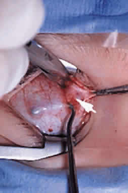
After the superior oblique tendon has been positively identified, it is gathered on a Stevens tenotomy or other muscle hook with a small tip. This maneuver is most readily accomplished by passing the hook over the tendon with its tip parallel to the sclera and pointed medially. Direct visualization of the tendon is essential, in that blind sweeping can be hazardous. A superior temporal vortex vein usually exists near the posterior insertion of the superior oblique tendon and the optic nerve is within reach. In addition, it is important to avoid passing the muscle hook into orbital fat because its release into the sub-Tenon's space predisposes the eye to develop an adherence syndrome postoperatively.50

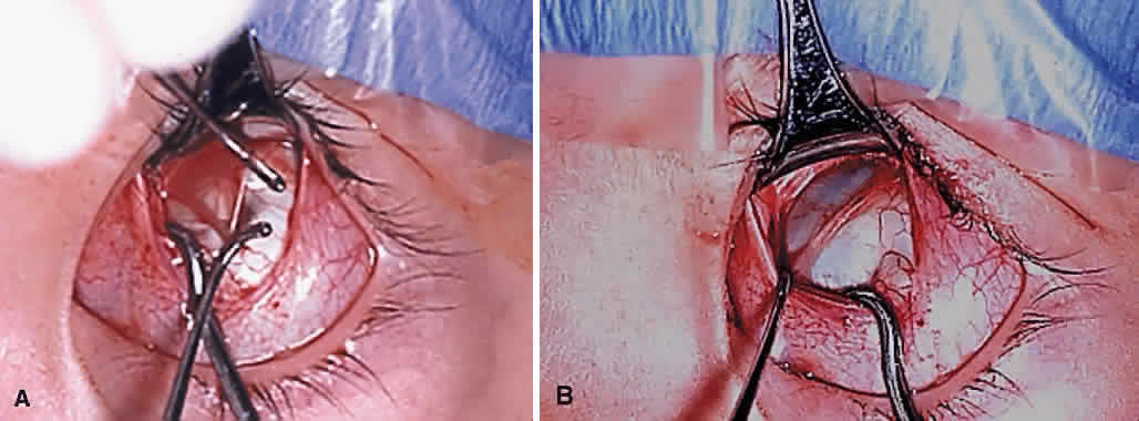
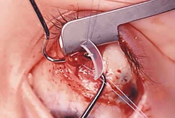
The superior oblique tendon is drawn forward through the surgical wound and attachments to Tenon's fascia and the superior rectus muscle are cut. A second pass of the muscle hook helps ensure that no fibers have been missed. Gross assessment of tendon laxity is then performed. A lax tendon requires a greater amount of tuck, whereas a normal tendon should be approached with caution, and a taut tendon should not be shortened at all. A Bishop tendon tucker is helpful to control the isolated tendon and permits the surgeon to quantitate the amount of tendon shortening conveniently. The tendon is drawn in the tucker until snug, but not tight. The loop of tendon is then sewn to itself using a nonabsorbable suture such as 5-0 braided Dacron. After this provisional tuck has been completed, the tendon is released into the orbit and a traction test is performed with the eye positioned in maximum adduction. It has been our experience that the ideal tuck results when the amount of tendon shortening produces its first resistance to elevation as the inferior limbus crosses an imaginary line between the medial and lateral canthus (Fig. 7).34,51 Care must be taken not to retropulse the globe into the orbit, because this exaggerates the duction limitation. An average tendon shortening of approximately 12 mm is required in congenital superior oblique muscle palsy (lax tendon) and 8 mm in acquired palsy (normal tendon). However, the optimum amount of tuck does not necessarily correlate with the size of the hypertropia in primary gaze position and intraoperative titration is essential. If the initial tuck is too tight or too loose based on results of traction testing, the tendon is retrieved from the orbit and the tuck is adjusted. The final tie-off is performed by passing the suture through and around the superior oblique tendon a second time after which it is tied securely. This minimizes the potential for tendon slippage through the tuck and provides additional security should a suture loop fail postoperatively. It is not necessary to sew the redundant tendon to the sclera. Conjunctival closure is performed at the surgeon's discretion. A running suture of 6-0 plain catgut is well tolerated and prevents the conjunctiva from gaping open postoperatively.

Fig. 7. Drawing of intraoperative traction test for titrating a superior oblique tendon tuck. The first resistance to elevation should be felt as the inferior limbus crosses an imaginary line (dashed line) between the medial and lateral canthus.

Tendon Plication/Imbrication

This procedure is similar to the superior oblique tendon tuck, except that a nonabsorbable suture is secured to the tendon 6 to 9 mm from its insertion and then passed through the sclera just beyond the insertion. Pulling these sutures tight folds the tendon over onto itself (“imbrication”) and advances the midportion of the tendon.46 Plication shares reversibility with the superior oblique tendon tuck, although it alters the normal anatomic insertion of the tendon and is more difficult to adjust intraoperatively.

Tendon Resection/Advancement

The superior oblique tendon can be resected or advanced in a manner similar to that used for rectus muscles (see Fig. 2C). However, the long and fanlike tendinous insertion makes this technique more challenging, even with optimal exposure, and typically results in narrowing and some anterior transposition of the scleral insertion. Furthermore, this procedure is less easily reversible than superior oblique tendon tuck and other nondisinsertion procedures. Therefore, superior oblique tendon resection is rarely, if ever, selected by most surgeons.

Harada-Ito Procedure

The Harada-Ito procedure is usually indicated in patients whose primary complaint is torsional diplopia (Fig. 2E, F and Fig. 8). This is encountered most often in adult patients with bilateral, posttraumatic superior oblique muscle palsy. Patients with congenital superior oblique palsy often have sensory adaptations that partially or completely compensate for years of excyclotropia. Although used primarily to correct symptomatic excyclotropia in patients with little or no vertical deviation in primary gaze position, it may also be effective in the treatment ocular torticollis with tilt-dependent nystagmus.52,53 The Harada-Ito procedure involves selective strengthening of the anterior fibers of the superior oblique tendon that are primarily responsible for incyclotorsion. It can be performed either by disinserting, advancing and anteriorly transposing the anterior tendon fibers (disinsertion technique) as described by Fells54 or by the classic Harada-Ito method, which redirects the anterior fibers as a “dog leg” without scleral disinsertion (Fig. 8).40 We prefer the classic method because it is more easily reversible. However, the disinsertion technique has the advantage of being readily modified for use with adjustable sutures.41,55 Superior oblique tendon tuck limited to the anterior fibers is an occasionally useful alternative when an intermediate amount of incycloduction is desired with some vertical correction.

Fig. 8. Surgical technique for the Harada-Ito procedure. A, Superior temporal fornix incision through the conjunctiva to expose the superior oblique tendon. B, Anterior fibers of the superior oblique tendon are engaged on a Stevens tenotomy hook. C, Nonabsorbable suture, such as 5-0 braided Dacron, is used to advance and transport the separated fibers in a ‘dog leg’ fashion anteriorly. (Locastro AJ, Kim SJ, Biglan AW: The treatment of cyclodiplopia with the Harada-Ito operation. Am Orthoptic J 42:111–119, 1992.)

To perform the classic Harata-Ito procedure, the superior oblique tendon is identified and isolated on a muscle hook in a fashion similar to a superior oblique tendon tuck (see previous discussion). A small muscle hook is used to separate between one third and one quarter of the most anterior tendon fibers for approximately 8 to 10 mm from their scleral insertion. A nonabsorbable, double-armed suture, such as 5-0 braided Dacron, is passed through the tendon defect and then through the sclera adjacent to the superior border of the lateral rectus muscle posterior to its scleral insertion. When the suture is secured to the sclera, the anterior fibers are advanced and anteriorly transposed to create a new functional insertion. The surgical effect can be titrated by altering both the tension and position of the suture using an intraoperative adjustment technique in cooperative patients or fundus examination under general anesthesia.56 Postoperative overcorrection can easily be addressed by cutting the scleral suture. If performed during the first several days after surgery, the transposed fibers of superior oblique tendon return to their normal anatomic position.

Tendon Transposition

Patients with a history of third (oculomotor) cranial nerve palsy and absent medial rectus muscle function often have persisting exotropia because of unopposed action of the ipsilateral lateral rectus muscle. If the vertical rectus muscles are paretic and transposition surgery is therefore inappropriate, the superior oblique muscle can be used as an adductor and elevator to stabilize the globe in primary gaze position. Jackson57 and later Costenbader58 suggested fracturing the trochlea and reattaching the superior oblique tendon to the sclera above the insertion of the medial rectus muscle. Scott subsequently described a simplification of this procedure, not involving trochleotomy, in which the reflected portion of the superior oblique tendon is disinserted from the sclera, rotated anteriorly, and reattached adjacent to the medial border of the superior rectus muscle.59 Some authors have reported favorable results when the superior oblique tendon transposition is accompanied by aggressive horizontal rectus muscle surgery,60 whereas others have found it less effective or accompanied by unacceptable paradoxical ocular movements.61

Expected Results

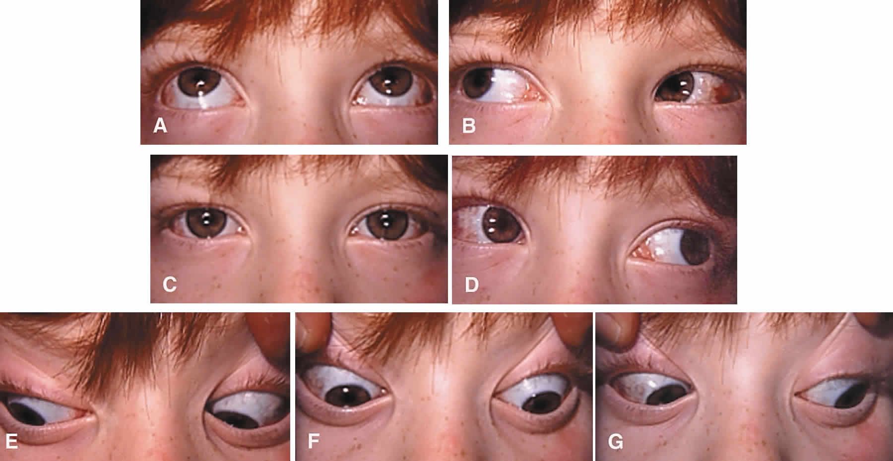
After tucking the superior oblique tendon, most patients have a mild-to-moderate elevation deficiency in adduction (Brown's syndrome).62 Elevation deficiency may be less pronounced after superior oblique tendon advancement or resection. Patients undergoing the Harada-Ito procedure do not characteristically have elevation defects in adduction postoperatively but may report subjective extorsion with corresponding incyclotropia on fundus examination.63 Following any shortening procedure of the superior oblique tendon, there may be tenderness in the trochlear region and avoidance of elevation in adduction.

When surgery for superior oblique muscle palsy is limited to one or more oblique muscles, a small overcorrection of less than 10 D is desirable in primary gaze position. Because results of Bielschowsky's head tilt test usually remains positive postoperatively, many patients temporarily adopt an opposite head tilt toward the side of the paretic muscle or a chin-up head posture to avoid diplopia or achieve fusion. If the patient wears glasses, a base-up Fresnel prism over the operated eye may be helpful during the early postoperative period. Surgical overcorrection, especially following treatment for a large-angle, congenital superior oblique muscle palsy, should not be reoperated hastily and rarely within 1 year. Almost all such patients improve spontaneously. Similar to the surgical treatment of intermittent exotropia, long-term undercorrection is much more common than permanent overcorrection. However, the surgeon should attempt to distinguish between a true surgical overcorrection and masked, bilateral superior oblique muscle palsy, which is likely to require additional treatment.17,20–22,64

Because of the heterogeneous nature of this population and differences in inherent prognosis, meaningful reoperation rates are difficult to calculate. Most patients undergoing superior oblique tendon tuck for appropriate indications will not require a second surgery. Successful outcomes (elimination of torticollis and manifest hypertropia in and around primary gaze position) may be as high as 90%.16,36,44,51 In general, patients with congenital or long-standing superior oblique muscle palsy characterized by inferior oblique muscle overaction and near normal superior oblique function have the best prognosis, regardless of the magnitude of the deviation in primary gaze position. Patients with bilateral or posttraumatic superior oblique muscle palsy tend to fare less well, perhaps because of poor vertical vergence amplitudes or limited fusion capability. It is likely (but unproven) that patients with partial superior oblique muscle dysfunction (paresis) have a better surgical prognosis than those with complete paralysis. Because the treatment indication for superior oblique tendon anterior transposition is third cranial nerve palsy rather than superior oblique muscle palsy, the expected surgical results should be considered separately. Although enhancing adduction forces on the eye may help stabilize the globe in primary gaze position, paradoxical adduction on attempted depression may be an undesired result.61 The tethering effect of the superior oblique tendon may also produce globe elevation in abduction.

COMPLICATIONS

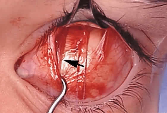
Serious complications of superior oblique muscle strengthening are infrequent. Intraoperative complications relate primarily to orbital hemorrhage from injury to a vortex vein or unintended entry into the orbital fat space, which results in an adherence syndrome with restricted ductions postoperatively. These can be avoided by good surgical exposure, use of loupe magnification with a surgical headlight, and deliberate and meticulous surgical technique. The superior oblique tendon can be surgically missed or inadvertently cut during dissection of the intermuscular septum. Anatomic variations including absence of the tendon are common.12 Confusion can also occur if the surgeon inadvertently engages the superior oblique tendon when first attempting to hook the superior rectus muscle.49 However, this is less common when the superior oblique tendon is approached temporally rather than nasally (Fig. 9). Surgical overcorrection occurs frequently, but it is fortunately not permanent in most patients. However, permanent overcorrection that results in a primary gaze position hypotropia is usually an unacceptable result and requires reoperation. It can be caused by excessive surgical dosage, spontaneous recovery of superior oblique muscle function, or failure to recognize bilateral superior oblique muscle palsy preoperatively.

Fig. 9. During a medial conjunctival approach, the superior oblique tendon can be inadvertently hooked with the superior rectus muscle and confused with the intermuscular septum. Both the right superior rectus muscle and right superior oblique tendon (arrow) have been engaged on the Jameson muscle hook (directed inferiorly).

Iatrogenic Brown's syndrome is an unavoidable complication of superior oblique muscle strengthening and occurs in almost 100% of patients undergoing superior oblique tendon tuck. It is virtually identical in clinical appearance to congenital Brown's syndrome, which is discussed later in this chapter (Fig. 10). In the absence of excessive tendon shortening, the limitation of elevation in adduction almost always improves with time and reoperation for this entity is rarely required. Tendon tucking medially (rather than temporally) to the superior rectus muscle is more hazardous because of proximity to the trochlea. Patients with persisting overcorrection in primary gaze position and at least moderate Brown's syndrome (no elevation above the horizontal meridian in adduction) are usually symptomatic and should have their tucks removed. However, adhesions between the superior rectus muscle, superior oblique tendon, and the globe are likely to be present and must be freed to normalize ductions. In general, tuck take down should be performed without simultaneous surgery on other cyclovertical muscles.

Fig. 10. Iatrogenic Brown's syndrome 1 month following a 10-mm right superior oblique tendon tuck and right inferior oblique muscle recession. Good alignment is present in primary position (left). However, the right eye elevates poorly in left lateral gaze (right). Ductions typically improve over time and tuck takedown is rarely required.

Back to Top
SUPERIOR OBLIQUE WEAKENING PROCEDURES
Superior oblique weakening procedures are most commonly used to treat Brown's syndrome, A-pattern strabismus associated with bilateral superior oblique overaction, and unilateral superior oblique muscle overaction (either primary or, rarely, secondary to ipsilateral inferior oblique muscle palsy) when associated with a hypotropia in primary gaze position.

INDICATIONS: BROWN'S SYNDROME

Brown's syndrome is a well-defined clinical disorder with several possible etiologies that can be either congenital or acquired.65–68 Brown's syndrome is characterized by limitation of elevation of the affected eye, particularly in adduction, typically caused by abnormalities of the trochlea and superior oblique tendon. Although elevation is typically normal or near normal in abduction, there is a characteristic “down shoot” of the affected eye when directed from primary position into adduction. Forced ductions are abnormal and usually characterized by an abrupt, absolute limitation of elevation in adduction. The elevation deficiency is made worse by retropulsion of the globe into the orbit. To ensure proper treatment, Brown's syndrome must be distinguished from other forms of monocular elevation deficiency such as superior rectus muscle palsy (sometimes called “double elevator palsy”), primary inferior rectus muscle fibrosis, inferior oblique muscle palsy (exceedingly rare), and pseudo-Brown's syndrome secondary to an orbital floor fracture or other causes of orbital restriction.69–71

Although stripping of the superior oblique tendon “sheath” has been advocated in the past, this surgical approach undoubtedly arose from a misunderstanding of the pathophysiology of this disorder. Most patients with congenital Brown's syndrome appear to have an intrinsic abnormality of the trochlea or proximal tendon limiting movement through a nonyielding trochlear tunnel.66 A few cases may be caused by abnormal leashes or bands extending from the trochlea to the globe, similarly limiting elevation of the eye.72 Other theories have also been proposed.7,65,73,74 Regardless of cause, effective treatment usually requires severing the superior oblique tendon between the trochlea and scleral insertion either by tenotomy or tenectomy.

Most patients who require corrective surgery have congenital Brown's syndrome. Acute onset Brown's syndrome is usually inflammatory or posttraumatic in etiology and may improve or resolve spontaneously. Congenital Brown's syndrome or chronic acquired Brown's syndrome that does not respond to steroids or other appropriate treatment may be treated surgically. The most common indication for surgical correction is a chin-up head posture or face turn away from the affected eye. This usually allows the patient to achieve fusion. Additional surgical indications include pain (typically over the trochlea), diplopia, and cosmetic disfigurement. It is not uncommon for parents of children with Brown's syndrome to believe the problem resides in the normal fellow eye, which appears to elevate excessively on attempted up gaze. Surgery is appropriate for patients with moderate-to-severe involvement, providing there is no reasonable expectation of spontaneous improvement.

INDICATIONS: A-PATTERN STRABISMUS

A-pattern strabismus is characterized by greater esotropia (or less exotropia) in up gaze and greater exotropia (or less esotropia) in down gaze. Although the difference can be as little as 10 PD, it is often much greater in magnitude, sometimes exceeding 100 PD in extreme down gaze. When a large A-pattern is associated with superior oblique muscle overaction, superior oblique muscle weakening is usually indicated. In most cases, the procedure of choice is bilateral superior oblique tendon tenotomy. Patients undergoing this operation should have unequivocal overaction of both superior oblique muscles. This finding is typically accompanied by a right hypertropia in right lateral gaze and a left hypertropia in left lateral gaze. Inferior oblique muscle underaction should be present and is associated with tight (but not restricted) superior oblique muscles on forced duction testing. If any doubt exists, objective incyclotropia on fundus examination is a useful way of confirming the presence of true superior oblique muscle overaction.75 Double Maddox rod testing may occasionally help document the presence of incyclotropia. Because bilateral superior oblique tendon tenotomy is a powerful operation, it is rarely appropriate when there are less than 40 PD of A-pattern or λ-pattern and should be avoided in the absence of inferior oblique muscle under action. Although a few patients undergo superior oblique tendon tenotomies as an isolated procedure, most also require surgery on one or more horizontal rectus muscles. It is therefore imperative that primary gaze position alignment be measured carefully, without the patient assuming a chin-up or chin-down head posture. Disregarding the patient's head position can lead to serious mismeasurement of the horizontal deviation.

Patients with asymmetric superior oblique muscle overaction often have a manifest vertical deviation in primary gaze position. In these cases, differential or even unilateral superior oblique muscle weakening may be indicated. Symmetric superior oblique weakening rarely corrects fully the primary gaze position hypertropia and leaves residual oblique muscle dysfunction postoperatively. The vertical deviation usually represents a “spillover” of the hypotropia of the more overacting superior oblique muscle into primary gaze position. If an esotropia is also present, measurements should be obtained with each eye fixating in primary gaze to confirm that the hypertropia does not reverse, which would indicate the need for bilateral surgery.

Asymmetric superior oblique muscle weakening can be accomplished using various techniques. As a general rule, the more extensive the dissection of the intermuscular septum surrounding the superior oblique tendon or the closer to the trochlea the tenotomy is performed, the greater the weakening effect will be. Tenotomy and tenectomy at the medial border of the superior rectus muscle are the most powerful operations. Less effective are tenotomy at the scleral insertion (disinsertion), recession or other tendon lengthening procedures, and posterior fiber tenectomy. However, increased effectiveness is generally associated with greater risk of postoperative complications.

OTHER INDICATIONS FOR SUPERIOR OBLIQUE MUSCLE WEAKENING

Patients occasionally present with strictly unilateral superior oblique muscle overaction with little or no associated A-pattern. This is commonly seen in congenital or acquired third cranial nerve palsy, but it may occur as a primary disorder or in association with presumed unilateral inferior oblique muscle palsy.76–78 Typically, 10 to 20 PD of hypotropia are present in primary gaze position, which increases in the field of action of the overacting superior oblique muscle and disappears in opposite lateral gaze positions. Fusion is often achieved with a compensatory face turn or chin up head posture. Ipsilateral superior oblique tendon tenotomy is the most reliable treatment for unilateral superior oblique muscle overaction. Lesser weakening procedures tend to be ineffective. Superior oblique tenectomy, which involves tendon excision, is likely to result in iatrogenic superior oblique muscle palsy.79

Patients with large-angle exotropia who wish to have their surgery limited to one eye may be candidates for simultaneous weakening of both superior and inferior oblique muscles.80 This situation occurs most commonly in sensory exotropia associated with severe amblyopia or structurally damaged eyes. Eliminating the abducting effect of the oblique muscles, especially if overacting, can augment the horizontal rectus muscle surgery. Between 50 and 100 PD of exotropia can be corrected without resorting to bilateral surgery or “supramaximal” surgical dosage that might produce unacceptable duction limitations postoperatively. However, both medial and lateral rectus muscles may need to be infraplaced to avoid postoperative hypertropia in the operated eye.

Superior oblique muscle myokymia is an uncommon, neuroophthalmologic disorder of unknown etiology characterized by rhythmic, small-amplitude vertical and torsional movements of the globe. Some cases are now suspected to be caused by vascular irritation of the trochlear nerve root exiting the brain stem.81 In patients symptomatic from diplopia or oscillopsia and not controlled on carbamazepine or other medical therapy, the superior and inferior oblique muscles can be simultaneously weakened. This usually consists of superior oblique tendon tenotomy or tenectomy combined with ipsilateral inferior oblique muscle weakening. Although reports in the literature are limited, simultaneous weakening of both oblique muscles appears to be effective in reducing symptoms.82,83 A “reverse” Harada-Ito procedure to weaken selectively the torsional fibers has also been reported.84 Trochlectomy may be appropriate in resistant cases.85

CONTRAINDICATIONS: BROWN'S SYNDROME

The most important contraindications to surgical treatment of Brown's syndrome are mild involvement without compensatory head posturing or vertical strabismus in primary gaze position and the likelihood of spontaneous resolution or response to nonsurgical treatment. These include all patients with recently acquired inflammatory disorders, which are often treated with systemic or local injections of corticosteroids. Brown's syndrome can also be the presenting sign of occult orbital tumors or sinusitis.86,87 Computed imaging of the orbits and paranasal sinuses is therefore indicated in patients presenting with acute onset Brown's syndrome of undetermined cause. Patients with occasionally manifest, intermittent duction limitations (e.g., “click” syndrome67), or those with mild congenital Brown's syndrome allowing elevation of the eye above the horizontal meridian in adduction are best left unoperated.

CONTRAINDICATIONS: A-PATTERN STRABISMUS

Patients lacking demonstrable superior oblique muscle overaction are rarely candidates for superior oblique muscle weakening, even in the presence of a large A-pattern. Care must be taken to distinguish true superior oblique muscle overaction from pseudosuperior oblique muscle overaction associated with exotropia in down gaze or Duane's syndrome. Patients with bifoveal fixation are at particular risk for developing torsional or vertical diplopia postoperatively.88,89 In most cases, they should be managed with vertical transposition of the horizontal rectus muscles. In addition, A-pattern exotropia without superior oblique muscle overaction can occur following inadvertent inclusion of the inferior oblique muscle in lateral rectus muscle surgery (Figs. 11 to 13).90 These patients require careful surgical exploration and release of the inferior oblique muscle. Caution should also be used in patients with dissociated vertical deviations, which may be exacerbated following superior oblique tendon tenotomy. They are effectively treated with simultaneous weakening of the superior rectus and superior oblique muscles.91

Fig. 11. A-pattern extrotropia following bilateral rectus muscle recessions for comitant, intermittent exotropia. Note the presence of pseudosuperior oblique muscle overaction (due to the A-pattern) and conspicuous absence of inferior oblique muscle underaction.

Fig. 12. Intraoperative photograph showing inferior oblique muscle (in small hook) incorporated into the recessed insertion of the left lateral rectus muscle (in Jameson hook).

Fig. 13. Same patient 1 month following release of left inferior oblique muscle. The A-pattern and pseudosuperior oblique muscle overaction has disappeared.

SURGICAL PROCEDURES

Free Tenotomy

The surgical approach to the superior oblique tendon has already been described. When operating for Brown's syndrome, it is imperative that forced ductions be performed at the beginning of the procedure to confirm the diagnosis. Using a toothed forceps, the eye is grasped at the 6-o'clock position near the corneoscleral limbus and an attempt is made to elevate the globe in adduction. The nonyielding superior oblique tendon can easily be palpated in restricting upward movement of the globe. Exaggerated traction testing is usually not necessary.47 Elevation is normal or nearly normal in abduction. Proptosing the globe forward improves passive ductions, whereas retropulsion into the orbit exacerbates the duction limitation.

Unless the superior oblique tendon is going to be recessed rather than tenotomized, a superior nasal surgical approach is technically the easiest. This allows the surgeon to engage the tendon where it is most anterior and cordlike and avoids the possibility of missing posterior fibers near the fanned out insertion, which will result in surgical failure. A temporal conjunctival incision can also be used to approach the tendon medial to the superior rectus muscle as described by Parks and Parker.46 Using this approach, the surrounding Tenon's fascia and intermuscular septum can be left nearly intact, which may limit retraction of the proximal end of the tendon. This is particularly desirable when a less powerful surgical effect is required.

Regardless of the site of conjunctival incision medial or temporal to the superior rectus muscle insertion, the superior oblique tendon is engaged on a muscle hook under direct visualization as it passes under the medial border of the superior rectus muscle and bends upward toward the trochlea. Although the tendon is located only several millimeters posterior to the superior rectus muscle insertion with the eye in primary gaze position, it slides posteriorly as the eye is rotated downward. If not readily identified, a limited dissection should be performed along the medial border of the superior rectus muscle. Removing the eyelid speculum and substituting a small retractor may be helpful in gaining surgical exposure. The tendon is engaged on a Stevens' tenotomy hook and verified by palpating over the trochlea. Redundant Tenon's fascia is carefully unloaded from the muscle hook, but extensive dissection should be avoided. The tendon is then cut and the free ends released into the orbit. Forced ductions are immediately repeated. Free movement of the eye confirms that complete transection of the tendon has been accomplished. However, it is not uncommon for forced ductions to improve temporarily before actual tendon transection because of intraoperative manipulation of the globe. This may be interpreted erroneously as a negative traction test result after incomplete tendon tenotomy and yield an unwelcome postoperative surprise when the Brown's syndrome or superior oblique muscle overaction returns immediately after surgery. When performing tenotomy for A-pattern strabismus, the surgical procedure is identical to that previously described for Brown's syndrome, although technically easier in the presence of a normal superior oblique tendon. The conjunctival incision can be made either medial or lateral to the superior rectus muscle. A lateral incision minimizes disruption of the intermuscular septum although identification of the tendon is more direct when approached medially (Figs. 14 and 15).

Fig. 14. Left superior oblique tendon tenotomy performed through a nasal conjunctival approach. A. A conjunctival incision is made medial to the superior rectus muscle insertion. B. The superior rectus muscle is engaged on a Jameson hook. C. The left superior oblique tendon is identified as a white, cordlike structure existing from beneath the medial rectus muscle and entering Tenon's capsule. D and E. Redundant Tenon's capsule is unloaded, but extensive dissection should be avoided. F. The superior oblique tendon is cut at the medial border of the superior rectus muscle; G. The superior nasal quadrant of the globe is carefully examined for missed tendon fibers. H. Complete tenotomy is confirmed by performing forced ductions.

Fig. 15. Right superior oblique tendon tenotomy performed through a temporal conjunctival approach. A. Conjunctival incision is made lateral to the superior rectus muscle insertion. B. The superior rectus muscle is engaged on a Green hook. C. The conjunctival opening is moved over the muscle hook and the medial border of the superior rectus muscle is exposed. The superior oblique tendon is visible exiting from under the superior rectus muscle (arrow). D and E. The superior oblique tendon is isolated on a muscle hook and Tenon's capsule carefully unloaded. F. Tenon's capsule carefully unloaded. G. The superior oblique tendon is cut adjacent to the medial border of the superior rectus muscle. H. The superior nasal quadrant of the globe is carefully examined for missed tendon fibers.

Scleral Disinsertion

The superior oblique tendon can also be disinserted from the sclera under the lateral border of the superior rectus muscle. The weakening effect is limited by fascial attachments of the superior oblique tendon to the undersurface of the superior rectus muscle. Therefore, superior oblique tendon disinsertion usually results in less overall weakening of the superior oblique muscle function than superior oblique tendon tenotomy medial to the superior rectus muscle. In addition, near its insertion, the tendon fans out across the sclera and may be difficult to identify, especially for the inexperienced surgeon.

Tenectomy

Superior oblique tendon tenectomy is performed in a similar fashion to superior oblique tenotomy, except that a 5- or 6-mm section of the reflected tendon is totally excised. Although perhaps more powerful in effect, it is not clear whether this operation has any advantage over tenotomy, even in cases of severe Brown's syndrome or A-pattern strabismus, and precludes surgical reanastomosis later on. Because of the additional fascial disruption, tenectomy probably increases the risk of producing iatrogenic superior oblique muscle palsy postoperatively.79

Posterior Tenectomy/Tenotomy

Tenotomy or disinsertion that is limited to the posterior superior oblique tendon fibers (leaving the anterior fibers intact) has been performed by some surgeons as an alternative to free tenotomy (Fig. 16).92,93 Although this procedure may be used to collapse a small or moderate A-pattern, it appears to be less effective in correcting the superior oblique muscle overaction seen on version testing. However, it is extremely useful as a minimal weakening procedure when superior oblique muscle overaction is mild, asymmetric, or in the context of a dissociated strabismus. One study showed that bilateral posterior tenotomy performed for Apattern strabismus resulted in a mean reduction of the A-pattern of 22 PD.94 Unilateral posterior tenect- omy was also shown to reduce hypotropia (in patients with unilateral superior oblique over action) by a mean of 7 PD. Posterior tenectomy or tenotomy is occasionally used to treat Brown's syndrome patients who are marginal surgical candidates with less than 5 PD of primary position hypotropia. Preserving the primarily torsional, anterior fibers of the superior oblique tendon may reduce the risk of postoperative torsional diplopia in patients with bifoveal fixation.

Fig. 16. A. Anterior 2 mm of the left superior oblique tendon are separated from the posterior tendon fiber using Stevens tenotomy hooks. Superior rectus muscle has been engaged on a Jameson muscle hook to hold eye in infraduction. B. Posterior superior oblique tendon fibers have been disinserted from the sclera and excised. Only the most anterior fibers remain intact.

Recession

Superior oblique tendon recession is an occasionally useful alternative to superior oblique tendon tenotomy.95–98 This operation is appropriate in patients requiring limited weakening of one or both superior oblique muscles. The usual indication for superior oblique tendon recession is asymmetric or unilateral superior oblique muscle overaction. Superior oblique recession has also been used to treat mild or moderate Brown's syndrome. In one small study, superior oblique recessions of 12 to 14 mm eliminated abnormal head position and restored ductions and versions without generating a secondary superior oblique muscle palsy.99 Recession is potentially reversible and offers the option of using an adjustable suture technique. The simplest surgical procedure is a “hang-back” technique in which the tendon is connected to the anterior half of the original insertion using an absorbable suture (Fig. 17).97 Unless combined with superior rectus muscle surgery, a superior oblique recession of at least 6 mm is required to produce a clinical effect. Allowing the tendon to retract to the medial border of the superior rectus muscle produces approximately 10 mm of recession. Tendon lengthening procedures, such as Z-tenotomy, are generally ineffective, except when they result in tendon transection.7

Fig. 17. “Hangback” technique for superior oblique tendon recession. Left superior oblique tendon has been secured with a double-armed polyglactin suture, which is then passed through the sclera at the anterior half of the original insertion. Tendon is shown near its normal anatomic position, but it will be allowed to retract under the superior rectus muscle (arrow) before the suture is tied. The superior rectus muscle has been engaged on a Jameson muscle hook to hold the eye in infraduction.

Spacers

Over the years, various attempts have been made to regulate the weakening caused by uncontrolled superior oblique tendon tenotomy. Nonabsorbable sutures have been used to bridge the space between the proximal and distal ends of the transected tendon. However, just as with hang-back recessions of the rectus muscles, it is not always clear how much weakening actually occurs and spontaneous reattachment of the ends of the transected tendon is always possible. For this reason, the use of rigid spacing materials (or “expanders”), such as a no. 240 (2.5 mm) or a no. 40 (2.0 mm) silicone band, have grown in popularity in an effort to grade more reliably the amount of superior oblique muscle weakening.100–102 When spacer surgery is planned, the surgical approach is the same as that used in superior oblique tendon tenotomy, except that two preplaced nonabsorbable sutures (such as 7-0 polypropylene) are placed 2 mm apart in the cordlike tendon before its transection. The sutures then secure the proximal and distal ends of the tendon to each end of the spacer, thus effectively lengthening the tendon between the trochlea and its anatomic insertion (Fig. 18). The surgical effect can thus, at least theoretically, be graded by altering the length of the spacing material. However, introduction of any foreign material into the orbit can result in restricted motility, particularly in down gaze, and it is subject to infection or extrusion.103 Although growing in popularity, the procedure is time consuming and the advantage over more conventional superior oblique weakening operations remains unproven.104 However, a recent study suggests that spacers may be effective in converting moderate-to-severe Brown's syndrome (termed Brown's syndrome “plus”) to mild Brown's syndrome.105

Fig. 18. A 4 to 7 mm section of a no. 240 silicone band can be used as a spacing material to lengthen the superior oblique tendon medial to the superior rectus muscle in lieu of superior oblique tendon tenotomy. Two double-armed sutures of 7-0 polypropylene (shown secured to the left superior oblique tendon on either side of the Jameson muscle hook) have been passed through the ends of the silicone spacer. After transection between the sutures, the cut ends of the tendon are tied to the spacer. Care must be taken to preserve Tenon's fascia and avoid direct contact of the silicone with the sclera, which predispose to an adherence syndrome postoperatively.

Orbital Procedures

Superior oblique tendon or trochlear luxation using a subperiosteal approach has been described as an alternative to conventional weakening procedures.106 Although this is a resurrection of a procedure previously described in the 1940s,9 it is not part of the standard surgical techniques of most strabismologists and is infrequently performed. It is therefore not possible to assess its effectiveness in a large number of patients.

EXPECTED RESULTS: BROWN'S SYNDROME

Surgical results in the treatment of Brown's syndrome are often disappointing with reoperation rates of up to 50%. Especially in severe cases, most patients who undergo superior oblique muscle weakening continue to have an elevation deficiency in the early postoperative period. This is probably desirable, because ductions tend to improve gradually over weeks to months. The presence of normal ductions immediately after surgery is worrisome and usually heralds the onset of inferior oblique muscle overaction with iatrogenic superior oblique muscle palsy. It is also not uncommon for patients who have essentially normal ductions in the operating room to present the day after surgery with a recurrent Brown's syndrome. The reasons for such failures are not always clear. However, if forced ductions remain positive, incomplete tenotomy of the superior oblique tendon should be suspected. Substantial under corrections do not spontaneously resolve and further treatment is necessary. Although multiple operations may be required, almost all patients can eventually be improved with surgery.

EXPECTED RESULTS: A-PATTERN STRABISMUS

The results of superior oblique tendon weakening depend on the operation chosen. Superior oblique tendon tenectomy medial to the superior rectus muscle is probably the most powerful, followed by superior oblique tendon tenotomy (medial more powerful than temporal), and then the various forms of more limited weakening procedures (e.g., recession, spacer). Superior oblique tendon posterior tenectomy, which leaves the anterior fibers intact, is the least effective operation, but it has the compensating benefit of surgical simplicity and fewer postoperative complications.

Superior oblique tendon tenotomy is a powerful and useful operation in appropriately selected patients. When performed bilaterally, it can collapse up to 100 D of A-pattern and is the only reliable treatment for large A-patterns associated with marked superior oblique muscle overaction. There is a similar, profound effect on excyclotropia. In general, the A-pattern tends to collapse around the middle rather than the apex of the “A.” Increased esodeviation in primary gaze position may occur in some cases.88,89,107 Although it is not uncommon to see residual superior oblique muscle overaction in the immediate postoperative period, this overaction may resolve over months to years. However, clinically significant asymmetry of the superior oblique muscle overaction usually persists postoperatively unless surgically addressed. Reoperations for either horizontal or vertical deviations are eventually needed in approximately 25% of cases.

COMPLICATIONS: BROWN'S SYNDROME

About one half of patients successfully operated with superior oblique tendon tenotomy eventually develop evidence of iatrogenic superior oblique muscle palsy. This can appear months or even years after surgery. Superior oblique tendon tenectomy, in particular, has been reported to have an unacceptable rate of superior oblique muscle palsy requiring reoperation.79 The frequency of this complication has motivated some surgeons to use more limited weakening procedures such as superior oblique tendon recession or spacer (which tend to be less effective) or concurrent, ipsilateral inferior oblique muscle recession (which may further embarrass elevation in adduction, particularly if residual Brown's syndrome is present).108 Because the surgeon cannot predict reliably which patients will develop superior oblique muscle palsy postoperatively, the most prudent course is usually to leave the inferior oblique muscle intact and warn patients of the potential for late overcorrection requiring reoperation.

Patients returning to the operating room for inadequate correction of Brown's syndrome should be managed aggressively. If the cause of the previous surgical failure cannot readily be determined (e.g., missed tendon fibers), it is often helpful to detach the superior rectus muscle from its insertion temporarily and explore the superior portion of the globe carefully. Occasionally, there is an anomalous superior oblique tendon insertion or congenital bands that cause the motility deficit. Traction sutures, used to hold the eye in elevation for several days postoperatively, may be helpful in resistant cases.109 A double-armed 5-0 polyglandular suture is passed through the sclera near the corneoscleral limbus inferiorly. Each end is then directed through the medial aspect of the upper eyelid and tied externally over a rubber or silicone peg (Fig. 19). An absorbable suture releases the eye spontaneously in 1 or 2 days. A nonabsorbable suture, if preferred, needs to be removed later. However, traction sutures are generally painful and probably increase the risk of iatrogenic superior oblique muscle palsy.

Fig. 19. Rubber catheter used to secure a traction suture in the left eye of a patient with recurrent Brown's syndrome. Suture has been passed through the sclera near the corneoscleral limbus at the 5-o'clock position. This fixates the eye in an elevated and adducted position when the suture is tightened and tied externally.

COMPLICATIONS: A-PATTERN STRABISMUS

Postoperative complications are usually related to surgical overcorrection or vertical deviations in primary gaze position. If bilateral superior oblique tendon tenotomies are performed in patients with only unilateral superior oblique muscle overaction, overcorrection associated with inferior oblique muscle overaction or iatrogenic superior oblique muscle palsy may occur. Small A-patterns can be converted to V-patterns. These patients may eventually require inferior oblique muscle weakening on the previously normal side. Iatrogenic Brown's syndrome, superior rectus muscle paresis, and ptosis have also been described.11,110 However, serious complications are rare with proper surgical technique. The most common operative error, especially for the inexperienced surgeon, is a missed or incompletely tenotomized tendon. However, this should occur in less than 5% of cases and should be suspected postoperatively in the presence of residual, unilateral superior oblique muscle overaction, persisting A-pattern, and a new vertical deviation in primary gaze position. Such patients do not improve spontaneously and reoperation is required. This complication can normally be detected by exaggerated traction testing in the operating room.47 Surgical exploration typically reveals an intact or incompletely tenotomized superior oblique tendon.

Small vertical deviations are common in primary gaze position postoperatively, but they do not normally represent surgical complications. Nevertheless, older children and adults with bifoveal fixation may be unable to achieve fusion and report vertical or torsional diplopia.88,89 Relief of symptoms may be obtained with torticollis or prism glasses in some cases. Because of their ability to avoid diplopia by suppressing the second image, younger children are not at similar risk for this complication. However, they are subject to loss of bifoveal fixation and development of amblyopia.

Back to Top
REFERENCES

1. Pollard ZF: Bilateral superior oblique muscle palsy associated with Apert's syndrome. Am J Ophthalmol 106:337, 1988

2. Biglan AW: Ophthalmologic complications of meningomyelocele: a longitudinal study. Trans Am Ophthalmology Soc 88:389, 1990

3. Botelho PJ, Giangiacomo JG: Autosomal-dominant inheritance of congenital superior oblique palsy. Ophthalmology 103:1508, 1996

4. Astle WF: Familial congenital fourth cranial nerve palsy. Arch Ophthalmol 103:532, 1985

5. Harris DJ, Memmen JE, Katz NNK et al: Familial congenital superior oblique palsy. Ophthalmology 93:88, 1986

6. Rush JA, Shafrin F: Ocular myasthenia presenting as superior oblique weakness. J Clin Neuroophthalmol 2:125, 1982

7. Parks MM: Doyne Memorial Lecture: The superior oblique tendon. Trans Ophthalmol Soc UK 97:288, 1977

8. Wheeler JM: Advancement of the superior oblique and inferior oblique ocular muscles. Am J Ophthalmol 18:1, 1935

9. Hughes WL, Bogart DW: Recession of the trochlea in overaction of the superior oblique. Am J Ophthalmol 25:911, 1942

10. Berke RN: Tenotomy of the superior oblique muscle for hypertropia: preliminary report. Arch Ophthalmol 38: 605, 1947.

11. Parks MM, Helveston EM: Direct visualization of the superior oblique tendon. Arch Ophthalmol 84:491, 1970

12. Fink WH: A study of the anatomical variations in the attachment of the oblique muscles of the eyeball. Trans Am Acad Ophthalmol Otolaryngol 52:500, 1947

13. Parks MM: Isolated cyclovertical muscle palsy. Arch Ophthalmol 60:1027, 1958

14. Robb RM: Idiopathic superior oblique palsies in children. J Pediatr Ophthalmol Strabismus 27:66, 1990

15. von Noorden GK, Murray E, Wong SY: Superior oblique paralysis. Arch Ophthalmol 104:1771, 1986

16. Maruo T, Iwashige H, Kubota N et al: Long-term results of surgery for superior oblique palsy. Jpn J Ophthalmol 40:235, 1996

17. Saunders RA, Roberts EL: Abnormal head posture in patients with fourth cranial nerve palsy. Am Orthoptic J 45:24, 1995

18. Wilson ME, Hoxie J: Facial asymmetry in superior oblique palsy. J Pediatr Ophthalmol Strabismus 30:315, 1993

19. Goodman CR, Chabner E, Guyton DL: Should early strabismus surgery be performed for ocular torticollis to prevent facial asymmetry?J Pediatr Ophthalmol Strabismus 32:162, 1995

20. Hermann JS: Masked bilateral superior oblique paresis. J Pediatr Ophthalmol Strabismus 18:43, 1981

21. Kraft SP, Scott WE: Masked bilateral superior oblique palsy: Clinical features and diagnosis. J Pediatr Ophthalmol Strabismus 23:264, 1986

22. Kushner BJ: The diagnosis and treatment of bilateral masked superior oblique palsy. Am J Ophthalmol 105: 186, 1988

23. Plager DA: Traction testing in superior oblique palsy. J Pediatr Ophthalmol Strabismus 27:136, 1990

24. Plager DA: Tendon laxity in superior oblique palsy. Ophthalmology 99:1032, 1992

25. Chan TK, Demer JL: Clinical features of congenital absence of the superior oblique muscle as demonstrated by orbital imaging. J AAPOS 3:143, 1999

26. Sato M: Magnetic resonance imaging and tendon anomaly associated with congenital superior oblique palsy. Am J Ophthalmol 127:379, 1999

27. Trobe JD: Cyclodeviation in acquired vertical strabismus. Arch Ophthalmol 102:717, 1984

28. Ruttum M, von Noorden GK: Adaption to tilting of the visual environment cyclotropia. Am J Ophthalmol 96: 229, 1983

29. Jampolsky A: Vertical strabismus surgery. In Dabezies O Jr (ed): Symposium on Strabismus, New Orleans, 1970. Trans New Orleans Acad Ophthalmol 366, 1971

30. Katz NNK: Denervation and extirpation of the inferior oblique muscle as the primary surgical procedure in the treatment of superior oblique palsy, pp 821–827. In Reinecke RD (ed): Strabismus. Vol 2. New York: Grune & Stratton, 1984

31. Knapp P: First Annual Richard G. Scobee Memorial Lecture: Diagnosis and surgical treatment of hypertropia. Am Orthopt J 21:29, 1971

32. May MA, Beauchamp GR, Price RL: Recession and anterior transposition of the inferior oblique for treatment of superior oblique palsy. Graefes Arch Clin Exp Ophthalmol 226:407, 1988

33. Mittleman D, Folk ER: The evaluation and treatment of superior oblique muscle palsy. Trans Am Acad Ophthalmol Otolaryngol 81:88, 1976

34. Saunders RA, Tomlinson E: Quantitated superior oblique tendon tuck in the treatment of superior oblique muscle palsy. Am Orthoptic J 35:81, 1985

35. Scott WE, Kraft SP: Classification and surgical treatment of superior oblique palsies: I. Unilateral superior oblique palsies. In Caldwell D (ed): J Pediatr Ophthalmol Strabismus. Trans New Orleans Acad Ophthalmol 1986:15-38.

36. Toosi SH, von Noorden GK: Effect of isolated inferior oblique muscle myectomy in the management of superior oblique muscle palsy. Am J Ophthalmol 88:602, 1979

37. Helveston EM, Krach D, Plager DA et al: A new classification of superior oblique palsy based on congenital variations in the tendon. Ophthalmology 99:1609, 1992

38. Reynolds JD, Biglan AW, Hiles DA: Congenital superior oblique palsy in infants. Arch Ophthalmol 102:1503, 1984

39. Elsas FJ: Vertical effect of the adjustable Harada-Ito procedure. J Pediatr Ophthalmol Strabismus 25:164, 1988

40. Harada M, Ito Y: Surgical correction of cyclotropia. Jpn J Ophthalmol 8:88, 1964

41. Metz HS, Lerner H: The adjustable Harada-Ito procedure. Arch Ophthalmol 99:624, 1981

42. Bachynski BN, Flynn JT: Direct trauma to the superior oblique tendon following penetrating injuries of the upper eyelid. Arch Ophthalmol 103:1510, 1985

43. Helveston EM, Ellis FD: Superior oblique tuck for superior oblique palsy. Aust J Ophthalmol 11:215, 1983

44. Helveston EM, Mora JS, Lipsky SN et al: Surgical treatment of superior oblique palsy. Trans Am Ophthalmol Soc 94:315, 1996

45. Helveston EM: Respecting the superior oblique tendon. Binocular Vis Strabismus Q 13: 239, 1998

46. Parks MM, Parker JE: Atlas of Strabismus Surgery, p 190. Philadelphia: Harper & Row, 1983

47. Guyton DL: Exaggerated traction test for the oblique muscles. Ophthalmology 88: 1035, 1981

48. McLean JM: Direct surgery of underacting oblique muscles. Trans Am Ophthalmology Soc 46:633, 1948

49. Raymond WR, Parks MM: Transection of the superior rectus muscle during intended superior oblique tenotomy: A report of three cases. Ophthalmic Surg 26:244, 1995

50. Wright KW: Color Atlas of Ophthalmic Surgery in Strabismus, p 6. Philadelphia: JB Lippincott, 1991

51. Saunders RA: Treatment of superior oblique palsy with superior oblique tendon tuck and inferior oblique muscle myectomy. Ophthalmology 93:1023, 1986

52. Prakash P, Arya AV, Sharma P et al: Torsional Kestenbaum in congenital nystagmus with torticollis. Ind J Ophthalmol 38:70, 1990

53. Conrad HG, deDecker W: Torsional Kestenbaum procedure: Evolution of a surgical concept. In Reinecke RD (ed): Strabismus, p 301. Vol 2. New York: Grune & Stratton, 1984

54. Fells P: Management of paralytic strabismus. Br J Ophthalmol 58:255, 1974

55. Ohtsuki H, Hasebe S, Hanabusa K et al: Intraoperative adjustable suture surgery for bilateral superior oblique palsy. Ophthalmology 101:188, 1994

56. Wright KW: Color Atlas of Ophthalmic Surgery in Strabismus, p 160. Philadelphia: JB Lippincott, 1991

57. Jackson E: Surgery of the eye. In: Weiner M, Scheie HG (eds): Surgery of the Eye, p 405. 3rd ed. New York: Grune & Stratton, 1952

58. Costenbader FD: Round table discussion. In: Allen JH (ed): Strabismus Ophthalmic Symposium II, p 502. St Louis: CV Mosby, 1958

59. Scott AB: Transposition of the superior oblique. Am Orthoptic J 27:11, 1977

60. Gottlob I, Catalano RA, Reinecke RD: Surgical management of oculomotor nerve palsy. Am J Ophthalmol 111: 71, 1991

61. Saunders RA, Rogers GL: Superior oblique transposition for third nerve palsy. Ophthalmology 89:310, 1982

62. Morris RJ, Scott WE, Keech RV: Superior oblique tuck surgery in the management of superior oblique palsies. J Pediatr Ophthalmol Strabismus 29:337, 1992

63. Locastro AJ, Kim SJ, Biglan AW: The treatment of cyclodiplopia with the Harada-Ito operation. Am Orthoptic J 42:111, 1992

64. Ellis FJ, Stein LA, Guyton DL: Masked bilateral superior oblique paresis: A simple overcorrection phenomenon? Ophthalmology 105:544, 1998

65. Sevel D: Brown's syndrome: A possible etiology explained embryologically. J Pediatr Ophthalmol Strabismus 18:2, 1981

66. Wilson ME, Eustis HS, Parks MM: Brown's syndrome. Surv Ophthalmol 34:153, 1989

67. White VA, Cline RA: Pathologic causes of the superior oblique click syndrome. Ophthalmology 106:1292, 1999

68. Brown HW: True and simulated superior oblique tendon sheath syndrome. Doc Ophthalmol 34:123, 1973

69. Coats DK, Paysse EA, Orenga-Nania S: Acquired Pseudo-Brown's syndrome immediately following armed valve glaucoma implant. Ophthalmic Surg Lasers 30:396, 1999

70. Jackson OB, Nankin SJ, Scott WE: Traumatic simulated Brown's syndrome: A case report. J Pediatr Ophthalmol Strabismus 16:160. 1979

71. Zipf RF, Trokel SL: Simulated superior oblique tendon sheath syndrome following orbital floor fracture. Am J Ophthalmol 75:700, 1973

72. Reinecke RD: Symposium on the superior oblique: Superior oblique surgery. Am Orthop J 24:23, 1974

73. Crawford JS: Surgical treatment of true Brown's syndrome. Am J Ophthalmol 81:289, 1976

74. Helveston EM, Merriam WW, Ellis FD et al: The trochlea: A study of the anatomy and physiology. Ophthalmology 89:124, 1982

75. Guyton DL: Clinical assessment of ocular torsion. Am Orthoptic J 33:7, 1983

76. Frey T: Isolated paresis of the inferior oblique. Ophthalmic Surg 13:936, 1982

77. Reese PD, Scott WE: Superior oblique tenotomy in the treatment of isolated inferior oblique paresis. J Pediatr Ophthalmol Strabismus 24:4, 1987

78. Scott WE, Nankin SJ: Isolated inferior oblique paresis. Arch Ophthalmol 95:1586, 1977

79. von Noorden GK, Olivier P: Superior oblique tenectomy in Brown's syndrome. Ophthalmology 89:303, 1982

80. Raab EL: Unilateral four-muscle surgery for large-angle exotropia. Ophthalmology 86:1441, 1979

81. Samii M, Rosahl SK, Carvalho GA et al: Microvascular decompression for superior oblique myokymia: First experience. J Neurosurg 89:1020, 1998

82. Brazis PW, Miller NR, Henderer JD et al: The natural history and results of treatment of superior oblique myokymia. Arch Ophthalmol 112:1063, 1994

83. de Sa LC, Good WV, Hoyt CS: Surgical management of myokymia of the superior oblique muscle. Am J Ophthalmol 114:693, 1992

84. Kosmorsky GS, Ellis BD, Fogt N et al: The treatment of superior oblique myokymia utilizing the Harada-Ito procedure. J Neuroophthalmol 15:142, 1995

85. Ruttum MS, Harris GJ: Superior oblique myectomy and trochlectomy in recurrent superior oblique myokymia. Graefes Arch Clin Exp Ophthalmol 226:145, 1988

86. Saunders RA, Stratas BA, Gordon RA et al: Acute-onset Brown's syndrome associated with pansinusitis. Arch Ophthalmol 108:58, 1990

87. Slavin ML, Goodstein S: Acquired Brown's syndrome caused by focal metastasis to the superior oblique muscle. Am J Ophthalmol 103:598, 1987

88. Parks MM: Management of overacting superior oblique muscles. In Caldwell D (ed): J Pediatric Ophthalmology and Strabismus: Transactions of the New Orleans Academy of Ophthalmology, pp 409–418. New York: Raven Press, 1986

89. Rubin SE, Nelson LB, Harley RD: A complication in weakening the superior oblique muscle in A-pattern exotropia. Ophthalmic Surg 15:134, 1984

90. Helveston EM, Alcorn DM, Ellis FD: Inferior oblique inclusion after lateral rectus surgery. Graefes Arch Clin Exp Ophthalmol 226:102, 1988

91. McCall LC, Rosenbaum AL: Incomitant dissociated vertical deviation and superior oblique overaction. Ophthalmology 98:911, 1991

92. Shin GS, Elliott RL, Rosenbaum AL: Posterior superior oblique tenectomy at the scleral insertion for collapse of A-pattern strabismus. J Pediatr Ophthalmol Strabismus 33:211, 1996

93. Yagasaki T, Nomura H, Koura T et al: Posterior tenotomy of the superior oblique at the scleral insertion for A-pattern deviations. Jpn J Ophthalmol 39:83, 1995

94. Vempali VMR, Lee JP: Superior oblique posterior tenotomy. J AAPOS. 2:147, 1998

95. Buckley EG, Flynn JT: Superior oblique recession versus tenotomy: a comparison of surgical results. J Pediatr Ophthalmol Strabismus 20:112, 1983

96. Drummond GT, Pearce WG, Astle WF: Recession of the superior oblique tendon in A-pattern strabismus. Can J Ophthalmol 25:301, 1990

97. Prieto-Diaz J: Management of superior oblique overaction in A-pattern deviations. Graefes Arch Clin Exp Ophthalmol 226:126, 1988

98. Romano P, Robalt P: Measured graduated recession of the superior oblique muscle. J Pediatr Ophthalmol Strabismus 20:134, 1983

99. Astle WF, Cornock E, Drummond G: Recession of the superior oblique tendon for inferior oblique palsy and Brown's syndrome. Can J Ophthalmol 28:207, 1993

100. Seawright AA, Gole AA: Results of treatment of superior oblique overaction by silicone expander technique. J Pediatr Ophthalmol Strabismus 35:33, 1998

101. Wright KW: Superior oblique silicone expander for Brown syndrome and superior oblique overaction. J Pediatr Ophthalmol Strabismus 28:101, 1991

102. Wright KW, Min BM, Park C: Comparison of superior oblique tendon expander to superior oblique tenotomy for the management of superior oblique overaction and Brown syndrome. J Pediatr Ophthalmol Strabismus 29:92, 1992

103. Wilson ME, Sinatra RB, Saunders RA: Downgaze restriction after placement of superior oblique tendon spacer for Brown syndrome. J Pediatr Ophthalmol Strabismus 32: 29, 1995

104. Lingua RW: Discussion: comparison of superior oblique tendon expander to superior oblique tenotomy for the management of superior oblique overaction and Brown syndrome. J Pediatr Ophthalmol Strabismus 29:98, 1992

105. Stager DR, Parks MM, Stager DR et al: Long-term results of silicone expander for moderate and severe Brown syndrome (Brown syndrome “plus”). J AAPOS 3:328, 1999

106. Mombaerts I, Koornneef L, Everhard-Halm YS et al: Superior oblique luxation and trochlear luxation as new concepts in superior oblique muscle weakening surgery. Am J Ophthalmol 120:83, 1995

107. Fierson WM, Boger WP, Diorio et al: The effect of bilateral superior oblique tenotomy on horizontal deviation in A-pattern strabismus. J Pediatr Ophthalmol Strabismus 17: 364, 1980

108. Parks MM, Eustis HS: Simultaneous superior oblique tenotomy and inferior oblique recession in Brown's syndrome. Ophthalmology 94:1043, 1987

109. Scott AB, Knapp P: Surgical treatment of the superior oblique tendon sheath syndrome. Arch Ophthalmol 88: 282, 1972

110. McNeer KW: Untoward effects of superior oblique tenotomy. Ann Ophthalmol 4:747, 1972

Back to Top