|
|
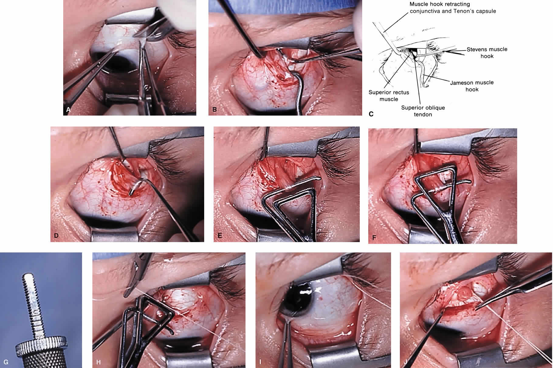
| Fig. 4. Superior oblique tendon tuck procedure in the left eye. A. A conjunctival incision is made temporal to the insertion of the superior rectus muscle. B. and C. With the superior rectus muscle engaged on a Jameson muscle hook, the globe is held in maximal depression and a Stevens tenotomy hook is passed under the superior oblique tendon near its scleral insertion. D. The superior oblique tendon is retrieved from the orbit and tendon laxity is assessed. E. The tendon is removed from the muscle hook and placed on a Bishop tendon tucker. F. The tucker is tightened until snug. G. The amount of tendon shortening (in mm) can be read directly above the screw knob of the tucker. H. Redundant tendon is sewn to itself with 5-0 braided Dacron suture forming a provisional tuck. I. The tendon is released into the orbit and the amount of tendon tuck evaluated using forced ductions. J. If satisfactory, sutures are cut and the conjunctival incision is closed at the surgeon's option. |