|
|
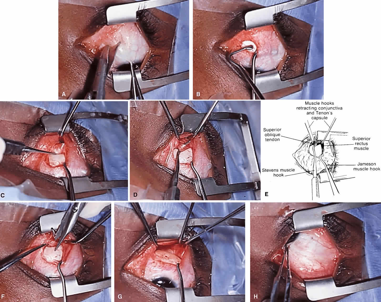
| Fig. 14. Left superior oblique tendon tenotomy performed through a nasal conjunctival approach. A. A conjunctival incision is made medial to the superior rectus muscle insertion. B. The superior rectus muscle is engaged on a Jameson hook. C. The left superior oblique tendon is identified as a white, cordlike structure existing from beneath the medial rectus muscle and entering Tenon's capsule. D and E. Redundant Tenon's capsule is unloaded, but extensive dissection should be avoided. F. The superior oblique tendon is cut at the medial border of the superior rectus muscle; G. The superior nasal quadrant of the globe is carefully examined for missed tendon fibers. H. Complete tenotomy is confirmed by performing forced ductions. |