Chapter 76
History of Photocoagulation
KENNETH R. JAEGERS and JAMES F. VANDER
Main Menu   Table Of Contents

Search

PRINCIPLES OF RETINAL PHOTOCOAGULATION
VARIABLES OF LASER TREATMENT
TREATMENT OF RETINAL VASCULAR DISEASES
RETINAL VENOUS OCCLUSIONS
MISCELLANEOUS PROLIFERATIVE DISORDERS
OTHER RETINAL VASCULAR ABNORMALITIES
TREATMENT OF RETINAL BREAKS
COMPLICATIONS
TREATMENT OF CHORIORETINAL DISEASES
REFERENCES

The effect of solar light on the retina has been known for centuries. After observing burns of the retina caused by watching an eclipse, Socrates warned against the danger of viewing the sun during an eclipse and suggested instead viewing its reflection in water. The first description of a central scotoma following solar burn of the retina dates back to Theophilus Bonetus, who practiced in Geneva during the 17th century.1 The French painter Degas may have sustained a solar retinal burn because he dated his visual impairment from a sunny day when he stood guard duty near a moat during the Franco-Prussian War in the 1860s.

Although we now think that solar maculopathy probably results from a photochemical effect, not a thermal or photocoagulative effect, it was the observation of this phenomenon that inspired the earliest experimental research on fundus photocoagulation. Both Czerny in 1867 and Deutschmann in 1882 used a concave mirror and a convex lens to focus sunlight through dilated pupils onto the retina of rabbits. After a few seconds of exposure, grayish burns of various sizes developed that gradually turned into pigmented scars.2,3 Maggiore performed the first experimental photocoagulation of the human retina in 1927.4 He focused sunlight for 10 minutes into an eye that was to be enucleated because of a malignant tumor.

In 1945, several patients sought medical attention after viewing a solar eclipse. Meyer-Schwickerath, after observing macular damage in these patients, theorized that focused sunlight might be used intentionally to create a potentially therapeutic chorio-retinal lesion.5 Clinical experience led him to the carbon-arc lamp and, eventually, to the xenon-arc lamp.

The xenon-arc lamp, which became available commercially in 1958, produces an emission spectrum similar to that of sunlight, emitting all wavelengths between 400 and 1600 nm. Hence, it produces a full-thickness burn without selective tissue targeting. The xenon-arc lamp burns steadily, and no adjustments are necessary during treatment. These were advances over earlier sources. Over the years, xenon has proved to be an extremely useful light source and has promoted the clinical application of light coagulation as an accepted therapeutic tool. However, it has been superseded by the argon blue-green laser, argon green laser, krypton red laser, dye lasers and, more recently, the diode laser.

In 1960, Theodore Maiman used a ruby crystal in the first ophthalmic laser.6 Flocks and Zweng used this laser in their clinical investigations.7–9 Although the ruby laser produces a pure wavelength of 694 nm and is highly coherent, its red wavelength makes adequate treatment of retinal vascular abnormalities difficult. This difficulty was overcome with the introduction of the argon blue-green laser in 1968.10,11 These wavelengths are fairly well absorbed by hemoglobin and therefore permit direct closure of vascular anomalies. The advent of the argon green laser, with less xanthophyll absorption, made macular photocoagulation safer. These were followed by the development of the krypton red laser.12–14 The tunable organic dye laser, developed during the early 1980s, offers a continuous spectrum of wavelengths from 560 to 640 nm.15,16 The practical introduction of the semiconductor diode laser to the field of ophthalmology occurred in the late 1980s. This is a compact, portable laser with emissions in the near-infrared range with wavelengths between 805 and 810nm.

All these lasers produce a photocoagulative tissue effect by transforming light energy into thermal energy, which produces the desired tissue effect in the photocoagulated tissue or, sometimes, in neighboring tissues by thermal conduction. An alternative method of inducing a laser effect on ocular tissues uses ionizing photodisruption. This is the method used by the neodymium:yttrium-aluminum-garnet (Nd:YAG) laser, and it has its principal applications in treating the tissues of the anterior segment. The excimer laser is photoablative, making use of ultraviolet radiation to produce tissue disruption in the absence of a thermal effect.

Back to Top
PRINCIPLES OF RETINAL PHOTOCOAGULATION
Lasers are grouped according to their active medium, which can reside in solid, liquid, gaseous, or semiconductor states. Argon and krypton lasers are examples of gas-ion lasers in which the active medium is an ionized rare gas. The ruby and Nd:YAG lasers are solid-state lasers. Semiconductor crystals of gallium arsenide (GaAs) or indium phosphide comprise the active media of diode lasers.

Although the technical aspects concerning the production of a laser beam depend on the type of laser being considered, the instruments routinely used in the treatment of retinal disorders all produce a collimated beam of monochromatic light that creates a specific tissue reaction in the photocoagulated retina or choroid. Light energy is absorbed to a variable degree, depending on its wavelength, by the pigments found in the fundus. These are melanin, hemoglobin, and macular xanthophyll. Intensity of any given photocoagulation burn is directly proportional to the duration and magnitude of the temperature increase.

Melanin, found in the retinal pigment epithelium (RPE) and the choroid, absorbs light best. Hemoglobin absorbs yellow, blue, and green wavelengths well. Red and infrared, however, are absorbed poorly. Xanthophyll is located primarily in the macula and resides largely in the inner and outer plexiform layers of the neurosensory retina. Blue light is well absorbed, whereas green, yellow, and red wavelengths are poorly absorbed. Some clinical implications of these findings include decreased absorption and therefore the need for increased power in lightly pigmented fundi, the advantage of krypton red and diode lasers in the presence of vitreous hemorrhage and cataract, and the contraindicated use of blue or argon blue-green laser when treating macular disorders.

The histopathologic characteristics after fundus photocoagulation have been studied extensively. Geeraets and colleagues and then Curtin and Norton showed that high-intensity light energy produces tissue damage involving all layers of the choroid, RPE, and sensory retina, whereas very low energy light intensity involves primarily the pigment epithelium and photoreceptor elements with sparing of the inner retinal layers.17,18 Zweng and different groups of coworkers in several studies noted that ruby laser photocoagulation burns cause early adhesion of the sensory retina to the pigment epithelium and choroid.7–9 In addition, after mild burns, they detected marked destruction and clumping of RPE and marked disruption of the outer half of the sensory retina. The histopathologic effects of argon, krypton and tunable dye lasers have been shown to vary depending to a large extent on the intensity of the burn created.19,20 In general, with longer wavelengths, deeper choroidal penetration and greater sparing of the inner retinal layers are achieved.

Back to Top
VARIABLES OF LASER TREATMENT

DELIVERY SYSTEMS AND INSTRUMENT SETTINGS

After the decision has been made in favor of laser treatment, the ophthalmologist is faced with multiple options including which type of laser to use, how to deliver the laser treatment, and which settings to employ. At present, there are three main delivery systems from which to choose: the slit-lamp biomicroscope, the laser indirect ophthalmoscope (LIO), and the endolaser probe.

The slit-lamp remains the delivery method of choice for most cases, especially those requiring precise laser burn application such as macular photocoagulation. If a slit-lamp system is selected, a contact lens is used. The specific lens chosen depends on the location of the treatment area within the fundus and the surgeon's preference. For panretinal photocoagulation (PRP), the wide-field view provided by a panfunduscopic lens is often helpful. The laser spot size produced with this lens is significantly magnified compared with a more traditional contact lens.

The LIO facilitates peripheral retinal photocoagulation and allows for treatment of patients who previously could not be treated with a slit-lamp system (e.g., pediatric or mentally retarded patients). It can be used to treat with argon, krypton, or diode systems. Using the LIO has become increasingly popular since its introduction in 1981.21 Coupled with the diode laser's portability and studies demonstrating its effectiveness,22 the LIO has largely replaced cryotherapy in the treatment of retinopathy of prematurity.

Laser treatment may also be delivered during vitrectomy surgery using endolaser probes. Newer probes combine laser capability with illumination alone or combined with aspiration functions.

After the type of delivery system has been chosen, variables such as the wavelength, power, spot size, and duration must be considered. The wavelength used is determined primarily by the disease being treated, although for many conditions more than one type of laser may be appropriate. In the past, the argon laser was a blue-green laser. Blue light, however, is scattered by cataractous lenses and absorbed by macular xanthophyll, characteristics that make it an undesirable choice for photocoagulation. Hence, the advent of the argon green laser that emits light that is well absorbed by melanin and hemoglobin but poorly absorbed by macular xanthophyll. Longer wavelengths such as krypton red or diode penetrate deeper into the choroid than green lasers and may therefore be more painful. However, these wavelengths are poorly absorbed by hemoglobin and are more effective in the presence of vitreous hemorrhage. These lasers are also better able to penetrate cataractous lenses.

Other parameters to be considered include the laser power, duration of each exposure, and the spot size. These are interrelated variables that are adjusted during treatment by the ophthalmologist when necessary. Settings depend on the condition being treated, the clarity of the media, and the background pigmentation of the fundus.

In general, topical anesthesia is adequate for most laser treatments. Periocular anesthetic injection may be necessary in cases in which extensive PRP is planned, in particularly sensitive patients, or in cases in which absolute akinesia is necessary, such as with juxtafoveal lesions.

Back to Top
TREATMENT OF RETINAL VASCULAR DISEASES

DIABETIC RETINOPATHY

Diabetic retinopathy is the leading cause of blindness in people 20 to 55 years of age. The most common cause of decreased vision in patients with diabetes is macular edema. Over the past 2 decades, multicenter randomized clinical trials evaluating laser treatment in the setting of diabetic retinopathy have been undertaken. These trials and the practical application of their recommendations are discussed in this chapter.

In 1976, the National Eye Institute completed the Diabetic Retinopathy Study (DRS), a collaborative study to evaluate the effectiveness of scatter PRP for proliferative diabetic retinopathy (PDR).23 More than 1700 patients with advanced retinopathy were enrolled in the study. One eye was arbitrarily assigned to treatment using either xenon arc or argon laser photocoagulation while the other eye was observed. This study showed that both argon and xenon scatter photocoagulation reduced the risk of severe vision loss by 50%.24,25 In this study, “severe vision loss” was defined as visual acuity worse than 5/200 at two consecutive follow-up visits 4 months apart. Because of fewer harmful side effects compared with those associated with xenon, argon is the preferred treatment modality.

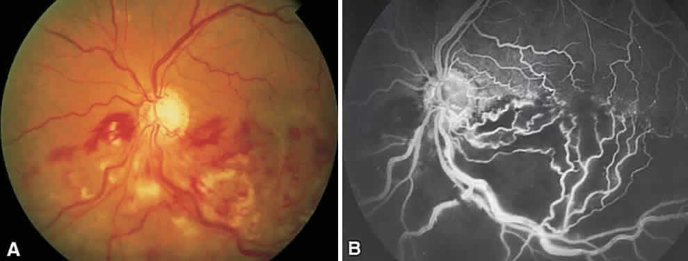
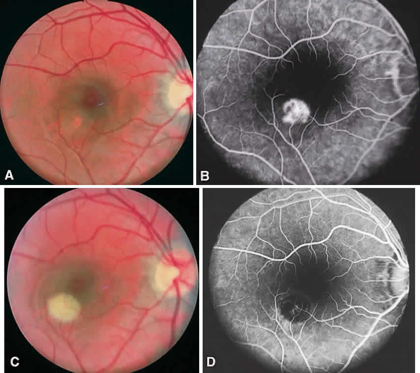
In the DRS, certain high-risk characteristics (HRCs) were identified as being the most accurate prognostic indicators of visual loss. HRCs include neovascularization on or within one disc diameter of the disc (NVD) that is equal to or greater than one fourth to one third of the disc area in extent or any NVD or retinal neovascularization else-where (NVE) associated with preretinal or vitreoushemorrhage (Fig. 1B). For eyes with these clinicalcharacteristics, prompt PRP is recommended. Forpatients with severe or severe nonproliferativediabetic retinopathy (NPDR), as well as for patientsin whom adequate follow-up cannot be ensured, early scatter therapy may be considered.26 The presence of rubeosis iridis or neovascular glaucoma in the setting of PDR, even without HRCs, may warrant PRP.27–29

Fig. 1. A. Florid neovascularization of the disc. Any such neovascularization that covers more than one fourth of the disc area meets high-risk characteristics and warrants prompt panretinal photocoagulation. B. Neovascularization elsewhere and preretinal hemorrhage. Panretinal photocoagulation is indicated.

The precise mechanism of formation of neovascular tissue remains uncertain, as does the mechanism by which PRP induces regression of this tissue. Traditionally, new vessel formation was thought to be a response to a possible angiogenic factor that may be produced by zones of ischemic retina.30 Destruction of these zones by PRP might then eliminate the source of this angiogenic stimulus. Although this approach may not accurately reflect the true pathophysiology involved, it does serve as a helpful model for understanding the proper methods of treatment for this condition. These concepts apply to other proliferative retinopathies as well including central retinal vein occlusion, branch retinal vein occlusion, SC sickle disease, sickle thalassemia, retinopathy of prematurity, Eales' disease, dominant familial exudative retinopathy, and X-linked dominant incontinentia pigmenti. Of note, pregnancy can exacerbate and accelerate all aspects of diabetic retinopathy. It is unusual for pregnant patients without retinopathy at the onset of pregnancy to pro-gress to PDR during their pregnancy. However, patients with preexisting PDR are at greatest risk of visual loss and need to be observed closely and treated aggressively.

Methods

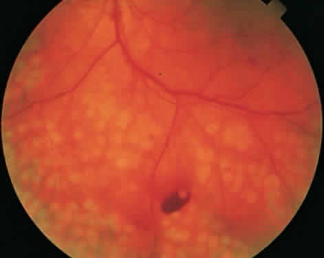
To complete scatter treatment, burns should be applied to the retina beginning at points on an oval defined as two disc diameters above, below, and temporal to the center of the macula, and one disc diameter nasal to the disc, and should extend peripherally at least to the equator (Fig. 2). This approach may help avoid inadvertent macular burns. A report by Blankenship suggests that sparing of the posterior aspect of the fundus may be possible without diminishing the beneficial effects of treatment.31 With the Goldmann lens, a 500-μm spot size is used, whereas with the panfunduscopic lens, a 200-μm spot is used. When vitreous hemorrhage is present, it may be necessary to reduce the size of the spot to 200 μm or to use a krypton or diode laser rather than an argon laser. The Krypton Argon Regression of Neovascularization Study found that krypton and argon were equally effective in inducing regression of NVD.32

Fig. 2. Panretinal photocoagulation. Initial burns surround the disc and macula to outline the most posterior extent of intended treatment. Subsequent treatment is placed only peripheral to this ring, thereby reducing the risk of inadvertent macular photocoagulation.

Care should be taken to avoid hitting any visible retinal vessels. The power setting should be such that a moderate white retinal coagulation is apparent. The power setting required to achieve this endpoint is recorded for future reference and is defined as the baseline power setting for the treatment session. It frequently needs to be higher for a panfunduscopic lens than for a three-mirror lens.

In general, application should be scattered uniformly, with the distance between burns being one burn diameter. It may be wise to treat inferiorly during the first session because vitreous hemorrhage, should it occur, tends to settle inferiorly. To minimize impairment of the temporal visual field, burns within four to five disc diameters of the disc on the nasal side should be arranged in rows parallel to the nerve fibers. Exposure time should be set at 0.1 or 0.05 second. In general, at least 1600 to 1800 burns should be applied. We usually treat over multiple sessions.

Occasionally PRP fails to induce regression of neovascularization and vision decreases. This can be a result of increased macular edema, focal bleeding from persistent neovascular fronds, retinal detachment, or neovascular glaucoma. Regression of HRCs occurs in 70% of cases within 3 weeks of treatment.33 Not surprisingly, patients who demonstrate a favorable early objective response to laser therapy have a significantly better visual prognosis than those who do not.34 In the case of increasing NVD or persistent NVD with bleeding, supplemen-tal treatment may be helpful. Peripheral retinal cryo-ablation may also be effective in causing regression of NVD or NVE. Cryotherapy is especially helpful in the setting of significant media opacities. About six applications are necessary in each quadrant. If vitreous hemorrhage has occurred and appears to be nonclearing, vitrectomy surgery may be indicated.

DIABETIC MACULAR EDEMA

Diabetic macular edema (DME) is a clinical diagnosis made with the slit-lamp biomicroscope. Intraretinal thickening is divided into focal and diffuse forms. Focal retinal edema is usually caused by specific leaking microaneurysms visible as pinpoint areas of leakage on fluorescein angiography (FA). The diffuse form of macular edema, conversely, usually represents a more widespread disruption of the inner blood-retina barrier. FA typically depicts large areas of leakage. It is important to remember that leakage of dye on FA does not always mean intraretinal thickening is present. Diagnosis of macular edema is based on the clinical examination of the retina.

The Early Treatment of Diabetic Retinopathy Study (ETDRS) was a multicenter, randomized, prospective clinical trial designed to address three main questions: Is laser photocoagulation effective in the treatment of DME? When in the course of diabetic retinopathy is it most effective to initiate PRP? Is aspirin effective in altering the course of diabetic retinopathy? The ETDRS demonstrated that laser photocoagulation for macular edema decreased the risk of moderate visual loss (defined as a doubling of the visual angle) by more than 50% especially in eyes with patterns of macular edema now known as clinically significant macular edema (CSME).35 Focal treatment also increased the chance of moderate visual gain.

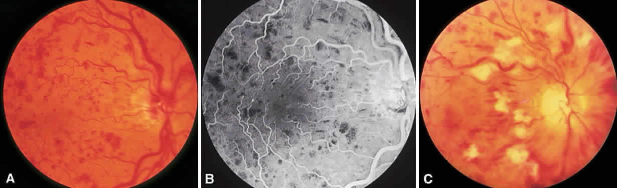
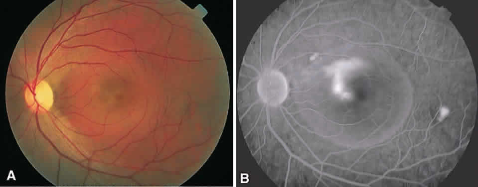
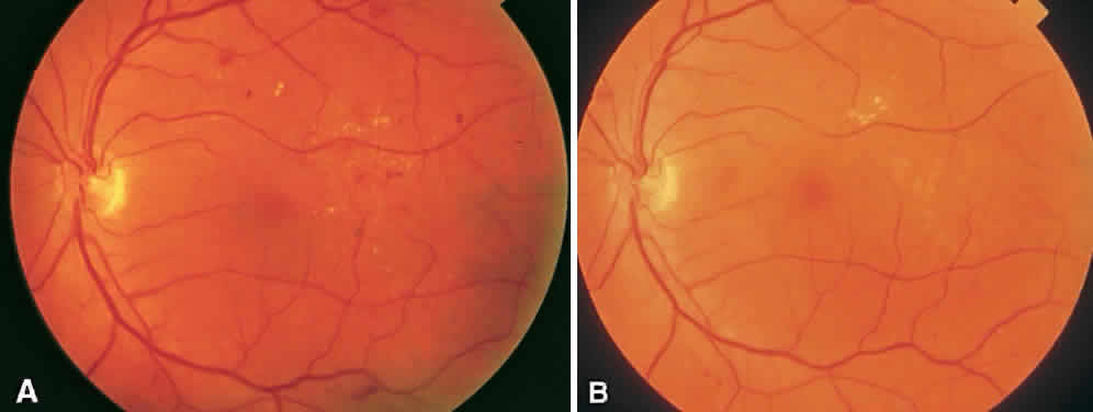
CSME was defined as retinal thickening within 500 μm of the center of the macula, intraretinal hard exudate within 500 μm of the center of the macula associated with adjacent retinal thickening, or retinal thickening greater than one disc area any part of which is within one disc diameter of the center of the macula (Fig. 3A). The beneficial effects of treatment demonstrated in this trial suggest that all eyes with CSME should be considered for focal photocoagulation (see Fig. 3B). Visual acuity was not a factor in determining the presence or absence of CSME and some eyes in the treated group had20/20 visual acuity. Many retinal specialists, how-ever, defer treatment in asymptomatic patients with20/20 visual acuity except when hard exudate is encroaching on the fovea.

Fig. 3. A. Microaneurysms, hard exudate, and clinically significant macular edema temporal to the fovea. B. Three years after laser photocoagulation, the edema has resolved and the laser burns are barely visible.

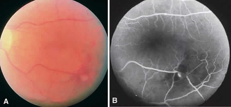
When CSME is present, fluorescein angiography is helpful in ruling out macular ischemia as a cause for decreased vision. If significant perifoveal capillary nonperfusion is present, then macular photocoagulation is associated with a higher risk of producing an immediate and permanent reduction in visual acuity. If photocoagulation is performed in such cases, care should be taken to avoid directly treating the few remaining perifoveal capillaries.

Methods

Various treatment strategies have been devised for photocoagulation of DME. Although the optimal method remains uncertain, most adhere to the basic principles described in the ETDRS. The argon green laser is widely used, although evidence now suggests that other wavelengths, such as dye yellow and orange, krypton red, and the diode, are comparable.36–39 A pretreatment fluorescein angiogram is generally used during photocoagulation to identify treatable lesions. Focal laser treatment involves direct laser treatment to all leaking microaneurysms.

When directly treating a microaneurysm, a 50- or 100-μm burn with 0.1-second duration is used, and one usually attempts to induce a slight change in the color of the lesion. Use of the 50-μm spot size may be associated with a higher risk of rupturing Bruch's membrane and resultant choroidal neovascularization (CNV), so very low energy levels should be used initially.40 Initial treatment is directed at lesions located within two disc diameters of the center of the macula, but not closer than500 μm from its center. However, if vision is lessthan 20/40 and follow-up examination reveals persistent CSME, additional treatment of lesions up to 300 μm from the center is recommended.

Areas of diffuse leakage or nonperfusion within two disc diameters of the center of the macula are treated with a grid pattern. Diode laser has been shown to be as effective as argon laser in the treatment of diffuse DME.41 The goal of treatment in such cases is to produce a burn of light to moderate intensity. A 100- to 200-μm spot size is used, and burns are typically spaced one burn-width apart. Burns may be placed in the papillomacular bundle but, again, not closer than 500 μm from the center of the macula at the first treatment. For patients with a combination of focal and diffuse edema, a modified grid technique may be employed.42

In addition to the development of CNV mentioned earlier, other complications can occur after focal macular photocoagulation. Permanent visual loss may ensue after treatment. Causes includehemorrhage, perifoveal capillary occlusion, enlargement of photocoagulation scars to involve the fovea, marked lipid exudation as edema resolves, or in-advertent foveal burns. Paracentral scotomataare a frequent side effect, especially after heavy treatment.

Some patients present with CSME in the presence of PDR with HRC. Because PRP can exacerbate preexisting macular edema, it is generally advisable to initiate focal or grid treatment prior to or along with PRP. Performing nasal PRP first and completing the PRP is additional sessions may help prevent a worsening of the macular edema.

Regarding the use of aspirin, the ETDRS found that aspirin use did not affect the progression of retinopathy. Aspirin did not increase the risk for vitreous hemorrhage, the duration of or severity of vitreous hemorrhage, nor the rate of pars plana vitrectomy. Visual outcomes were not affected by aspirin use.43 Therefore, there is no ocular contradiction to the use of aspirin in patients with diabetic retinopathy.

Back to Top
RETINAL VENOUS OCCLUSIONS

BRANCH RETINAL VEIN OCCLUSION

Branch retinal vein occlusions (BRVOs) usually occur at arteriovenous crossings, with the arteriole typically crossing anterior to the vein.44 Visual loss may be a result of macular hemorrhage or edema, capillary nonperfusion, RPE changes or vitreous hemorrhage (Fig. 4). The Branch Vein Occlusion Study (BVOS) was a prospective, randomized multicenter clinical trial designed to address the following questions: Is macular argon laser photocoagulation beneficial in preserving or improving central visual acuity in eyes with 20/40 or worse vision secondary to macular edema related to a branch vein occlusion? Can peripheral scatter argon laser photocoagulation prevent the development of retinal neovascularization? Can peripheral scatter laser treatment prevent vitreous hemorrhage from retinal neovascularization? This study demonstrated conclusively the effectiveness of argon laser photocoagulation for the treatment of macular edema and retinal neovascularization (Fig. 5).45,46 After 3 years of follow-up, the gain of at least two lines of visual acuity from baseline maintained for two consecutive visits was significantly greater in treated eyes. As a result, laser photocoagulation is now recommended for patients with macular edema from branch vein occlusion. In addition, Roseman and Olk have shown that krypton red is effective for treating macular edema and neovascularization from BRVO in eyes with cataracts, vitreous hemorrhage, or extensive intraretinal hemorrhage.47 Visual improvement, however, is unlikely in cases with marked capillary nonperfusion and severe visual loss.

Fig. 4. A. Hemiretinal vein occlusion. B. Fluorescein angiogram reveals severe capillary nonperfusion.

Fig. 5. A. Fluorescein angiogram of a superotemporal branch retinal vein occlusion with marked retinal ischemia. The localized hyperfluorescence in the papillomacular bundle represents retinal neovascularization. B. Three years after sector laser photocoagulation to area of retinal nonperfusion.

If, after 3 months, visual acuity is 20/40 or worse, laser treatment is indicated. Grid treatment with a spot size of 100 to 200 μm and 0.1-second duration is placed in areas of leakage as shown on FA. There should be sufficient clearing of retinal hemorrhage before FA or laser because visual acuity may increase and FA may be more useful in terms of evaluating the macula for evidence of capillary nonperfusion. Recommendations concerning intensity of burns, spacing between laser burns, and treating near the fovea as well as potential complications are similar to those for laser treatment of DME. Eyes are reevaluated in 4 months and retreated if persistent macular edema accounts for the decrease in visual acuity.

The BVOS also assessed the role of scatter photocoagulation in preventing formation of retinal neovascularization and subsequent vitreous hemorrhage in patients with ischemic occlusions.47 It was noted that scatter argon laser photocoagulation to the affected segments, as determined by color photography and FA, extending no closer than two disc diameters to the center of the fovea, significantly decreased the risk of developing retinal neovascularization and lessened the occurrence of vitreous hemorrhage. Eyes at greatest risk for the development of retinal neovascularization were those with ischemic branch vein occlusions defined as eyes having more than five disc areas of capillary nonperfusion on FA. The risk of hemorrhage impairing vision in untreated patients is fairly low, however, and as a result no difference was found in terms of final visual acuity between treated and untreated groups. The study therefore recommends that scatter laser treatment be deferred until retinal neovascularization is present. The methods, as well as risks, of treatment are similar to those relating to PRP in patients with diabetes, except that treatment is limited to the affected area.

CENTRAL RETINAL VEIN OCCLUSION

Central retinal vein occlusions (CRVOs) are caused by thrombosis formation within the vein at the level of the lamina cribrosa. Clinical findings in the setting of a CRVO include intraretinal hemorrhages in all four quadrants, increased dilation and tortuosity of the retinal veins, macular edema, nerve fiber layer infarcts, and disc edema. Acutely, the decrease in visual acuity is secondary to macular changes such as macular edema, hemorrhage and capillary nonperfusion. Long-term visual loss may be secondary to macular capillary nonperfusion, chronic macular edema, arteriole occlusion, tractional or exudative retinal detachment and neovascular glaucoma.48,49 Anterior segment neovascularization is much more common with CRVOs than BRVOs.

CRVOs are usually described as “ischemic” or “nonischemic” based on clinical examination and FA findings (Fig. 6). Roughly 75% to 80% of CRVOs are nonischemic. However, up to 34% of these will progress to the ischemic type usually within 1 year.46,47,50,51 An eye with a nonischemic CRVO typically has minimal capillary nonperfusion on FA, relatively few intraretinal hemorrhages, and a better visual prognosis than the ischemic variety. Ischemic CRVOs, conversely, have widespread capillary nonperfusion and are associated with a worse visual prognosis with increased risk of rubeosis iridis and neovascular glaucoma.52

Fig. 6. Nonischemic (A and B) and ischemic (C) central retinal vein occlusion.

The Central Vein Occlusion Study (CVOS) was a prospective, randomized, multicenter clinical trial designed to evaluate the role of PRP in promoting regression of anterior segment neovascularization resulting from ischemic CRVO, to determine the efficacy of macular grid laser photocoagulation for treating CRVO-related macular edema associated with visual acuity of 20/50 or worse, and to define the natural history of CRVO.53

The CVOS demonstrated that, although prophylactic PRP does decrease the risk of developing anterior segment neovascularization, prompt regression of neovascularization was more likely in eyes that had not received prior laser photocoagulation.54 Therefore, the study recommends observing ischemic CRVOs closely and treating with PRP at the first sign of iris or angle neovascularization rather than treating prophylactically. PRP may be warranted in the setting of acute ischemic CRVO if there is a high likelihood of poor patient compliance with follow-up. Unlike the BVOS, the CVOS did not demonstrate any benefit to laser photocoagulation for macular edema.55 Although laser therapy did decrease the amount of leakage on FA, there was no difference in visual acuity between treated and untreated eyes at any point in follow-up.

An innovative treatment for nonischemic CRVO has previously been described.56 By using lasers to create an anastomotic connection between a retinal and a choroidal vein, a bypass to the obstruction can be created. Modifications to the initial technique have improved the rate of successful creation of chorioretinal anastomosis to 54%.57 Of the patients with a functioning anastomosis, 84% had visual improvement with an average improvement of 4.3 lines. BVOs may also benefit from such therapeutic intervention.58 The principle complication of this technique is the development of neovascularization, which can be subretinal, intraretinal or intravitreal. A pilot study using transscleral diode laser to create a chorioretinal anastomosis is under way.

All patients with a CRVO should be examined at monthly intervals for the first 6 months. Particular attention should be given to anterior segment structures such as the iris, especially the pupillary margin, and the anterior chamber angle, with the practitioner looking specifically for neovascularization. Thus, an undilated examination of the anterior segment including gonioscopy is essential in following these patients. This close follow-up is especially important in eyes with symptoms of less than 1 month's duration, initial visual acuity less than 20/200, numerous intraretinal hemorrhages, and extensive capillary nonperfusion because these eyes have a much higher risk of developing anterior segment neovascularization.59,60

Back to Top
MISCELLANEOUS PROLIFERATIVE DISORDERS
A multitude of other disorders can produce significant retinal ischemia with subsequent neovascular proliferation. Some, as in the ocular ischemic syndrome, are associated with decreased ocular blood supply. In this condition, PRP may help control rubeosis iridis, neovascular glaucoma, and posterior segment neovascularization.61–63 Other entities such as talc retinopathy and radiation retinopathy may produce changes warranting PRP.64,65 Use of focal and grid lasers may be beneficial in treating radiation-induced macular edema.66

Numerous other retinal vascular disorders can lead to posterior or peripheral retinal neovascularization. Experience using photocoagulation for the management of conditions such as proliferative sarcoid retinopathy or Eales' disease is certainly limited, and no controlled studies exist to verify the efficacy of treatment for such patients. Nevertheless, when progressive retinal ischemia and neovascularization develop, photocoagulation of areas of retinal nonperfusion is a rational approach and frequently leads to neovascular regression.

SICKLE CELL RETINOPATHY

Proliferative sickle cell retinopathy is most common in the SC form of this disorder. The initial finding is peripheral arteriolar occlusions, which cause retinal ischemia.67–71 Arteriovenous connections develop in the temporal periphery and lead to the typical sea fan formation and ultimately to vitreous hemorrhage. In the past, it was recommended that these arteriovenous communications be treated focally in an attempt to eliminate the feeder vessel entering the frond. This form of treatment poses some degree of risk, however, because it is not uncommon for the high-intensity burns used to occlude feeder vessels to rupture Bruch's membrane. As a result, peripheral scatter photocoagulation to areas of ischemia has gained favor as the treatment method of choice.72 Local scatter treatment is recommended for the compliant patient who will return for regular follow-up examinations. Otherwise, 360-degree scatter should be considered.

RETINOPATHY OF PREMATURITY

In 1988, the CRYO-ROP study first reported on the efficacy of peripheral retinal cryotherapy for “threshold” retinopathy of prematurity (ROP).73 Although cryotherapy is an effective treatment in many cases, the treatment occasionally may be technically difficult to perform and the complications, especially systemic ones, can be severe.74 Laser treatment is much less traumatic. The development of the laser indirect ophthalmoscope as a laser delivery system offers an alternative method of peripheral retinal treatment for these infants. McNamara and colleagues, in a randomized controlled clinical trial, showed that scatter photocoagulation to the peripheral avascular retina using the indirect laser system to deliver argon laser irradiation was as effective as cryotherapy in preventing visually significant structural retinal changes.75 The Laser ROP Study Group also concluded that laser photocoagulation was as effective as cryotherapy at inducing regression of threshold ROP.76 Additionally, laser photocoagulation appears to be effective in eyes with zone I threshold ROP whereas the CRYO-ROP study failed to show a significant benefit for treatment of these eyes.77–79 The argon and diode lasers appear equally effective in causing regression.80 The diode laser's portability makes it the preferred laser in many cases. The diode is also able to penetrate the tunica vasculosa lentis better than the argon. Seiberth and partners have demonstrated the effectiveness of transcleral diode laser therapy in the management of threshold ROP.81

Back to Top
OTHER RETINAL VASCULAR ABNORMALITIES

RETINAL TELANGIECTASIA (COATS' DISEASE)

In 1908, Coats published the first of two articles in which he described a condition characterized by retinal vascular changes and exudation.82 Retinal telangiectasia or Coats' disease occurs in healthy young males, although females are occasionally affected. Of these cases, 90% are unilateral. The superior temporal quadrant is most commonly involved. The hallmark of Coats' disease is the development of “lightbulb” telangiectasis in the retinal periphery, which can lead to subretinal exudation that has an affinity for the posterior pole.

Untreated leakage usually has a poor prognosis, with eyes developing macular lipid exudation, exudative retinal detachment, and secondary glaucoma. Although the spectrum of clinical severity is wide and not all eyes need treatment, when lipid is threatening to or does involve the macula treatment is indicated. Treatment is directed at obliterating the abnormal telangiectatic vascular changes by photocoagulation or cryotherapy. Posterior lesions are more easily treated with laser than cryotherapy. Although cryotherapy has traditionally been the treatment of choice in the management of Coats' disease, laser photocoagulation can be useful.83,84 Cryotherapy is probably superior if large areas of exudate are present beneath the telangiectatic areas, because it is difficult to achieve an adequate photocoagulation reaction when underlying exudate is present. Occasionally, subretinal fluid may need to be drained before an adequate tissue reaction can be obtained. Multiple treatment sessions are often necessary.

The prognosis with treatment is best when only one or two quadrants are affected and diminishes significantly when the disease involves more than 180 degrees of the retinal periphery. With the successful obliteration of the abnormal vasculature, exudate usually begins to absorb in about 6 to 8 weeks. As long as 10 to 12 months may elapse, however, before it is entirely gone. Even after complete obliteration of all abnormal vessels and resorption of all exudate, follow-up examinations are mandatory. If new exudate begins to appear later, it can be assumed that new vascular abnormalities have developed. This disease has been noted to recur as long as 5 years after an apparent cure.83 The disease course may be especially severe in younger patients and they therefore need to be treated more aggressively.

ARTERIAL MACROANEURYSM

Arterial macroaneurysms are frequently encountered in patients with systemic hypertension but may also occur without apparent antecedent cause. They are often associated with surrounding exudation and, in some cases, present because of significant hemorrhage. Visual loss is usually secondary to macular edema, hard exudate, or hemorrhage. Hemorrhage from an arterial macroaneurysm may be located beneath, within, or anterior to the retina. FA is helpful in establishing the diagnosis, because the macroaneurysm usually retains fluoresceinand appears as a discrete hyperfluorescent spot(Fig. 7).85

Fig. 7. Retinal macroaneurysm. Focal dilatation along the inferotemporal arcade and a corresponding focal area of hyperfluorescence can be seen on this fluorescein angiogram. Macroaneurysms are usually arterial but can be venous.

Treatment of these lesions is controversial.86 Many resolve spontaneously after bleeding, and treatment is generally reserved for those that have an exudative component threatening the macula. The optimal method of treatment is also uncertain. Direct treatment of the macroaneurysm runs the risk of inducing hemorrhage or an arterial obstruction. If such treatment is attempted, very low energy levels and a large spot size should be used. Light treatment with a tight grid pattern around the macroaneurysm may be all that is necessary to arrest or reverse the exudative complications of this lesion. Overall, for eyes with macroaneurysms with a primarily hemorrhagic component, observation may be best because these tend to bleed only once. When macular edema and hard exudate are increasing and threatening the macula, laser therapy is probably indicated.

RETINAL CAPILLARY HEMANGIOMA

Retinal capillary hemangiomas are benign vascular hamartomas inherited as an autosomal dominant disease and are bilateral in 50% of cases. Importantly, 25% of patients with retinal capillary hemangiomas have von Hippel-Lindau disease, a phakomatosis associated with central nervous system hemangioblastomas, with cysts of the liver, pancreas, and kidney, and with renal cell carcinoma.87 A complete medical evaluation as part of the initial examination and continued follow-up is mandatory in these patients.

The retinal capillary hemangioma itself appears as an orange-red lesion usually located in the equatorial or preequatorial region. The lesion may vary from that too small to detect ophthalmoscopically to those several disc diameters in size. Classically, the tumor is associated with dilated and tortuous afferent and efferent vessels. Although the tumor may remain stable for years or even regress spontaneously, it can also leak significantly, which leads to lipid exudation and exudative retinal detachment. As with Coats' disease, lipid in the macula should lead the examiner to perform a careful examination of the peripheral retina.

Although treatment is clearly indicated for lesions causing macular edema or hard exudate in or threatening the macula, management of small asymptomatic lesions is more controversial. Given the relatively poor prognosis of large tumors, some prefer to treat all lesions as soon as they are discovered.

Early attempts to eradicate these lesions included radiation and diathermy. The advent of photocoagulation provided another means for successful treatment of small and medium tumors. Photocoagulation offers significant advantages over the other modes of treatment. It requires no conjunctival incision and causes no scleral necrosis. The technique involves mild photocoagulation burns to the surface of the angioma. These are just intense enough to cause blanching on the surface of the tumor. The treatments are frequently spread out over several sessions and have as their major complicationshemorrhage and secondary exudative retinal detachment.88

Larger lesions are best managed with feeder vessel photocoagulation. Dye yellow wavelengths should be used in these instances because of their maximal absorption by hemoglobin.89 Cryotherapy is also effective and may be the treatment of choice in patients with hazy media.90,91

Other vascular anomalies such as idiopathic juxtafoveal retinal telangiectasis, retinal cavernous hemangioma and racemose hemangioma only occasionally warrant laser therapy.

Back to Top
TREATMENT OF RETINAL BREAKS

CREATING CHORIORETINAL ADHESIONS

Before addressing the treatment of retinal breaks, some discussion of the different types of retinal breaks that may occur is warranted. A retinal break is defined as any full-thickness retinal defect. Retinal holes are breaks without vitreoretinal traction whereas a tear is a break with or caused by vitreoretinal traction. A horseshoe tear is caused by persistent vitreal traction pulling the flap anteriorly. In an operculated tear, conversely, a piece of retina has been torn completely free. A symptomatic break is associated with photopsias and entopsias.

The incidence of retinal breaks in the general population is 5% to 7%.92,93 Treatment, either by laser photocoagulation or transcleral cryotherapy, aims at creating a chorioretinal adhesion that will prevent liquid vitreous from passing through the break, thereby causing a retinal detachment. Thus, it is important to know which breaks need treatment and which can be safely managed with observation alone. The presence or absence of symptoms is important in making this decision. Although asymptomatic breaks in patients without a history of retinal disease are at low risk for progressing to retinal detachment,94 patients with a retinal break who are experiencing symptoms may well need treatment. This is especially the case in acute, symptomatic horseshoe tears that will progress to retinal detachment between 25% and 90% of the time.95–97 Thus, the type of break is also important in the decision-making process. Most horseshoe tears and dialyses need treatment whether or not the patient is having symptoms. Atrophic holes and lattice degeneration only occasionally need to be treated.98,99 Operculated holes in a patients with symptoms should probably be treated.

Either laser photocoagulation or cryotherapy can be used to treat retinal breaks. Transcleral cryotherapy is useful in the presence of media opacity. In the presence of clear media, however, laser photocoagulation, delivered at the slit-lamp biomicroscope or through the LIO, has become the treatment modality of choice. Laser treatment of posterior breaks avoids cutting cryotherapy. Laser also avoids liberation of RPE cells as seen with cryotherapy.100 Breaks that may be treated with laser therapy or cryotherapy are horseshoe tears that are either totally flat or have only minimal subretinal fluid (i.e., less than two disc diameters in area) under the edges. Breaks with bridging vessels can be treated in a similar fashion, but they may bleed subsequently and should be watched closely afterward.

Adequate treatment of the break necessitates encircling it with two or three rows of laser application. Settings are adjusted to produce a 200- to 500-μm burn of moderate intensity. Treatment of flap tears should be extended to the ora serrata except in cases with posterior breaks where the entire break should be well surrounded. Inadequate anterior treatment is the most common cause of treatment failure. Close follow-up is essential because up to 14% of eyes develop new breaks at some point after treatment of a retinal tear.101,102

Breaks in lattice degeneration occur primarily as round holes within the lattice or as horseshoe tears at the edge of the lattice. The latter are more serious and warrant treatment if the patient is symptomatic or has a history of detachment in the contralateral eye. When laser treatment is given for lattice with break formation, we prefer to encircle the lattice by placing two or three rows of laser burns in sound retina around the area. The settings are generally the same as described for treatment of retinal breaks. A retinal detachment is referred to as subclinical if subretinal fluid extends at least one disc diameter from the break but no more than two disc diameters posterior to the equator. Because 30% of such retinal detachments progress,97 treatment is indicated, usually with a scleral buckling procedure or pneumatic retinopexy. In some cases, delimiting this type of detachment with photocoagulation is satisfactory. Again, such treatment is likely to fail unless carried to the ora serrata at each end of the detachment.

DEGENERATIVE RETINOSCHISIS

Degenerative retinoschisis is considered to be a posterior extension of peripheral cystoid degeneration resulting in a split in the outer plexiform layer of the neurosensory retina. It is usually bilateral and inferotemporal. It should be differentiated from rhegmatogenous retinal detachment. Byer has demonstrated that clinical retinal detachment is rare.103 He therefore recommends prophylactic treatment only in the setting of a symptomatic and progressive schisis-retinal detachment.

Patients are occasionally seen to develop outer-layer retinal breaks, but these rarely become associated with symptomatic retinal detachment secondary to the schisis. As a result of Byer's report, when outer-layer retinal breaks are present, we do not recommend treatment. If clinically symptomatic retinal detachment occurs, then scleral buckling is indicated.

Back to Top
COMPLICATIONS
Laser photocoagulation may be associated with complications involving anterior and posterior segment structures, singly or in combination. In addition to these objective findings, the patient may notice subjective changes in her or his vision as well. There is also good evidence that the treating ophthalmologist may personally suffer color vision changes after chronic exposure to argon blue light.104 Corneal damage may range from a small corneal abrasion to an inadvertent thermal burn. Iris burns can lead to iritis and posterior synechiae.105 Lenticular burns and transient loss of accommodative amplitudes have also been reported.106,107

PRP can induce elevation in intraocular pressure, the peak of which usually occurs in the first 24 hours after treatment.108 Although the anterior chamber is usually open, occasionally choroidal effusion or detachment causes forward rotation of the ciliary body and secondary angle closure glaucoma. Secondary glaucoma due to choroidal detachment after PRP is probably related to the amount of treatment given during a session.109 The glaucoma is usually transient and can almost always be treated medically. PRP may also be associated with visual field loss, loss of color vision, nyctalopia, and worsening of preexisting macular edema. Blankenship and others have discussed methods to minimize the risk of PRP-induced macular edema.31,110,111 These methods include treating CSME before starting PRP if possible and dividing PRP into multiple treatment sessions.

Both PRP and intense focal macular laser can cause CNV. The neovascular complex grows through an iatrogenic break in Bruch's membrane.112 The risk of this complication can be minimized by not using small, short-duration high-power burns, which tend to puncture Bruch's membrane. Laser scar enlargement can also occur. If the original burn is close enough to the fovea, an increasingly disturbing scotoma or a frank decrease in visual acuity may occur. This is especially noted in treatment of CNV related to high myopia. Inadvertent macular/foveal photocoagulation has also occurred. RPE tears and subretinal hemorrhage are other possible complications. Fortunately, the risk of encountering these problems can be minimized by using the appropriate laser settings, careful attention to the position of the fovea and dividing PRP into multiple treatment sessions.

Back to Top
TREATMENT OF CHORIORETINAL DISEASES

MACULAR DEGENERATION

In the United States, age-related macular degeneration (AMD) is the most common cause of severe visual loss in people older than 65 years of age. Most patients with AMD who suffer severe vision loss have CNV.113 AMD-related CNV tends to be a bilateral disorder and, over 5 years, CNV develops in nearly 60% of fellow eyes that were initially free of neovascularization.114 CNV represents new and abnormal blood vessel growth either between the RPE and Bruch's membrane or in the subretinal space. Typically patients with CNV present complaining of blurred or distorted vision and, on examination, are found to have evidence of neovascularization such as subretinal fluid, hemorrhage, or exudation. In cases where CNV is apparent or suspected, FA alone or combined with indocyanine green angiography should be performed (Fig. 8). The results of these studies help determine treatment options that include observation, laser photocoagulation, photodynamic therapy (PDT), transpupillary thermotherapy (TTT), laser prophylaxis, and dye-enhanced laser treatment.

Fig. 8. A. Choroidal neovascularization. B. Classic CNVM on fluorescein angiography. C. Clinical appearance immediately after laser treatment shows intense retinal whitening. D. Angiographic appearance 2 weeks after photocoagulation shows good closure of CNVM.

The Macular Photocoagulation Study (MPS) was a randomized, prospective multicenter clinical trial that documented that laser photocoagulation was preferable to observation in the management of select patients with well-defined CNV from AMD, ocular histoplasmosis, and idiopathic causes.115–118 Unfortunately, using the MPS criteria, only 15% of patients with AMD-related CNV are eligible for laser treatment and one half of these are subfoveal on presentation. Of nonsubfoveal membranes treated with laser, over 50% recur, usually on the foveal side of treatment. Recurrence of CNV is associated with a poor visual prognosis.119 In the MPS, the major outcome variable, severe visual loss, was defined as a loss of six or more lines from initial or baseline visual acuity.

The MPS categorized CNV based on its location relative to the center of the foveal avascular zone as depicted by FA. Extrafoveal lesions are CNV200 μm or more from the center of the foveal avas-cular zone, whereas juxtafoveal lesions are between1 and 199 μm from the center. Subfoveal membranesextend through or underneath the foveola. For ex-trafoveal CNV related to AMD, severe vision loss occurred in 64% of untreated eyes versus 46% of treated eyes after 5 years of follow-up.115 Fifty-four percent of treated eyes developed recurrent CNV, which was typically subfoveal. For juxtafoveal lesions, severe vision loss after 5 years was noted in 65% of untreated eyes versus 55% of treated eyes.116 As in the extrafoveal group, the major cause of vision loss was persistent and recurrent CNV. The MPS has demonstrated a treatment benefit of laser treatment over observation for subfoveal recurrent CNV.118 Although occult CNV may benefit from lasers in certain circumstances, in the MPS the treatment benefit was greatest for eyes that had only classic CNV.120,121 Later, the MPS evaluated laser treatment for subfoveal CNV in the subfoveal118 and subfoveal recurrence122 studies. Both studies reported a treatment benefit, but only if the CNV was relatively small. The MPS also evaluated laser treatment of idiopathic CNV and CNV related to ocular histoplasmosis. Outcomes in both groups were better than for AMD-related CNV with a decreased incidence of severe visual loss and fewer recurrences.123,124

METHODS

Recognizing that CNV can grow at the rate of70 μm/day,125 the MPS recommends projection of a FA no more than 72 hours old to guide laser treatment. This helps ensure complete treatment of the CNV. There is a higher rate of recurrence if theinitial treatment fails to completely cover the CNV.126Although topical anesthesia usually suffices, if the patient cannot fixate with the fellow eye or if there is too much eye movement, retrobulbar anesthesia is indicated. A test burn is usually given in a safe location to determine the appropriate power setting. We typically begin with a 200-μm spot size, 200-mW power, and 0.2- to 0.5-second duration. The perimeter of the lesion is outlined first with overlapping burns with treatment extending 100 μm beyond the CNV. Then, the center is treated. Burns should be heavy, white, and confluent.122,124 There appears to be no difference between argon green and krypton red wavelengths in terms of final outcomes.118 The dye yellow laser is also effective. A posttreatment FA is obtained 2 to 3 weeks later. Persistent or recurrent CNV can be managed according to the clinical and angiographic findings.

NEW TREATMENT MODALITIES

Although laser photocoagulation of AMD-related CNV is useful in selected situations, most patients with CNV are not candidates for conventional treatment. Because of these limitations, new treatment modalities are under investigation. One of these, PDT, is currently being used in the management of subfoveal CNV.127–132 Unlike conventional laser treatment that results in the thermal destruction of normal as well as abnormal tissues, PDT uses nonthermal laser light to activate a photosensitizing agent that initiates a free radical cascade resulting in selective occlusion of the CNV without damaging the overlying neurosensory retina. This selective tissue destruction with PDT is achieved by sequestration of the photosensitizer in the target tissue, which is then irradiated with low energy light. The photosensitizing dye is injected intravenously 15 minutes before laser treatment using the diode laser. A subthreshold treatment is delivered such that the treating ophthalmologist cannot appreciate a change in the clinical appearance of the patient's fundus.

Several photosensitizing agents are under investigation including verteporfin (Visudyne), tin ethyl etiopurpurin (SnET2/Purlytin), and luteium texaphyrin (Lu-Tex). The Treatment of Age-Related Macular Degeneration with Photodynamic Therapy (TAP) Study is using verteporfin as the photosensitizing agent. The 12-month data from this study, which is being conducted in 609 patients at 22 centers in North America and Europe, indicate that verteporfin therapy reduces the risk of vision loss compared with placebo for some patients with AMD who also have subfoveal CNV.133 Predominately classic CNV lesions (where the area of classic CNV was greater than or equal to 50% of the area of the entire lesion at baseline) had a significant treatment benefit. Lesions in which the classic component was less than 50% of the entire lesion had no benefit from treatment. Purlytin has been proven effective in phase I/II trials and recruitment for a phase III trial has been completed.

Other therapies on the horizon include using conventional laser in new ways. Examples include TTT, dye-enhanced laser photocoagulation, and using laser prophylactically to prevent the development of CNV. TTT delivers heat to the choroid and RPE through a dilated pupil using a modified diode laser, and has been reported to be effective in the treatment of choroidal melanomas.134,135 TTT delivers less thermal energy compared with traditional laser. This may result in less damage to adjacent tissues, an important factor when treating CNV near or under the fovea. Reichel and coworkers performed TTT on 16 eyes in 15 patients deemed untreatable by MPS standards.136 Most of these patients experienced either stabilization or improvement of vision. A randomized, prospective clinical trial is currently under way to evaluate the ultimate role of TTT in the treatment of occult, subfoveal CNV associated with AMD.

Dye-enhanced laser photocoagulation, like PDT, is based on the administration of an exogenous dye that is concentrated in the target tissue. Using a laser with an emission wavelength similar to the wavelength of absorption of the dye permits relatively specific tissue targeting. Indocyanine green has a wavelength of absorption of 805 nm, which is close to the diode laser's emission wavelength of 810 nm. Thus, intravenous administration of in-docyanine green dye before diode laser therapy may allow selective treatment of CNV with less energy.137,138

Olk and associates have demonstrated a statistically significant improvement in visual acuity in eyes with nonexudative AMD treated prophylactically with diode laser macular grid photocoagulation.139 The Prophylactic Treatment of AMD Trial (PTAMD) is currently in progress comparing subthreshold treatment with the diode laser to observation in eyes with nonexudative AMD.

CENTRAL SEROUS CHORIORETINOPATHY

Central serous chorioretinopathy (CSR) is an idiopathic condition typically characterized by one or more serous detachments of the retina. Small serous detachments of the RPE are sometimes located within the larger area of neurosensory detachment. Although classically associated with young men, its incidence in older patients and women may not be as low as originally thought.140 Concomitant pregnancy or steroid therapy may exacerbate findings and can even lead to exudative retinal detachment.141–143 FA findings usually include a pinpoint area of hyperfluorescence at the level of the RPE (Fig. 9B). Later frames reveal pooling of dye under the neurosensory detachment. The classic “smokestack” configuration of leakage is only found in 7% to 10% of patients.

Fig. 9. Central serous chorioretinopathy of the left eye. A. The circumscribed serous detachment of the neurosensory retina in the macula can be appreciated by the color change from the surrounding retina. B. Late venous phase of the fluorescein angiogram demonstrates a retinal pigment epithelial (RPE) detachment centrally with “escape” of the fluorescein under the sensory detachment, resulting in a classic “smokestack” appearance. A second RPE detachment is seen temporal to the macula. The rounded silhouette of the serous detachment also can be appreciated in the angiogram film. (Courtesy of Retina Service, Wills Eye Hospital, Philadelphia.)

Most patients experience spontaneous resolution of subretinal fluid with subsequent improvement in visual acuity, typically over a 2- to 4-month period. However, many patients may note permanent subjective visual changes including alterations in color vision, decreased contrast sensitivity, and subtle visual field changes.144,145 Recurrences are common and usually occur within one disc diameter of the original leakage site.146 Roughly 5% of patients will undergo multiple recurrences that lead to RPE decompensation and a significant, permanent decrease in visual acuity 147,148.

Although the differential diagnosis of CSR includes Harada's disease, optic nerve pits, posterior scleritis, rhegmatogenous retinal detachment, and choroidal tumors, the most important disease to consider is AMD with associated CNV formation. The presence of drusen in an older patient should make one suspicious of CNV. Thus, it is essential to remember that especially in patients older than age 45, what appears to be a pigment epithelial detachment may actually be early CNV.149 If there is any doubt about whether the lesion in question is a pigment epithelial detachment or CNV, careful observation with close follow-up is essential.

Only laser photocoagulation has been consistently shown to be effective in treating CSR. Indications for treatment vary and, given the favorable natural history of untreated CSR, most cases go without treatment. Treatment may be considered for patients who have not improved after four months of observation, who have immediate visual requirements or who have bilateral, recurrent, or chronic disease. Although laser hastens visual recovery, ultimate visual acuity is similar in treated and untreated eyes.150–153 However, one study of chronic CSR without RPE decompensation found laser treatment superior to observation in terms of final visual acuity and incidence of recurrence.154 Patients with diffuse, chronic RPE changes may benefit from grid pattern of laser treatment.147

Treatment is guided by FA findings. Very light laser is applied directly to focal leaking spots visible on FA. Typical settings include 100- to 200-μm spot size, 0.1-second duration, and 100-mW power. The power may be increased just enough to blanch the RPE. The patient should be rechecked in 2 to 3 weeks and, if after 4 to 6 weeks no improvement has occurred, repeated FA and retreatment may be considered. Patients should be given an amsler grid and instructed to report any changes. Complications, although uncommon, include paracentral scotomata and iatrogenic CNV formation.

Back to Top
REFERENCES

1. Hamm H. Zentralskotom nach Sonnenblendung [dissertation]. University of Hamburg, Hamburg, Germany, 1947

2. Czerny. Berl Wien Akad Wiss 1912;11:56

3. Deutschmann R. Über die Blendung der Netzhaut durch direktes Sonnenlicht. Graefes Arch Ophthalmol 1882;28:241

4. Maggiore L. Soc Ital Oftal Rome 1927

5. Meyer-Schwickerath G. Indications and limitations of light coagulation of the retina. Trans Am Acad Ophthalmol Otolaryngol 1959;63:725

6. Maiman TH. Stimulated optical radiation in ruby. Nature 1960;187:493

7. Flocks M, Zweng HC. Laser coagulation of ocular tissues. Arch Ophthalmol 1964;72:604

8. Zweng HC, Flocks M, Peabody RR. Histology of human ocular laser coagulation. Arch Ophthalmol 1966;76:11

9. Zweng HC, Little HL, Peabody RR. Laser Photocoagulation and Retinal Angiography. St Louis: CV Mosby, 1969

10. L'Esperance FA Jr. An ophthalmic laser photocoagulation system: Design, construction and laboratory investigations. Trans Am Ophthalmol Soc 1968;66:827

11. L'Esperance FA Jr. Clinical applications of argon laser photocoagulation. Trans Ophthal Soc UK 1970;89:557

12. Yannuzi LA, Shakin JL. Krypton red laser photocoagulation of the ocular fundus. Retina 1982;2:1

13. Singerman LJ. Red krypton laser therapy of macular and retinal vascular diseases. Retina 1982;2:15

14. L'Esperance FA Jr. Clinical photocoagulation with the krypton laser. Arch Ophthalmol 1972;87:693

15. L'Esperance FA Jr. Clinical applications of the organic dye laser. Ophthalmology 1985;92:1592

16. Brancato R, Menchini U, Pece A et al. Clinical applications of the tunable dye laser. Lasers Ophthalmol 1986;1:115

17. Geeraets W J, Williams RC, Chan G et al. The relative absorption of thermal energy in retina and choroid. Invest Ophthalmol 1962;1:340

18. Curtin VT, Norton EWD. Early pathological changes of photocoagulation in the human retina. Arch Ophthalmol 1963;69:744

19. Marshall J, Bird AC. A comparative histopathological study of argon and krypton laser of the human retina. Br J Ophthalmol 1979;63:657

20. Brooks HL, Eagle RC, Schroeder RP et al. Clinicopathologic study of organic dye laser in the human fundus. Ophthalmology 1989;96:822

21. Mizuno K. Binocular indirect argon laser photocoagulator. Br J Ophthalmol 1981;65:425

22. McNamara JA, Tasman W, Vander JF et al. Diode laser photocoagulation for retinopathy of prematurity: Preliminary results. Arch Ophthalmol 1992;110:1714

23. Diabetic Retinopathy Study Research Group: Preliminary reports on effects of photocoagulation therapy. Am J Ophthalmol 1976;81:383

24. Diabetic Retinopathy Study Research Group: Photocoagulation treatment of proliferative diabetic retinopathy: The second report of Diabetic Retinopathy Study findings. Trans Am Acad Ophthalmol Otolaryngol 1978;85:82

25. The Diabetic Retinopathy Study Research Group: Photocoagulation treatment of proliferative diabetic retinopathy: Clinical application of Diabetic Retinopathy Study (DRS) findings. DRS Report 8. Ophthalmology 1981;88:583

26. Early Treatment Diabetic Retinopathy Study Group: Early photocoagulation for diabetic retinopathy. ETDRS Report 9. Ophthalmology 1991;98:766

27. Jacobson DR, Murphy RP, Rosenthal AR. The treatment of angle neovascularization with panretinal photocoagulation. Ophthalmology 1979;86:1270

28. Murphy RP, Egbert PR. Regression of iris neovascularization following panretinal photocoagulation. Arch Ophthalmol 1979;97:700

29. Tasman W, Magargal LE, Augsburger JJ. Effects of argon laser photocoagulation on rubeosis iridis and angle neovascularization. Ophthalmology 1980;87:400

30. Michaelson IC. The mode of development of the vascular system of the retina: With some observations on its significance for certain retinal diseases. Trans Ophthalmol Soc UK 1948;68:137

31. Blankenship GW. A clinical comparison of central and peripheral argon laser panretinal photocoagulation for proliferative diabetic retinopathy. Ophthalmology 1988;95:170

32. Krypton Argon Regression of Neovascularization Study Group. Randomized comparison of krypton versus argon scatter photocoagulation for diabetic disc neovascularization. The KARNS Report 1. Ophthalmology 1993;100:1655

33. Doft BH, Blankenship GW. Retinopathy risk factor regression after laser panretinal photocoagulation for proliferative diabetic retinopathy. Ophthalmology 1984 91:1453

34. Vander JF, Duker JS, Benson WE et al. Long-term stability and visual outcome after favorable initial response of proliferative diabetic retinopathy to panretinal photocoagulation. Ophthalmology 1991;98:1575

35. Early Treatment Diabetic Retinopathy Study Research Group: Results from the ETDRS. Ophthalmology 1991;98(Suppl 5):739

36. Olk RJ. Argon green (514 nm) versus krypton red (647 nm) modified grid laser photocoagulation for diffuse diabetic macular edema. Ophthalmology 1990;97:1101

37. Pugesgaard T, Laursen AB. Modified grid pattern treatment of diabetic perifoveal edema by orange dye laser photocoagulation. Acta Ophthalmol 1988;66:286

38. Avci R, Hendrikse F. Grid dye-yellow (577 nm) laser photocoagulation for diffuse diabetic macular edema. Lasers Light Ophthalmol 1992;5:1

39. Ulbig MW, McHugh DA, Hamilton AMP. Diode laser photocoagulation for diabetic macular oedema. Br J Ophthalmol 1995;79:318

40. Lewis H, Schachat AP, Haimann MH et al. Choroidal neovascularization after laser photocoagulation for diabetic macular edema. Ophthalmology 1990;97:503

41. Akduman L, Olk RJ. Diode laser (810 nm) versus argon green (514) modified grid photocoagulation for diffuse diabetic macular edema. Ophthalmology 1997;104:1433

42. Olk RJ. Modified grid argon (blue-green) laser photocoagulation for diffuse diabetic macular edema. Ophthalmology 1986;93:938

43. Chew EY, Klein ML, Murphy RP et al and the Early Treatment Diabetic Retinopathy Study Research Group. Effects of aspirin on vitreous/preretinal hemorrhage in patients with diabetes mellitus. ETDRS Report 20. Arch Ophthalmol 1995;113:52

44. Zhao J, Sastry SM, Sperduto RD et al. Arteriovenous crossing patterns in branch retinal vein occlusion. Ophthalmology 1993;100:423

45. Branch Vein Occlusion Study Group: Argon laser photocoagulation for macular edema in branch vein occlusion. Am J Ophthalmol 1985;99:218

46. Branch Vein Occlusion Study Group: Argon laser scatter photocoagulation for prevention of neovascularization and vitreous hemorrhage in branch vein occlusion: A randomized clinical trial. Arch Ophthalmol 1986;104:34

47. Roseman RL, Olk RJ. Krypton red laser photocoagulation for branch retinal vein occlusion. Ophthalmology 1987;94:1120

48. Brown GC. Central retinal vein obstruction with lipid exudate. Arch Ophthalmol 1989;107:1001

49. Weinberg D, Jampol LM, Schatz H et al. Exudative retinal detachment following central and hemicentral retinal vein occlusions. Arch Ophthalmol 1990;108:271

50. Minturn J, Brown GC. Progression of nonischemic central retinal vein occlusion to the ischemic variant. Ophthalmology 1986;93:1158

51. Chen JC, Klein ML, Watzke RC et al. Natural course of perfused central retinal vein occlusion. Can J Ophthalmol 1995;30:21

52. Hayreh SS. Classification of central retinal vein occlusion. Ophthalmology 1983;90:458

53. Central Vein Occlusion Study Group. Baseline and early natural history report. Arch Ophthalmol 1993;111:1087

54. Central Vein Occlusion Study Group: A randomized clinical trial of early panretinal photocoagulation for ischemic central vein occlusion. Ophthalmology 1995;102:1434

55. Central Vein Occlusion Study Group. Evaluation of grid pattern photocoagulation for macular edema in central retinal vein occlusion. Ophthalmology 1995;102:1425

56. McAllister IL, Constable IJ. Laser-induced chorioretinal venous anastomosis for treatment of non-ischemic central retinal vein occlusion. Arch Ophthalmol 1995;113:456

57. McAllister IL, Douglas JP, Constable IJ et al. Laser-induced chorioretinal anastomosis for non-ischemic central retinal vein occlusion: Evaluation of complications and their risk factors. Am J Ophthalmol 1998;126:219

58. Fekrat S, Goldberg MF, Finklestein D. Laser-induced chorioretinal venous anastomosis for non-ischemic central or branch retinal vein occlusion. Arch Ophthalmol 1998;116:43

59. The Central Vein Occlusion Study Group. Natural history and clinical management of central retinal vein occlusion. Arch Ophthalmol 1997;115:486

60. Quinlan PM, Elman MJ, Bhatt AK et al. The natural course of central retinal vein occlusion. Am J Ophthalmol 1990;110:118

61. Johnston ME, Gonder JR, Canny CLB. Successful treatment of the ocular ischemic syndrome with panretinal photocoagulation and cerebrovascular surgery. Can J Ophthalmol 1988;23:114

62. Carter JE. Panretinal photocoagulation for progressive ocular neovascularization secondary to occlusion of the common carotid artery. Ann Ophthalmol 1984;16:572

63. Sivalingam A, Brown GC, Magargal LE. The ocular ischemic syndrome. III. Visual prognosis and the effect of treatment. Int Ophthalmol 1991;15:15

64. Tse DT, Ober RR. Talc retinopathy. Am J Ophthalmol 1980;90:624

65. Augsburger JJ, Roth SE, Magargal LE et al. Panretinal photocoagulation for radiation-induced ocular ischemia. Ophthalmic Surg 1987;18:589

66. Kinyoun JL, Zamber RW, Lawrence BS et al. Photocoagulation treatment for clinically significant radiation macular oedema. Br J Ophthalmol 1995;79:144

67. Cohen S, Fletcher ME, Goldberg MF et al. Diagnosis-management of ocular complications of sickle hemoglobinopathies. Part 1. Ophthalmic Surg 1986;17:57

68. Cohen S, Fletcher ME, Goldberg MF et al. Diagnosis-management of ocular complications of sickle hemoglobinopathies. Part 2. Ophthalmic Surg 1986;17:110

69. Cohen S, Fletcher ME, Goldberg MF et al. Diagnosis-management of ocular complications of sickle hemoglobinopathies. Part 3. Ophthalmic Surg 1986;17:184

70. Cohen S, Fletcher ME, Goldberg MF et al. Diagnosis-management of ocular complications of sickle hemoglobinopathies. Part 4. Ophthalmic Surg 1986;17:312

71. Cohen S, Fletcher ME, Goldberg MF et al. Diagnosis-management of ocular complications of sickle hemoglobinopathies. Part 5. Ophthalmic Surg 1986;17:369

72. Jampol LM, Farber M, Rabb MF et al. An update on techniques of photocoagulation treatment of proliferative sickle cell retinopathy. Eye 1991;5:260

73. Cryotherapy for Retinopathy of Prematurity Cooperative Group. Multicenter trial of cryotherapy for retinopathy of prematurity: Preliminary results. Arch Ophthalmol 1988;106:471

74. Brown GC, Tasman W, Naidoff M et al. Systemic complications associated with retinal cryoablation for retinopathy of prematurity. Ophthalmology 1990;97:855

75. McNamara JA, Tasman W, Brown GC et al. Laser photocoagulation for stage 3+ retinopathy of prematurity. Ophthalmology 1991;98:576

76. Laser ROP Study Group. Laser therapy for retinopathy of prematurity. Arch Ophthalmol 1994;112:154

77. Fleming TN, Runge PE, Charles ST. Diode laser photocoagulation for prethreshold posterior retinopathy of prematurity. Am J Ophthalmol 1992;114:589

78. Capone A Jr, Diaz-Rohena R, Sternberg P Jr et al. Diode laser photocoagulation for zone 1 threshold retinopathy of prematurity. Am J Ophthalmol 1993;116:444

79. Hammer ME, Pusateri TJ, Hess JB et al. Threshold retinopathy of prematurity: Transition from cryopexy to laser treatment. Retina 1995;15:486

80. Benner JD, Morse LS, Hay A et al. A comparison of argon and diode photocoagulation combined with supplemental oxygen for the treatment of ROP. Retina 1993;13:222

81. Seiberth V, Linderkamp O, Vardarli I. Transscleral versus transpupillary diode laser photocoagulation for the treatment of threshold retinopathy of prematurity. Arch Ophthalmol 1997;115:1270

82. Coats G. Forms of retinal diseases with massive exudation. R Lond Ophthalmol Hosp Rep 1908;17:440

83. Ridley ME, Shields JA, Brown GC et al. Coats' disease: Evaluation of management. Ophthalmology 1982;89:1381

84. Silodor SW, Augsburger JJ, Shields JA et al. Natural history and management of advanced Coats' disease. Ophthalmic Surg 1988;19:89

85. Lewis RA, Norton EWD, Gass JDM. Acquired arterial macroaneurysms of the retina. Br J Ophthalmol 1976;60:21

86. Rabb MF, Gagliamo DA, Teske MP. Retinal arterial macroaneurysms. Surv Ophthalmol 1988;33:73

87. Hardwig P, Robertson DN. Von Hippel-Lindau disease: A familial, often lethal, multi-system phakomatosis. Ophthalmology 1984;91:263

88. Bonnet M. Laser treatment for retinal angiomas in von Hippel's disease. In Gitter KA, Schatz H, Yannuzzi LA et al (eds). Laser Photocoagulation of Retinal Disease: From the International Laser Symposium of the Macula. San Francisco: Pacific Medical Press, 1988:243

89. Blodi CF, Russell SR, Pulido JS et al. Direct and feeder vessel photocoagulation of retinal angiomas with dye yellow laser. Ophthalmology 1990;97:791

90. Annesley WH, Leonard BC, Shields JA et al. Fifteen year review of treated cases of retinal angiomatosis. Trans Am Acad Ophthalmol Otolaryngol 1977;83:446

91. Shields JA. Response of retinal capillary hemangioma to cryotherapy. Arch Ophthalmol 1993;111;551

92. Foos RY. Retinal holes. Am J Ophthalmol 1978;86:354

93. Byer NE. Clinical study of retinal breaks. Trans Am Acad Ophthalmol Otolaryngol 1965;69:1064

94. Byer NE. The natural history of asymptomatic retinal breaks. Ophthalmology. 1982;89:1033

95. Benson WE. Prophylactic therapy of retinal breaks. Surv Ophthalmol 1977;22:41

96. Neumann E, Hyams S. Conservative management of retinal breaks: A follow-up study of subsequent retinal detachment. Br J Ophthalmol 1972;56:482

97. Davis MD. Natural history of retinal breaks without detachment. Arch Ophthalmol 1974;92:183

98. Byer NE. Lattice degeneration of the retina. Surv Ophthalmol 1979;23:213

99. Byer NE. Long-term natural history of lattice degeneration of the retina. Ophthalmology 1989;95:1396

100. Campochiaro PA, Kaden IG, Vidaurri-Leal J et al. Cryotherapy enhances intravitreal dispersion of viable retinal pigment sells. Arch Ophthalmol 1985;103:434

101. Goldberg RE, Boyer DS. Sequential retinal breaks following an initial retinal break. Ophthalmology 1980;88:10

102. Smiddy WE, Flynn HWJ, Nicholson DH et al. Results and complications in treated retinal breaks. Am J Ophthalmol 1991;112:623

103. Byer N. Long-term natural history study of senile retinoschisis with implications for management. Ophthalmology 1986;93:1127

104. Arden GB, Berninger T, Hogg CR et al. A survey of color discrimination in German ophthalmologists. Ophthalmology 1991;98:567

105. Irvine WD, Smiddy WE, Nicholson DH. Corneal and iris burns with the laser indirect ophthalmoscope. Am J Ophthalmol 1990;110:311

106. Cartwright MJ, Blair CJ, Stratford TP. Krypton laser-induced lens opacity as a complication of retinal photocoagulation. Ann Ophthalmol 1990;22:463

107. Braun CI, Benson WE, Remaley NA et al. Accommodative amplitudes in the Early Treatment Diabetic Retinopathy Study: ETDRS report no. 21. Retina 1995;15:275

108. Blondeau P, Pavan PR, Phelps CD. Acute pressure elevation following panretinal photocoagulation. Arch Ophthalmol 1981;99:1239

109. Huamonte F, Peyman GA, Goldberg MF et al. immediate fundus complications after retinal scatter photocoagulation. Ophthalmic Surg 1974;93:287

110. Ferris FL III, Podgor MJ, Davis MD. Diabetic Retinopathy Study Research Group: Macular edema in diabetic retinopathy study patients. Diabetic Retinopathy Study Report 12. Ophthalmology 1987;94:754

111. Doft BH, Blankenship GW. Single versus multiple treatment sessions of argon laser panretinal photocoagulation for proliferative diabetic retinopathy. Ophthalmology 1982;89:772

112. Chandra SR, Bresnick GH, Davis MD et al. Choroidovitreal neovascular ingrowth after photocoagulation for proliferative diabetic retinopathy. Arch Ophthalmol 1980;98:1593

113. Ferris FL III, Fine SL, Hyman L. Age-related macular degeneration and blindness due to neovascular maculopathy. Arch Ophthalmol 1984;102:1640

114. Macular Photocoagulation Study Group. Risk factors for choroidal neovascularization in the second eye of patients with juxtafoveal or subfoveal choroidal neovascularization secondary to age-related macular degeneration. Arch Ophthalmol 1997;115:741

115. Macular Photocoagulation Study Group. Argon laser photocoagulation for neovascular maculopathy after five years: Results from randomized clinical trials. Arch Ophthalmol 1991;109:1109

116. Macular Photocoagulation Study Group. Laser photocoagulation for juxtafoveal choroidal neovascularization: Five-year results from randomized clinical trials. Arch Ophthalmol 1994;112:500

117. Macular Photocoagulation Study Group. Laser photocoagulation of subfoveal neovascular lesions in age-related macular degeneration: Results of a randomized clinical trial. Arch Ophthalmol 1991;109:1220

118. Macular Photocoagulation Study Group. Laser photocoagulation of subfoveal recurrent neovascular lesions in age-related macular degeneration: Results of a randomized clinical trial. Arch Ophthalmol 1991;109:1232

119. Macular Photocoagulation Study Group. Recurrent choroidal neovascularization after argon laser photocoagulation for neovascular maculopathy. Arch Ophthalmol 1986;104:503

120. Regillo CD, Benson WE, Maguire JI et al. Indocyanine green angiography and occult choroidal neovascularization. Ophthalmology 1994;101:280

121. Macular Photocoagulation Study Group. Occult choroidal neovascularization: Influence on visual outcome in patients with age-related macular degeneration. Arch Ophthalmol 1996;114:400

122. Macular Photocoagulation Study Group. Subfoveal neovascular lesions in age-related macular degeneration: guidelines for evaluation and treatment in the Macular Photocoagulation Study. Arch Ophthalmol 1991;109:1242

123. Macular Photocoagulation Study Group. Argon laser photocoagulation for ocular histoplasmosis: Results of a randomized clinical trial. Arch Ophthalmol 1983;101:1347

124. Macular Photocoagulation Study Group. Argon laser photocoagulation for idiopathic histoplasmosis: Results of a randomized clinical trial. Arch Ophthalmol 1983;101:1358

125. Vander JF, Morgan CM, Schatz H. Growth rate of subretinal neovascularization in age-related macular degeneration. Ophthalmology 1989;96:1422

126. Macular Photocoagulation Study Group. Argon laser photocoagulation for neovascular maculopathy: Three year results from randomized clinical trials. Arch Ophthalmol 1986;104:694

127. Hussain D, Miller J, Michaud N et al. Intravenous infusion of liposomal benzoporphyrin derivative for photodynamic therapy of experimental choroidal neovascularization. Arch Ophthalmol 1996;114:978

128. Obana A, Gohto Y, Kaneda K. Selective occlusion of choroidal neovascularization by photodynamic therapy with a water-soluble photosensitizer, ATX-S-10. Lasers Surg Med 1999;24:209

129. Thomas E, Rosen R, Murphy R et al. Visual acuity stabilizes after a single treatment with SnET2: Photodynamic therapy in patients with subfoveal choroidal neovascularization. Invest Ophthalmol Vis Sci 1999;40S:401

130. Rivellese MJ, Baumal CR. Photodynamic therapy of eye disease. Laser Surg Rev 1999;30:653

131. Baumal CR, Puliafito CA, Pieroth L et al. Photodynamic therapy of experimental choroidal neovascularization with tin ethyl etiopurpurin. Invest Ophthalmol Vis Sci 1996;37(Suppl):S122

132. Schmidt-Erfurth U, Miller J, Sickenberg M et al. Photodynamic therapy of subfoveal choroidal neovascularization: Clinical and angiographic examples. Graefes Arch Clin Exp Ophthalmol 1998;236:365

133. Treatment of Age-Related Macular Degeneration with Photodynamic Therapy (TAP) Study Group. Verteporfin (Visudyne) therapy of subfoveal choroidal neovascularization in age-related macular degeneration: One-year results of two randomized clinical trials. TAP report #1. Arch Ophthalmol 1999;117:1329

134. Shields CL, Shields JA, DePotter P et al. Transpupillary thermotherapy in the management of choroidal melanoma. Ophthalmology 1996;103:1642

135. Shields CL, Shields JA, Cater J et al. Transpupillary thermotherapy for choroidal melanoma: Tumor control and visual results in 100 consecutive cases. Ophthalmology 1998;105:581

136. Reichel E, Berrocal AM, Ip M et al. Transpupillary thermotherapy of occult subfoveal choroidal neovascularization in patients with age-related macular degeneration. Ophthalmology 1999;106:1908

137. Guyer DR, Duker JS, Puliafito CA. Indocyanine green angiography and dye-enhanced diode laser photocoagulation. Semin Ophthalmol 1992;7:172

138. Reichel E, Puliafito CA, Mones JM et al. Digital indocyanine angiography-guided laser photocoagulation of poorly defined subfoveal choroidal neovascularization. Ophthalmic Surg 1994;25:195

139. Olk RJ, Friberg TR, Stickney KL et al. Therapeutic benefits of infrared (810 nm) diode laser macular grid photocoagulation in prophylactic treatment of nonexudative age-related macular degeneration. Ophthalmology 1999;106:2082

140. Spaide RF, Campeas L, Haas A et al. Central serous chorioretinopathy in younger and older adults. Ophthalmology 1996;103:2070

141. Polak BC, Baarasma GS, Snyers B. Diffuse retinal pigment epitheliopathy complicating systemic corticosteroid treatment. Br J Ophthalmol 1995;79:922

142. Gass JDM, Little H. Bilateral bullous exudative retinal detachment complicating idiopathic central serous chorioretinopathy during systemic corticosteroid therapy. Ophthalmology 1995;102:737

143. Gass JDM. Central serous chorioretinopathy and white subretinal exudation during pregnancy. Arch Ophthalmol 1991;109:677

144. Mutlak JA, Dutton GN, Zeini M et al. Central visual function in patients with resolved central serous retinopathy: A long-term follow-up study. Acta Ophthalmol 1989;67:532

145. Jalali S, Guptz A, Jain IS et al. Visual prognosis in central serous choroidopathy: Residual amsler grid changes. Can J Ophthalmol 1991;26:270

146. Gilbert CM, Owens SL, Smith PD et al. Long-term follow-up of central serous chorioretinopathy. Br J Ophthalmol 1984;68:815

147. Yannuzzi LA, Slakter JS, Kaufman SR et al. Laser treatment of diffuse retinal pigment epitheliopathy. Eur J Ophthalmol 1992;2:103

148. Jalkh AE, Jabbour N, Avila MP et al. Retinal pigment epithelial decompensation. I. Clinical features and natural course. Ophthalmology 1984;91:1544

149. Schatz HJ, Yannuzzi LA, Gitter KA. Subretinal neovascularization following argon laser photocoagulation treatment for central serous chorioretinopathy: Complication or misdiagnosis? Trans Am Acad Ophthalmol Otolaryngol 1977;83:893

150. Ficker L, Vafidis G, While A et al. Long-term follow-up of a prospective trial of argon laser photocoagulation in the treatment of central serous retinopathy. Br J Ophthalmol 1988;72:829

151. Leaver P, Williams C. Argon laser photocoagulation in the treatment of central serous retinopathy. Br J Ophthalmol 1979;63:674

152. Brancato R, Scialdone A, Pece A et al. Eight-year follow-up of central serous chorioretinopathy with and without laser treatment. Graefes Arch Clin Exp Ophthalmol 1987;225:166

153. Yap EY, Robertson DM. The long-term outcome of central serous chorioretinopathy. Arch Ophthalmol 1996;114:689

154. Burumcek E, Mudum A, Karacorlu S et al. Laser photocoagulation for persistent central serous chorioretinopathy: Results of long-term follow-up. Ophthalmology 1997;104:612

Back to Top