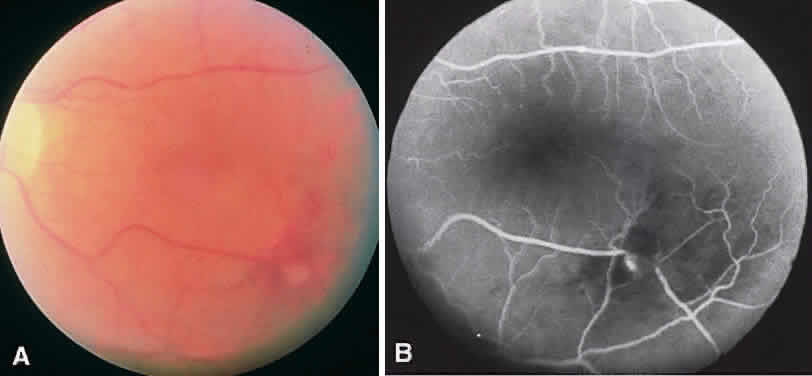
Fig. 7.
Retinal macroaneurysm. Focal dilatation along the inferotemporal arcade and a corresponding focal area of hyperfluorescence can be seen on this fluorescein angiogram. Macroaneurysms are usually arterial but can be venous.