|
|
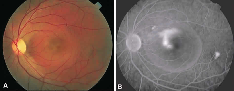
| Fig. 9. Central serous chorioretinopathy of the left eye. A. The circumscribed serous detachment of the neurosensory retina in the macula can be appreciated by the color change from the surrounding retina. B. Late venous phase of the fluorescein angiogram demonstrates a retinal pigment epithelial (RPE) detachment centrally with “escape” of the fluorescein under the sensory detachment, resulting in a classic “smokestack” appearance. A second RPE detachment is seen temporal to the macula. The rounded silhouette of the serous detachment also can be appreciated in the angiogram film. (Courtesy of Retina Service, Wills Eye Hospital, Philadelphia.) |