Chapter 61
Macular Holes and Epiretinal Macular Membranes
ALAN R. MARGHERIO and PAUL V. RAPHAELIAN
Main Menu   Table Of Contents

Search

MACULAR HOLES
RETINAL DETACHMENTS ASSOCIATED WITH MACULAR HOLES
EPIRETINAL MACULAR MEMBRANES
REFERENCES

MACULAR HOLES
Macular holes were considered rare, however, recent data have shown that macular holes are a common cause of unilateral visual loss.1 The first known reported case of a macular hole was by Knapp, in 1869.2 This was followed by reports from other investigators. These early reports were related to traumatic cases only. The first nontraumatic case was documented by Kuhnt.3 Results of pathologic studies followed with the classic paper by Coats in 1907.4 Some macular holes are associated with trauma or myopia, however, most are idiopathic. Idiopathic holes are most commonly seen in Caucasian women in the seventh decade of life who have no apparent predisposing conditions.5–8

DIFFERENTIAL DIAGNOSIS

Stage 1 and early stage 2 macular holes can be difficult to diagnose. They have often been confused with epiretinal macular membranes, pseudomacular holes, lamellar macular holes, macular cysts, cystoid macular edema, vitreomacular traction, adult vitelliform degeneration, bull's-eye maculopathy, and idiopathic juxtafoveal telangectasia.

Macular holes usually progress through several stages. A staging method for the development of idiopathic macular holes was originally described by Gass.9 In 1995, he refined his staging classification based on more recent surgical and pathologic data. A modified summary is presented here. Stage 1 occurs when the foveal depression is either decreased or absent, and a yellow ring or spot is present. Stage 2 is marked by early, full-thickness hole formation that is less than 400 μm in diameter. Stage 3 is reached when the hole is fully developed, greater than 400 μm in diameter without a Weiss ring. When the vitreous detaches and a Weiss ring is present, the hole is at stage 4.

Macular holes can resolve spontaneously. This most commonly occurs in stage 1 but has been reported for stage 2 holes as well. The resolution occurs when the posterior hyaloid separates. Traumatic macular holes have also been reported to resolve on their own. Because early holes and traumatic holes may resolve, many surgeons feel it is wise to observe them for a few months. If vision deteriorates or the hole progresses, vitreous surgery is indicated.

The visual acuity of patients with macular holes is variable. Typically, the acuity correlates with the stage of hole development. In stage 1 and early stage 2 holes, the acuity usually falls between 20/25 and 20/60. Late stage 2 and stage three holes fall between 20/70 and 20/200. Stage 4 holes and chronic macular holes (holes greater than 1 year's duration) have visual acuities between 20/400 and counting fingers.

Macular holes are most commonly unilateral.5 However, patients with unilateral macular holes, regardless of stage, should be informed that a hole can develop in their other eye. The incidence of hole formation in the fellow eye in patients with unilateral macular holes is 7%.5 Symptoms of impending holes should be explained to these patients. Symptoms include visual distortion, decreased visual acuity, and changes observed with home Amsler grid testing.

DIAGNOSIS

To diagnose a macular hole accurately, all available methods should be used to rule out other clinically similar conditions. First, the fundus should be examined carefully to determine if there appears to be a clinical posterior vitreous separation, if there is evidence of vitreomacular traction, or if an operculum is present. Next, using a very fine slit beam, the macula should be examined carefully to determine if there is any tissue bridging the apparent macular hole. Also, it should be noted if the edges of the hole are everted or irregular and if there are small, yellowish bodies near the base of the suspected hole. These signs lead to the diagnosis of a true macular hole rather than a pseudohole or other simulating lesion.10

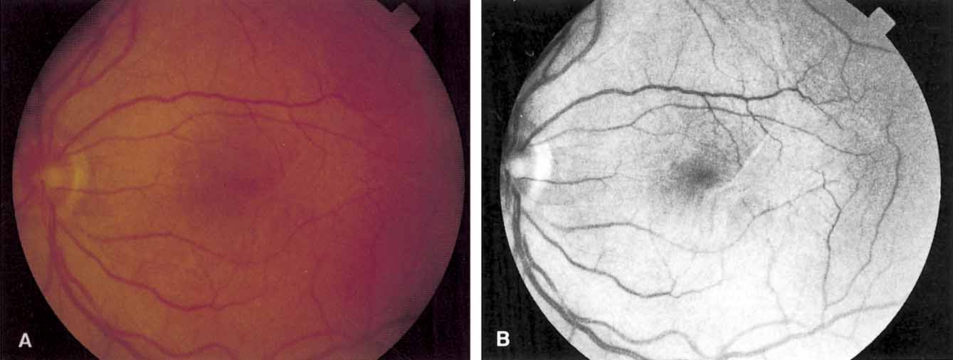
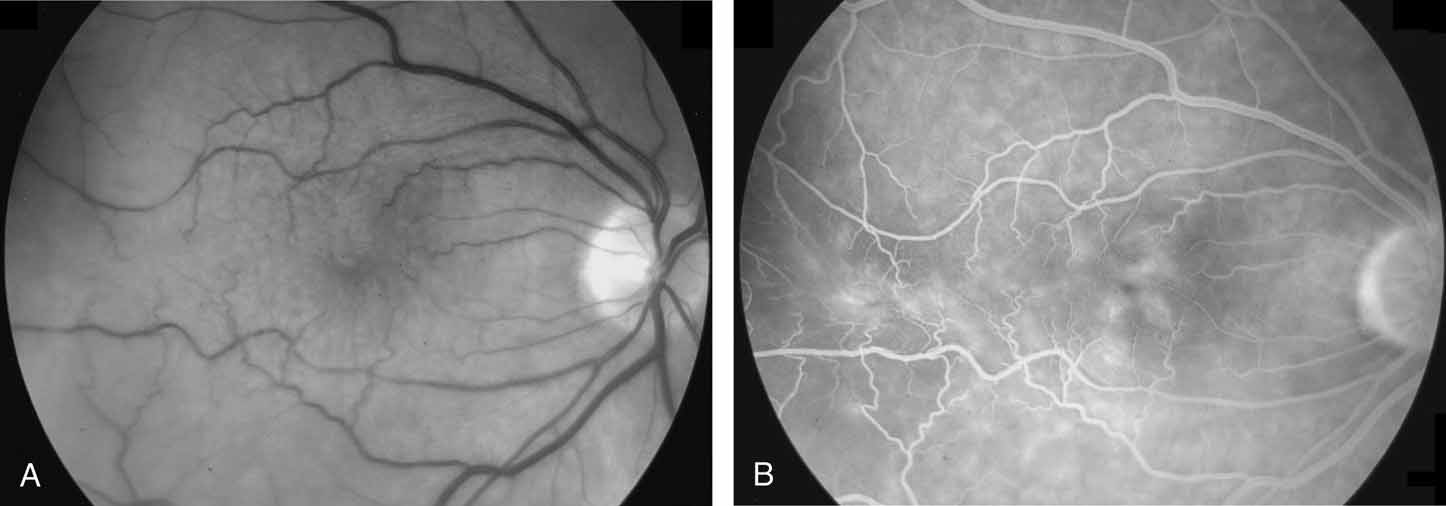
Fundus photography and fluorescein angiography are useful tools in the diagnosis of a macular hole. Stereo color photography helps document the stage, size, and configuration of the hole so that any changes may be detected easily. However, it is important to obtain similar stereo images for serial comparison. Monochromatic photography with use of 490- to 610-nm filters is another useful tool. Monochromatic photos help identify subtle vitreoretinal interface changes.11 Fluorescein angiography, although useful in differentiating a true macular hole from a simulating lesion, can be misleading. The increased transmission of choroidal fluorescence that is associated with macular holes can reverse spontaneously.12 It may also reverse after successful surgical treatment.13

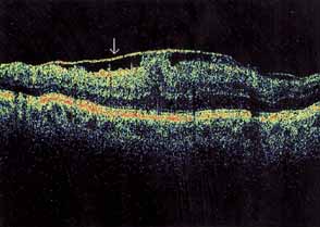
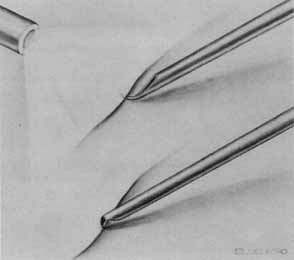
Functional testing has been used to help differentiate pseudoholes or other lesions from true macular holes. The Watzke-Allen sign was one of the first such functional tests.14 This test is performed by presenting a narrow slit beam over the suspected macular hole. The patient is then asked to describe what he or she perceives. If the patient describes a break in the line, a full thickness hole should be suspected (Fig. 1). Another method of performing this test is to pass the slit beam slowly over the macula from varying angles. If the patient perceives a break in the line, at any time, the test is considered to be positive (i.e., there is a full-thickness macular hole).

Fig. 1 A. Slit lamp light is focused on the retina using the widest possible slit that fits within margins of the macular cyst or hole. B. Appearances of slit beam when focused on macular cyst or hole of patient. (Margherio R, Trese MT, Margherio A, et al: Surgical management of vitreomacular traction syndromes. Ophthalmology 96:1437, 1989)

Another functional test may be performed by aiming the beam of the argon or dye laser. With visualization of the macular region, microperimetry is performed over the macular area. This is accomplished by presenting the 50-μm aiming beam, set at low intensity, to the area of the suspected macular hole and surrounding retina. Cases with full-thickness holes will demonstrate an absolute scotoma in the area of the hole. There will be a relative scotoma at the elevated rim of the neurosensory retina. With other lesions, either no scotoma or only a relative scotoma is seen. In describing the relative scotoma, the patient will usually say that the spot is “present, but different.” The Amsler grid has not been found to be of much value in differentiating true holes from pseudoholes because generally the absolute scotoma in patients with full-thickness macular holes is quite small.

The scanning laser ophthalmoscope (SLO) and the focal electroretinogram have been described as useful for differentiating true holes from other lesions.15–18 Laser biomicroscopy has also been used by some investigators to help demonstrate the fine retinal architecture at the fovea. With this technique, the vitreoretinal interface may be seen with greater detail. This is partly because of the enhanced illumination and the use of monochromatic green light.19,20 Also, some authors believe that a carefully performed B-mode ultrasound can help define the vitreoretinal relationships better than a clinical examination alone.

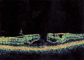
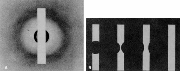
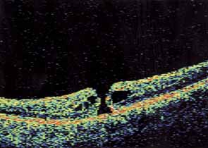
Optical coherence tomography or OCT is a relatively new tool used to diagnose macular lesions. It provides a unique view of the vitreoretinal architecture. OCT is particularly useful in diagnosing lamellar holes. With improvements in resolution and more widespread use, OCT will lead to new advances in the diagnosis and pathogenesis of macular holes (Fig. 2).

Fig. 2 Optical coherencd tomography of stage 2 macular hole with a visual acuity of 20/70.

PATHOGENESIS

The evidence incriminating vitreomacular traction in the pathogenesis of idiopathic macular holes is based on the correlation of clinical and surgical observations with known histopathology and OCT findings. Gass9 proposed that idiopathic macular holes begin from a tractional dehiscence of the umbo with minimal loss of photoreceptors. The tractional forces may be tangential, anteroposterior, or circumferential.21 These forces may resolve with a spontaneous vitreomacular separation22 but in the majority of cases the continuing tractional force will result in a full-thickness macular hole.

Early investigators focused on anteroposterior vitreomacular traction. Schepens,22 in 1955, was the first investigator to relate anteroposterior vitreous traction to the production of macular holes. Subsequently, other authors have made similar observations.6,23,24 Later studies indicated that in most cases, tangential traction plays a major role in the development of idiopathic full-thickness holes.13,25–27

On the basis of histopathologic studies, Foos28 demonstrated the presence of a vitreofoveal attachment that may be involved in the formation of macular holes. The opercula of macular holes obtained during vitrectomy for macular holes have been examined histologically. Macular hole opercula are rarely composed of true retinal tissue.29 The absence of cellular and fibrocellular fragments in the vitreous specimens obtained suggests that mechanisms other than cellular proliferation are important in the generation of the tractional forces required to create a macular hole.

Current theory, based on OCT, biomicroscopy, histology, and surgical experience, suggests that the posterior hyaloid applies traction to the foveola/umbo and causes it to go on stretch. The umbo dehisces because it is the thinnest point in the fovea. Then, according to the hydration theory proposed by Tornambe,30 the middle and inner retina absorbs vitreous fluid at the exposed edges of the hole and begins to swell. The hole enlarges because of a lateral extension of fluid into the outer plexiform layer. There is no mechanical loss of photoreceptors. Moreover, there is no microdetachment beyond the cuff of the hole. Once the inner retina is breached, the macular hole progresses from stage two to three by hydration of the fovea and perifoveal macula. Eventually, the posterior hyaloid separates completely and stage 4 is reached.

Additional support of the macular hole hydration theory may be derived from a report from Chung and Spaide,31 in which emulsified silicone oil migrated (or was absorbed) into the middle layers of the macula, around a successfully closed hole. The authors suggest that internal limiting membrane (ILM) peeling may have allowed the emulsified oil to infiltrate the retina into the macula at the exposed areas of the peel. However, the oil may have entered the middle retina layers through the exposed edges of the macular hole in a fashion similar to vitreous fluid proposed by the hydration theory.

TREATMENT

Stage 1: Impending Macular Holes

The role of vitrectomy for the prevention of full-thickness macular holes is still uncertain. The possible benefits of mechanically removing vitreous traction must be weighed against the known risks. These include retinal detachment, infection, nuclear sclerosis, and the creation of a full-thickness macular hole secondary to surgical manipulation.13,27,32,33 Surgical objectives for stage 1 macular holes consist of removing all tangential and anteroposterior vitreous traction on the foveal region. This must be accomplished without creating a full-thickness hole secondary to forces related to the surgical manipulation.

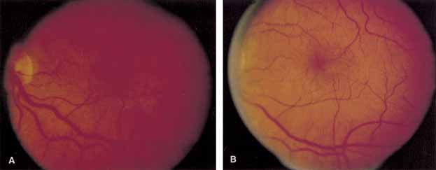
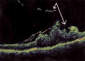
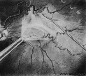
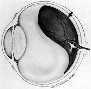
Some authors have commented on the importance of a posterior vitreous detachment in the pathogenesis of a macular hole.34,35 It is difficult to determine the vitreoretinal relationship preoperatively, even with careful slit-lamp evaluation. OCT testing can sometimes be helpful. However, the vitreomacular relationships are more accurately determined intraoperatively with use of oblique intraocular illumination and by noting the effect of gentle tractional forces on the macula during the vitrectomy. In some cases, what was thought to be a posterior vitreous separation preoperatively was actually found to be a large, optically empty space (Fig. 3).

Fig. 3 Large syneretic cavity simulating a complete posterior vitreous separation but with thin layer of posterior hyaloid still attached to posterior pole (arrow). (Margherio RR: Epiretinal macular membranes. In Albert R, Jakobiec EA, eds. Principles and Practice of Ophthalmology, Vol 2. Philadelphia: WB Saunders, 1993. Copyright 1990:919–925, S.W. Cochran)

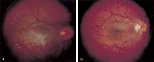
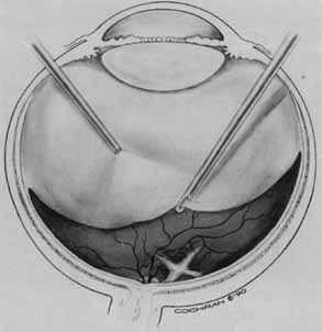
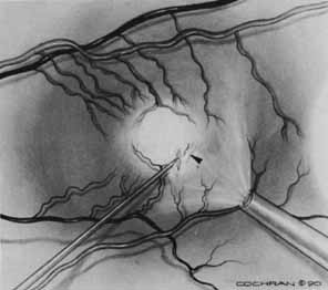
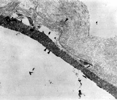
Tissue surgically peeled from the macular region in cases of impending macular hole has been found to be clinically consistent with posterior hyaloid. This finding was supported by electron microscopic examination of the tissue (Fig. 4).13,36 This observation is supported by the work of Kishi and Shimizu.37 They noted a large, optically empty space that appeared to be a complete posterior vitreous detachment in eyes with advanced liquefaction of the vitreous. They termed this area the posterior precortical vitreous pocket (PPVP). They found this pocket in 48 of 84 eyes with either an incomplete or no posterior vitreous detachment, and in 19 of 36 eyes with a posterior vitreous detachment. They noted that in eyes with advanced liquefaction of the vitreous, a large PPVP appeared to be a complete posterior vitreous detachment. In all of their postmortem cases, the posterior layer of the PPVP was found to be a thin layer of cortical vitreous. The presence of this PPVP strengthens the hypothesis that contraction of remaining attached cortical vitreous causes tangential traction on the macula, which gives the clinical appearance of an idiopathic macular cyst or hole.13,25,26,38 These impending holes or cysts' have been noted to resolve with spontaneous or surgical stripping of the membranes.13,25

Fig. 4 Transmission electron micrograph shows folded collagen (arrowhead) conshstent with posterior hyaloid (morphologically type 2 collagen). Adherent to the denser posterior surface is a cellular pseudopod (double arrowheads). The cell body is not present in this grid. (Margherio R, Trese MT, Masgherio A, et al: Surgical management of vitreomacular traction syndromes. Ophthalmology 96:1437, 1989)

In the management of eyes considered to be at high risk for the development of full-thickness macular holes, emphasis must be placed on careful follow-up. Patients demonstrating these characteristics should be followed on a monthly or bimonthly basis. Of the patients followed in this manner, approximately one-third will develop a spontaneous vitreomacular separation with an improvement in symptoms. However, if the vision continues to deteriorate to the 20/50 to 20/70 level, surgical intervention should be considered because many of these eyes are likely to progress to full-thickness holes.39 Moreover, better visual acuity results can be achieved with early intervention.

Vitreous Surgery for Full-Thickness Holes

Prior to the landmark paper of Kelly and Wendel,40 full-thickness macular holes were considered to be untreatable. Because there appeared to be an irretrievable loss of foveal tissue, it was assumed that treatment would be of little or no benefit. Once they proved that full-thickness holes could be closed with subsequent improvement in vision, histopathology followed, with proof that photoreceptors were spared in macular hole formation.

Vitreous surgery for macular holes has been refined over the last few years. A standard three-port core vitrectomy is performed. The posterior hyaloid is detached from the retina with the vitrectomy instrument switched to suction mode, at a level of 200 mm Hg. The vitrector port is then placed near the nasal edge of the optic nerve and suction is applied. The posterior hyaloid is engaged and the vitrector tip is pulled anteriorly to promote a posterior hyaloidal separation. A hyaloidal separation is only achieved when a Weiss ring is visualized. After successful detachment of the posterior hyaloid, a more peripheral vitrectomy is performed to provide maximal space for gas tamponade.

An air–fluid exchange is performed with the aid of a soft-tipped cannula. Confirmation of posterior hyaloidal separation can be determined at this time. The soft-tipped cannula is placed near the optic nerve and suction is applied. If the tip engages residual hyaloid, it will bend toward the retina in a fishhook fashion. If residual hyaloid is noted, the vitreous cavity is refilled with fluid and another attempt is made to separate the posterior hyaloid. After the air fluid exchange is completed, 5 to 15 minutes are allowed to elapse for fluid reaccumulation resulting from surface tension. The residual fluid is removed with the soft-tipped cannula and the sclerotomies are trimmed free of vitreous and closed. The air in the vitreous cavity is exchanged with either 16% C3F8 or 20% SF6. The patient is instructed to remain in a face-down position for up to 14 days.

ILM peeling has been reported to be a beneficial adjunct to macular hole surgery. Many surgeons have reported high hole closure rates and excellent visual results.41,42 However, peeling the ILM is the most difficult maneuver in macular hole surgery. New instruments have been designed to aid the surgeon. Vital dyes such as indocyanine green and trypan blue have also been used to help identify the ILM for peeling.43,44

We have found that the ILM does not have to be peeled for surgical success in most cases.45 Moreover, in 2002, Spaide46 reported three cases of successful macular hole surgery with limited vitrectomy over the macular hole without ILM peeling. Similarly, in 2003, Dori et al.47 reported a 100% closure rate without ILM peeling in 50 eyes with stage 2 holes. Ninety-eight percent of these eyes had visual acuity results of 20/50 or better.

Additional complications may occur if ILM peeling is performed. ILM peeling-associated complications include phototoxicity, retinal hemorrhage, RPE defects, and nerve fiber layer defects. Moreover, several recent reports have suggested that indocyanine green, used as an adjunct to ILM peeling, may cause delayed photochemical damage to the RPE.48,49 Indocyanine green has been shown to be present in the fundus and optic disc for up to 12 months after use.50–52 Haritoglou et al.53 found that indocyanine green may alter the cleavage plane in the innermost layers of the retina that may result in less improvement in vision and unexpected visual field defects. Therefore, because of the increased potential for complications, we recommend ILM peeling only for stage 4 macular holes, chronic macular holes, previous surgical failures, and late reopened macular holes.

Laser

Photocoagulation has been used as an adjunct to help repair macular holes. Outpatient laser treatment, combined with fluid gas exchange has been used successfully to close macular holes after initial surgical failures. In 1998, Ohana and Blumenkrantz54 used slit-lamp–based yellow laser to close 13 of 15 holes. Ikuno et al.55 used one spot of argon green to the RPE at the base of the hole to close 12 of 13 holes. Intraoperative endolaser has also played a role in treating macular holes. Woog-Ki et al.56 used one spot of very light endolaser to the RPE at the base of the macular hole and closed 8 of 8 holes. Kwok et al.57 used endophotocoagulation to the base of the hole to help close macular holes in 3 of 4 associated with myopic retinal detachments.

We have reserved the use of endolaser to stage 4 holes, chronic holes, initial surgical failures, and late reopened holes in which the ILM is not peeled or is only partially peeled. We know from experience that these cases are likely to fail if the ILM is not peeled or an endolaser is not used. We use an argon green endolaser at very low power and short duration. A test spot is placed in the macula in a safe zone near an arcade vessel. Laser power is slowly increased until a faint grey blanching of the RPE is achieved. The probe is then placed over the hole and very close to the RPE to minimize the spot size. One spot is then applied to the RPE through the opening in the hole. We have had excellent success with this technique. The application of laser to the RPE at the base of the hole may stimulate the RPE to pump out the intraretinal fluid proposed by Tornambe's macular hole hydration theory.30

Complications

Complications associated with macular hole surgery, in addition to those that may occur with any intraocular procedure, include elevated intraocular pressure, peripheral retinal tears, retinal detachment, lens opacities, visual field defects, vitreous hemorrhage, and late reopenings.58 Some of these conditions will resolve spontaneously or with treatment; others may result in permanent loss of vision. Most of these eyes will develop visually significant nuclear sclerotic lens opacities within 1 year.58–61

CHRONIC MACULAR HOLES

Chronic macular holes are generally considered to be stage 3 or stage 4 holes that have been present for more than 1 year. These holes, possibly due to long-term intraretinal fluid accumulation, epiretinal membrane formation, and RPE atrophy are more difficult to close compared to acute macular holes. Fortunately, because of increased awareness of macular holes by the general ophthalmic community, chronic holes are becoming far more rare. Visual improvement may occur with successful hole closure. However, these improvements are generally not as pronounced as those seen with acute macular holes. Roth et al.62 were able to close 9 of 11 chronic holes in their series, with a mean postoperative vision of 20/100. ILM peeling, adjuvants, and endolaser may assist in macular hole closure in this challenging subgroup.

TRAUMATIC MACULAR HOLES

Traumatic macular holes are much less common than idiopathic macular holes. Most develop from significant blunt trauma to the eye. These holes are commonly preceded by Berlin's edema. Traumatic macular holes may close spontaneously,63,64 therefore, these holes should be observed for 3 to 6 months before surgery is contemplated. Surgical closure rates for traumatic holes are similar to idiopathic macular holes. Autologous plasmin enzyme has been used as an adjuvant to help close pediatric and adult cases.65,66 Adult traumatic holes have also been successfully closed without the use of adjuvants.67

Back to Top
RETINAL DETACHMENTS ASSOCIATED WITH MACULAR HOLES
Retinal detachments secondary to macular holes are exceedingly rare, but they pose a challenging clinical problem. The incidence has been reported to range between 0.6% and 4%.68,69 Most rhegmatogenous detachments that appear to be the result of macular holes are actually secondary to peripheral holes. In the presence of a retinal detachment involving the macula, the normal thinning of the retina in the foveal region may give the appearance of a full-thickness macular hole when none is present. If a detachment involving a peripheral hole is combined with a suspected macular hole, the detachment can be treated in a standard fashion, ignoring the hole.68,70,71 In most cases, the suspected macular hole will close after successful retinal reattachment.

Retinal detachments associated with true macular holes are seen most frequently in highly myopic eyes.7,72 These detachments may only extend out from the macular hole to the major arcade vessels or slightly beyond. The association with high myopia probably accounts for the higher reported incidence of detachments caused by macular holes in Asians and other races with a large number of highly myopic people. Morita et al.,7 in a review of 209 eyes with retinal detachment secondary to macular holes, noted the factors relating to retinal detachment in macular holes to be: the degree of refractive error, the severity of myopic chorioretinal change, and the presence of posterior staphyloma.

Many methods for repairing detachments secondary to macular holes have been reported. These techniques can be categorized as either transscleral or transvitreal. The transscleral methods include macular diathermy or cryopexy, macular buckling, sling procedures, scleral resection, or some combination of these with equatorial scleral buckling.68,73–75 Transvitreal techniques include vitrectomy with gas tamponade, or vitrectomy with silicone tamponade.76–81

With the advent of vitreous surgery, membrane stripping, and long-acting gases, most methods of mechanically indenting the macula as the initial procedure of choice have been abandoned. Most of these mechanical methods caused complications because of the embarrassment of the retinal vasculature and physical damage to the optic nerve or its blood supply. Also, treatment in this region with diathermy or cryotherapy, in addition to causing extensive damage to retinal photoreceptors, could damage the optic nerve and adjacent structures. Kuriyama and colleagues82 evaluated the results obtained with both the transvitreal and transscleral methods in 250 eyes. They found that the initial results obtained with the transscleral methods were better (83% versus 56% for transvitreal methods), but that the final success rate of 95% was the same regardless of which approach was selected initially.

Today, most retinal surgeons choose pars plana vitrectomy and gas tamponade as the initial procedure to treat detachments associated with macular holes.76–81 This technique has the best visual prognosis. A careful and thorough peeling of the cortical vitreous and posterior hyaloid is accomplished with use of a soft-tipped cannula or vitrector. Any epiretinal membrane in the area should be peeled. Drainage of subretinal fluid can be accomplished through the existing macular hole or through a posterior retinotomy. A gas-fluid exchange is then performed with one of the long-acting gases. The patient is instructed to maintain a face-down position for 2 weeks. If a recurrence of the detachment occurs, the possible causes of the failure must be carefully evaluated. Endolaser to the RPE at the base of the hole in conjunction with repeat vitrectomy with gas fluid exchange has recently been shown to be effective in highly myopic, macular hole-related detachments.57

SUMMARY

Our identification, understanding, and treatment of macular holes has advanced tremendously since this chapter was first written. Optical coherence tomography has allowed us to examine macular holes in ways that were not possible just 10 years ago. We expect many more exciting advances to be made with OCT in the future as OCT technology improves and more macular holes are examined. The role of ILM peeling in the treatment of macular holes is still controversial, as is the use of indocyanine green dye. Vitreoretinal surgeons have demonstrated that all macular holes can be closed, with subsequent improvement in vision. Early macular holes have the best prognosis for 20/40 or better vision. With early diagnosis and prompt treatment, more patients will regain excellent vision.

Back to Top
EPIRETINAL MACULAR MEMBRANES
Epiretinal membranes can be divided into those that are idiopathic and those that are secondary to another clinical condition. The proliferation of epiretinal membranes on the inner retinal surface along the internal limiting membrane was first described by Iwanoff in 1865.83 Epiretinal membranes are relatively common. In 1971, Roth and Foos84 reported that in a series of autopsy eyes, epiretinal membranes were present in 2% of patients at age 50 and in over 20% of patients at age 75. Clarkson et al.,85 in a review of some 1612 postmortem eyes, noted epiretinal membranes in 0.7% of eyes that had not undergone previous ocular surgery. Pearlstone86 reported an incidence of 6.4% in 1000 consecutive routine eye exams in patients older than age 50, with 20% being bilateral. Other authors report bilateral involvement in 10% to 20% of cases.87–89

The wrinkling of the retinal surface caused by epiretinal membranes in the macular region has been termed surface wrinkling retinopathy,84 macular pucker,90–92 cellophane maculopathy,24,93 wrinkling of the ILM,94 preretinal macular fibrosis,94,95 epiretinal macular membrane, primary retinal folds,96 internoretinal fibrosis,97 idiopathic preretinal macular gliosis,88,98,99 undetected central retinal vein occlusion,100 and silkscreen retinopathy. The term macular pucker has most frequently been applied to the condition when retinal surface wrinkling occurs after reattachment of a rhegmatogenous retinal detachment. This condition complicates 4% to 8% of otherwise successful primary retinal reattachments.101,102

The presence of epiretinal membranes in the macular region has been associated with numerous clinical conditions, including retinal tears and holes, previous retinal detachment repair, other intraocular surgery, cryopexy, photocoagulation, nonproliferative retinovascular disorders, trauma, ocular inflammatory disorders, certain proliferative retinopathies, vitreous hemorrhage, congenital disorders,103 as well as idiopathic or spontaneous development. The membranes develop as a result of the proliferation of cells and collagen on the surface of the retina. The clinical appearance depends to some degree on cell type, membrane thickness, and presence or absence of vessels. Vinores et al.104 found a correlation between the disease process and the amount of extracellular matrix, with proliferative vitreoretinopathy having a thicker membrane than post detachment macular pucker, which in turn was thicker than an idiopathic macular pucker. These ultrastructural findings correlate well with the clinical appearance of the various entities.

CLINICAL FEATURES

Patients with epiretinal membranes peripheral to the macula are generally asymptomatic. However, when the membranes involve the macula or are perimacular, the type and degree of symptoms experienced will vary. Symptoms depend on the membrane thickness, the degree of retinal distortion caused by the overlying membrane, the presence or absence of significant traction, which can cause a microdetachment of the posterior pole, and the presence or absence of edema in the macular and perimacular regions. Thin epiretinal membranes usually cause few symptoms. The condition appears to be relatively stable or slowly progressive, with only a small number (approximately 5%) obtaining a vision of 20/200 or worse.87,105,106 In more advanced cases there is a reduction in vision, micropsia, metamorphopsia, Amsler grid distortion, and occasionally monocular diplopia. Spontaneous separation of an epiretinal macular membrane, although uncommon, can occur. After separation, there is a general decrease in symptoms and a concomitant improvement in visual acuity.96,107–109

The clinical appearance is variable and may present as only a mild sheen or glint in the macular region that can best be seen with red-free or monochromatic green or blue light (Fig. 5). In more severe cases, there is increased vascular tortuosity and the perimacular vessels are seen to be pulled toward an epicenter, with striae and heterotopia of the macula. The superior and inferior arcuate vessels are also closer together and straighter than in an uninvolved eye. Other findings that may be present include small intraretinal hemorrhages, cystic changes in the macula, macular edema, and cotton-wool spots.110 Pseudoholes or macular cysts have been noted in up to 8% of idiopathic cases (Fig. 6).25,88 Thin membranes may be completely translucent, whereas thicker membranes are frequently opaque or pigmented and generally obscure details of the underlying fundus (Fig. 7).110–112 The thicker and occasionally pigmented membranes are often seen after retinal detachment surgery, severe inflammatory conditions, and trauma. An apparent posterior vitreous separation has been reported by most authors to exceed 75% in cases of idiopathic epiretinal membranes.84–88,93,104,105,113–117 It is sometimes difficult to accurately determine the vitreoretinal relationships preoperatively.

Fig. 5 A. An idiopathic epiretinal membrane in a 65-year-old man. Visual acuity was 20/25. B. Black-and-white fundus photograph taken with green 540-nm filter. Note increased definition of epiretinal membrane. (Margherio RR: Epiretinal macular membranes. In Albert R, Jakobidc EA, eds. Principles and Practice of Ophthalmology, Vol. 2. Philadelphia: WB Saunders, 1993:919–925)

Fig. 6 A. Idiopathic epiretinal membrane with macular pseudohole in a 35-year-old woman. B. Postoperative appearance with disappearance of macular pseudohole and improvement in vision from 20/100 to 20/40. (Margherio RR: Epiretinal macular membranes. In Albert R, Jakobiec EA, eds. Principles and Practice of Ophthalmology, Vol. 2. Philadelphia: WB Saunders, 1993:919–925)

Fig. 7 A. Preoperative appearance of thick epiretinal membrane in 10-year-old boy after trauma. B. Postoperative appearance after removal of epiretinal tissue. Vision improved from counting fingers to 20/100. (Margherio RR: Epiretinal macular membranes. In Albert R, Jakobiec EA, eds. Principles and Practice of Ophthalmology, Vol. 2. Philadelphia: WB Saunders, 1993:919–925)

Clinical testing, in addition to visual acuity, most commonly involves fluorescein angiography and ocular coherence tomography. Fluorescein angiography can show retinal vascular tortuosity, straightening, and leakage, as well as cystoid macular edema (Fig. 8). OCT typically demonstrates retinal folding, increased macular thickness, cystoid macular edema, traction macular retinal detachment, and both lamellar or macular hole formation (Figs. 9, 10, and 11). Sine Amsler chart testing may help quantify metamorphopsia in eyes with macular distortion.118 Abnormal macular function has been shown using both focal and multifocal electroretinography.119,120

Fig. 8 A. Black and white photograph taken with a green 540-nm filter, showing dragging and distortion of the macular vessels. B. Fluorescein angiogram. Note distortion of macula and vessels, with associated vascular leakage and cystoid macular edema.

Fig. 9 Optical coherence tomography of left macula with epiretinal membrane (arrow) and visual acuity of 20/100. Note retinal thickening and folding.

Fig. 10 Optical coherence tomography of left macula in eye with visual acuity of 20/40. Note epiretinal membrane with vitreous adherence (arrow), macular hole, and intraretinal edema around the hole.

Fig. 11 Optical coherence tomography of thickened and folded right macula in eye with 20/300 visual acuity. Note traction retinal detachment (small arrow) and posterior vitreous detachment with avulsed piece of epiretinal membrane (large arrows).

PATHOLOGY

The cellular origin of epiretinal membranes has long been debated, and almost every possibility has been considered. In 1865, Iwanoff83 implicated the endothelial cell in the formation of the membranes.83 Manschot,121 in 1958, thought that the membrane was an extension of the Müller cell processes, whereas Wolter122 considered the cells to originate from fibroblasts in the vascular connective tissue. Smith123 suggested that the cells in the membrane originated from the pigmented or nonpigmented cells of the pars ciliaris, retinal pigment epithelial (RPE) cells, mesodermal elements of the vascular system, normal vitreous cells, inflammatory cells within the vitreous, or retinal glial cells. In 1962, Kurz and Zimmerman124 believed that the cells originated from migration of RPE cells. Ashton125 later suggested that they originated from a transformation of vascular mesenchymal cells into fibroblasts, and in 1969, Von Gloor126 proposed that hyalocytes were the cells of origin.

The development of vitreous surgery has provided an opportunity to advance our understanding of the clinicopathologic relationships of epiretinal membranes. Constable127 reported on the histopathology of membranes obtained by open-sky vitrectomy. He found proliferating fibroblasts interspersed with extracellular material and clumps of pigment. Since then, numerous authors have attempted to characterize the cells of origin based on the suspected etiology of the membrane obtained by pars plana vitrectomy. The general consensus was that membranes in eyes that had retinal breaks or previous detachments were composed of cells of RPE origin, and that cells of glial origin predominated in the thin idiopathic epiretinal membranes.36,104,124–134 Some studies have demonstrated a high incidence of RPE type cells even in the idiopathic epiretinal membranes.135–138 This is supported by Vinores and colleagues,104 who studied the ultrastructural and electron immunocytochemical characterization of cells in epiretinal membranes. Their work suggests that both RPE cells and retinal glial cells are most likely to be the major participants in the pathogenesis of epiretinal membranes.104 Foos139 suggested that the glial cells found in the thin, idiopathic membranes were derived from the glial cells of the superficial retina, which had migrated through breaks in the internal limiting lamina to proliferate on the retinal surface. This hypothesis was subsequently supported by Bellhorn and associates.140 The dispersion of RPE cells on the retinal surface has been demonstrated clinically and experimentally in cases of retinal breaks and detachment.141–143 However, the finding of RPE cells in idiopathic membranes is more difficult to explain. Smiddy and colleagues136 suggest that the RPE cells gain access to the retinal surface by various methods, including migration through occult breaks, inactivation of developmental rests of RPE cells already on the surface of the retina, transformation from other cell types, and transretinal migration. Stern and co-workers144 suggested that the contractive forces of the membranes were related to their constituent cell types and were not dependent on intercellular collagen, as suggested by previous investigators.145

While the exact mechanism or combination of mechanisms is yet to be elucidated, recent studies have shed some light on the biochemical processes involved with epiretinal membrane proliferation. Armstrong and colleagues146 have demonstrated the presence of both vascular endothelial growth factor and tumor necrosis factor alpha in surgically excised membranes. Tissue-type plasminogen activator, plasminogen, and urokinase, which are involved in extracellular matrix breakdown, also have been found in epiretinal membranes.147,148 Recently, chemokine receptors mediating cell migration were shown to be expressed on both retinal pigment epithelium and epiretinal membranes.149 Consequently, the sequence of vascular endothelial growth factor-mediated activation of the signal transduction pathway, leading to extracellular matrix breakdown and RPE cell migration, may be involved in the formation and proliferation of some epiretinal membranes.

TREATMENT

The majority of patients with epiretinal macular membranes have symptoms that are mild and either nonprogressive or slowly progressive and treatment is rarely indicated. In a few cases, the membrane may spontaneously release, with a marked decrease in symptomatology and improvement in acuity. For patients with significant symptoms and substantially reduced visual acuity, bimanual vitrectomy with epiretinal membrane peeling can diminish the severity of the symptoms and improve acuity in 75% or more of cases.

There is no effective treatment available for mild forms of epiretinal macular membranes. In 1978, Machemer150 first reported on the surgical management of advanced cases of epiretinal macular membranes using the technique of pars plana vitrectomy. Subsequent authors reported on larger series and attempted to identify preoperative factors that appeared to influence the postoperative visual prognosis.25,113,114,133,134,151–171

A conventional three-port pars plana vitrectomy technique is now well-established in the removal of epiretinal membranes. Technique and instrumentation have improved over the years, almost eliminating complications such as iatrogenic retinal tears and detachments. The problems of membrane recurrence172 and accelerated nuclear sclerosis still remain unsolved. Also, although improvement in acuity is achieved in most cases, it may be less than expected, and it is rare to completely eliminate metamorphopsia. This appears to be especially true in eyes with relatively good preoperative vision.167,173 Therefore, when vision is better than 20/70, surgery should be approached with caution.

The surgical technique for removing epiretinal macular membranes includes first performing a pars plana vitrectomy (Fig. 12). After vitrectomy, the tip of a 20-gauge microvitreoretinal blade is bent approximately 80 degrees. (Alternatively, a 23-gauge 1.5-inch needle can be bent toward its lumen. The needle is then attached to a tuberculin syringe and filled with balanced salt solution in order to prevent the introduction of air bubbles into the vitreous.) Dissection is preferentially carried out with the tip of the blade facing the light source to avoid working in a shadow (Fig. 13). If there is an obvious edge to the membrane, the tip of the blade can be slid under it and the membrane can be removed (Fig. 14). This should be done with tangential rather than anteroposterior force. As the membrane is lifted off the surface of the neurosensory retina, great care must be used to avoid creating retinal tears. This is particularly true when peeling the membrane off the fovea. After the membrane has been lifted off the posterior pole, it can be removed from the eye by use of vitreous forceps or a vitrector.

Fig. 12 A pars plana vitrectomy is completed. (Margherio RR: Epiretinal macular membranes. In Albert R, Jakobiec EA, eds: Principles and Practice of Ophthalmology, Vol. 2. Philadelphia: WB Saunders, 1993:919–925)

Fig. 13 By keeping the blade directed toward source of endoillumination, working in the “shadow” is avoided and visualization improved during dissection of epiretinal membrane. (Margherio RR: Epiretinal macular membranes. In Albert R, Jakobiec EA, eds. Principles and Practice of Ophthalmology, Vol. 2. Philadelphia: WB Saunders, 1993:919–925)

Fig. 14 In cases with an obvious edge, the tip of the bent 20-gauge microvitreoretinal blade or 23-gauge needle engages the membrane and gently peels it from the retinal surface. (Margherio RR: Epiretinal macular membranes. In Albert R, Jakobiec EA, eds. Principles and Practice of Ophthalmology, Vol. 2. Philadelphia: WB Saunders, 1993:919–925)

For eyes in which there is no obvious edge to the epiretinal membrane, the oblique illumination of the light pipe may be used to cast a “shadow” of the retinal vessels on the retinal pigment epithelium. This will readily enable identification of the area of maximum elevation of the neurosensory retina. The bent blade is then used over a small retinal vessel in that area to open a small hole in the membrane. It can then be removed as previously described (Fig. 15).

Fig. 15 Opening is created in epiretinal membrane with tip of bent blade. Dissection is performed over small retinal vessel to guard against creating a retinal hole. Arrow shows area of maximum vascular shadowing, which identifies area of greatest elevation of the neurosensory retina from the retinal pigment epithelium. (Margherio RR: Epiretinal macular membranes. In Albert R, Jakobiec EA, eds. Principles and Practice of Ophthalmology, Vol. 2. Philadelphia: WB Saunders, 1993:919–925)

Recently, trypan blue has been used to help intraoperative visualization of epiretinal membranes and the ILM.44,174,175 A concentration of 0.06% trypan blue was found to be a safe intravitreal dose for staining membranes in a study by Veckeneer and colleagues,176 and 0.2% in a study by Teba and associates.177 Unlike trypan blue, indocyanine green selectively stains the ILM, not epiretinal membranes.178 Various preparations, concentrations, and exposure times of indocyanine green have been used, either with a balanced salt solution-filled eye or under air, to improve epiretinal membrane visualization and peeling by staining the adjacent uncovered internal limiting membrane.

After initial removal of the membrane, an attempt should be made to determine whether a multilayered membrane is present. Membrane staining may be helpful in this regard. Additional layers must be sequentially peeled, or the expected visual and anatomic results will be compromised. Incomplete removal of a multilayered membrane can sometimes be confused with a recurrence. After complete removal of the membrane, the peripheral retina is inspected for tears and/or detachments, which, if present, can be subsequently repaired.

RESULTS

After surgical removal of epiretinal macular membranes, there is a tendency toward normalization of the appearance of the retina in most cases, although there is almost never a complete resolution of the vascular tortuosity and retinal striae. Similarly, vision is improved in most eyes, but a return to normal acuity is rare even in eyes without previous underlying pathology. In Margherio's25 series of 328 eyes, there was an overall improvement of at least two lines in 74%, with 24% being unchanged and 2% becoming worse. In light of absent preexisting macular pathology, better visual results were generally found in the 184 idiopathic cases. Other series, however, report greater improvement in nonidiopathic than in idiopathic cases.173

Numerous authors have attempted to define prognostic indicators of postoperative vision in vitrectomy for treatment of epiretinal membranes of the macula. Indicators that have been mentioned include preoperative visual acuity, duration of diminished acuity prior to surgery, the presence of preoperative cystoid macular edema, age of the patient, thickness of the epiretinal membrane, idiopathic versus nonidiopathic epiretinal membranes, and the presence of RPE window defects on fluorescein angiography. Trese et al.171 reported that transparent membranes had a better visual prognosis than did opaque membranes. They also believed that the presence of cystoid macular edema was a poor prognostic sign. Rice and colleagues,173 however, found that eyes with thin, transparent membranes had a poorer prognosis than eyes with thicker membranes. McDonald and Aaberg159 found no association between the transparency of the membrane and the final postoperative vision. Ferencz and associates,153 in a study of 167 cases of idiopathic epiretinal membranes, found no association between the transparency of the membrane and the final visual outcome. Likewise, they found no association between the presence or absence of cystoid macular edema or a RPE defect and the final visual outcome. Only preoperative vision had a predictive value with regard to the ultimate visual prognosis.153 Maguire and colleagues179 reviewed the preoperative fluorescein angiographic features of 229 cases of idiopathic epiretinal membranes. They found that postoperative visual improvement was greatest in eyes with the most severe degree of macular edema. No such association was noted with the degree of retinal vascular distortion or contraction of the foveal avascular zone.179 Other studies have suggested that the presence of cystoid macular edema is a poor prognostic sign.114,114,160,163,171,173,180 However, larger series have shown no relationship between the presence of preoperative cystoid macular edema and the ultimate visual prognosis.25,153,158,167 There has been recent discussion as to whether it is necessary to additionally peel the ILM. Park and associates181 compared short-term results for a total of 44 eyes with idiopathic macular pucker undergoing epiretinal membrane peeling either with or without associated peeling of the internal limiting membrane. Staining of membranes was not utilized, and a statistically significant visual acuity benefit between the two groups was not demonstrated. Intraoperative staining of epiretinal and ILMs can facilitate membrane visualization and removal. Histologic specimens of peeled epiretinal membranes stained with trypan blue have both included174 and lacked44 ILM. No added visual benefit from the use of trypan blue has yet been demonstrated, and its long-term safety is unknown.

Similar to trypan blue, indocyanine green allows improved visibility of the internal limiting membrane, and consequently overlying epiretinal membrane. Sorcinelli,182 in a series of 28 eyes with idiopathic and secondary epiretinal membranes, used indocyanine green to stain the membranes during vitrectomy. He reports that the membrane was stained in all cases, and visual acuity at a mean 6-month follow-up was improved an average of two lines, without clinical evidence of indocyanine green toxicity.182 However, there are reports of potential RPE toxicity and traumatic retinal damage with its use. Sippy and associates183 found that cultured RPE cell enzymatic activity was reduced when exposed to indocyanine green. A subsequent study by Engelbrecht49 demonstrated RPE atrophy at the site of previous macular holes in eyes that underwent vitrectomy and internal limiting membrane peeling with the aid of indocyanine green. Recently, Tadayoni and associates52 have shown persistence of indocyanine green fluorescence of the optic disc and macula up to 7 months after assisted peeling of the ILM for macular holes. However, macular fluorescence was not present after the peeling of epiretinal membranes not associated with a macular hole. Possibly the intact retina provides a barrier against indocyanine green-related RPE cell toxicity.52 Paques and co-workers,184 using a rabbit model, suggest that retrograde diffusion of indocyanine green in retinal ganglion cells is a causative mechanism for optic disc fluorescence.

Trauma to the retina after indocyanine green-stained membrane peeling also can occur. All 10 ILM specimens peeled with the aid of indocyanine green by Gandorfer and colleagues,185 and examined by transmission electron microscopy, contained parts of Müller cells. Haritoglou and co-workers53 also had similar histologic results in a recent study, with unstained membranes having fewer retinal elements compared to stained membranes. In their series, indocyanine green was used for 20 of 48 consecutive eyes with idiopathic macular pucker undergoing vitrectomy. Visual acuity did not improve statistically in the eyes which were exposed to indocyanine green, unlike in eyes undergoing membrane peeling without staining, and visual field deficits were documented.53 Consequently, it is unclear at this time whether the intraoperative use of either trypan blue or indocyanine green to assist membrane visualization and peeling is visually beneficial.

COMPLICATIONS

The most frequently occurring intraoperative complications with pars plana vitrectomy for epiretinal membrane surgery include vitreous hemorrhage and peripheral or posterior iatrogenic retinal breaks. The vitreous hemorrhage is generally small and is easily removed with the vitrectomy instrument. Posterior breaks are rare and occur in less than 1% of cases in most series.25,114,152,156,164 In some series, however, the occurrence of posterior breaks has been as high as 9%.168 The retinal tears are treated with photocoagulation or transscleral cryosurgery.

Postoperative complications can include the development of accelerated nuclear sclerosis, retinal tears or detachment, rubeosis iridis, and endophthalmitis. Retinal detachments can generally be repaired without further loss of vision. Rubeosis and endophthalmitis have occurred in less than 0.5% of cases.25,134 The most troublesome postoperative complication has been the development of accelerated nuclear sclerosis, which has ranged from 12% to over 60%.25,134,160,163,167,168 The reasons for this are unclear, but possible factors are the influence of the operating microscope light or endoilluminator, the chemical composition of infusion fluid, infusion via an anterior infusion port, and/or change in intraocular temperature.25,186

The membrane recurs in less than 5% of idiopathic cases. In eyes with membranes from known etiologies, recurrence is much higher, approaching 100% in eyes of young patients who develop epimacular membranes after trauma or inflammatory disease.25,172

SUMMARY

Epiretinal membranes are relatively common. They occur in 2% to 6% of patients. The incidence of idiopathic membranes increases with age and may approach 20% by age 70.

Many cell types have been implicated in the origin of epiretinal macular membranes. RPE cells and retinal glial cells are most likely the major participants in the pathogenesis of epiretinal membranes. The epidemiology of the membranes includes proliferative diseases, trauma, inflammation, and retinal surgery, as well as those membranes that are idiopathic. The clinical appearance is variable and depends on cell type, membrane thickness, presence of vessels, and etiology. In most cases, the condition is relatively stable or slowly progressive, with only 5% progressing to 20/200 or worse. Membranes involving the macular or perimacular regions can cause a reduction in vision, metamorphopsia, micropsia, or occasionally monocular diplopia. The contractile forces of the membranes appear to be more closely related to their cellular constituents than to the extracellular collagen matrix. The contraction of epiretinal membranes may be implicated in the development of macular cysts and/or holes. Occasionally, the membrane may spontaneously separate with relief of symptoms.

Bimanual pars plana vitrectomy surgery can successfully remove epiretinal macular membranes in virtually all advanced cases. After surgical removal, vision improves in approximately 75% of cases. However, a return to normal vision is unlikely. Postoperatively, retinal complications such as tears and detachment are rare in the idiopathic cases but are somewhat more common in cases associated with other etiologies. The most frequent postoperative complication is the development of accelerated nuclear sclerosis, which occurs in 12% to 68% of cases. The reasons for this have yet to be defined. Epiretinal membranes recur in approximately 5% of cases.

Back to Top
REFERENCES

1. Rahmani B, Tielsch JM, Katz J, et al: The cause specific prevalence of visual impairment in an urban population: The Baltimore Eye Survey. Ophthalmology 103:1721, 1996

2. Knapp H: Uber isolierte zerreissungen der aderhaut in folge von traumen auf dem augapfel. Arch Aug Ohrenhdilk 1:6, 1869

3. Kuhnt H: Tber eine eigentumliche veranderung der netzhaut ad maculam (retinitis atrophicans sive rareficans centralis). Z Augenheilk 3:105, 1900

4. Coats G: The pathology of macular holes. R Lond Ophthalmol Hosp Rep 17:69, 1907

5. Chew E, Sperduto R, Hiller R, et al: Clinical course of macular holes. Arch Ophthalmol 117:242, 1999

6. Margherio RR, Schepens CL: Macular breaks. I. Diagnosis, etiology and observations. Am J Ophthalmol 74:219, 1972

7. Morita H, Ideta H, Ito K, et al: Causative factors of retinal detachment in macular holes. Retina 11:281, 1991

8. Morgan CM, Schatz H: Involutional macular thinning, a pre-macular hole condition. Ophthalmology 93:153, 1986

9. Gass, JD: Reappraisal of biomicroscopic classification of stages of development of a macular hole. Am J Ophthalmol 119:752, 1995

10. Kraushar MF: Foveal diseases. Ann Ophthalmol 18:354, 1986

11. Ortiz RG, Lopez PF, Lambert M et al: Examination of macular vitreoretinal interface disorders with monochromatic photography. Am J Ophthalmol 113; 243, 1992

12. Lewis H, Cowan G, Straatsma R: Apparent disappearancd of a macular hole associated with development of an epiretinal membrane. Am J Ophthalmol 102:172, 1986

13. Margherio RR, Trese MT, Margherio AR, et al: Surgical management of vitreomacular traction syndromes. Ophthalmology 96:1437, 1989

14. Watzke RC, Allen L: Subjective slitbeam sign for macular disease. Am J Ophthalmol 68:449, 1969

15. Sjaada RN, Frank DA, Glaser BM, et al: Resolution of an absolute scotoma and improvement of relative scotomata after successful macular hole surgery. Am J Ophthalmol 116:129, 1993

16. Acosta F, Lashkari K, Reynaud X, et al: Characterization of functional changes in macular holes and cysts. Ophthalmology 98:1820, 1991

17. Birch DG, Jost BF, Fish GE: The focal electroretinogram in fellow eyes of patients with idiopathic macular holes. Arch Ophthalmol 106:1558, 1988

18. Smith RG, Brimlow GM, Lea SJH, et al: Evoked responses in patients with macular holes. Doc Ophthalmol 75:135, 1990

19. Ogura Y, Shahidi M, Mori MT, et al: Improved visualization of macular hole lesions with laser biomicroscopy. Arch Ophthalmol 109:957, 1991

20. Kiryu J, Ogura Y, Shahidi M, et al: Enhanced visualization of vitreoretinal interface by laser biomicroscopy. Ophthalmology 100:1040, 1993

21. Chauhan D, Antcliff R, Rai P, et al: Papillofoveal traction in macular hole formation: the role of optical coherence tomography. Arch Ophthalmol 118:32, 2000

22. Schepens CL: Fundus changes caused by alterations of the vitreous body. Am J Ophthalmol 39:631, 1955

23. Reese AB, Jones IS, Cooper WC: Macular changes second`ry to vitreous traction. Trans Am Ophthalmol Soc 64:123, 1966

24. Maumenee AE: Further advances in the study of the macula. Arch Ophthalmol 78:151, 1967

25. Margherio RR, Cox MS, Trese MT, et al: Removal of epimacular membranes. Ophthalmology 92:1975, 1985

26. Johnson RN, Gass DM: Idiopathic macular holes: observations, stages of formation and implications for surgical interwention. Ophthalmology 95:917, 1988

27. Smiddy WE, Michels RG, Glaser BM, et al: Vitrectomy for impending idiopathic macular holes. Am J Ophthalmol 105:371, 1988

28. Foos RY: Vitreoretinal juncture: topographic variations. Invest Ophthalmol 11:801, 1972

29. Sadda S, Campochiaro P, de Juan E, et al: Histopathological features of vitreous removed at macular hole surgery. Arch Ophthalmol 117:478, 1999

30. Tornambe PE: Macular hole genesis: the hydration theory. Retina 23:421, 2003

31. Chung J, Spaide R: Intraretinal silicone oil vacuoles after macular hole surgery with interoal limiting membrane peeling. Am J Ophthalmol 136:766, 2003

32. Jost BF, Hutton WL, Fuller DG, et al: Vitrectomy in eyes at risk for macular hole formation. Ophthalmology 97:843, 1990

33. deBustros S: Vitrectomy for prevention of macular hole study. Arch Ophthalmol 109:1057, 1991

34. Akiba J, Quiroz MA, Trempe CL: Role of posterior vitreous detachment in idiopathic macular holes. Ophthalmology 97:1610, 1990

35. Hikichi T, Akiba J, Trempe CL: Effect of the vitreous pocket. Arch Ophthalmol 116:273, 1993

36. Trese MT, Margherio RR, Hartzer M et al: Macular cysts associated with subtle epimacular membranes. Vitreoretinal Surg Technology May/June:2, 1990

37. Kishi S, Shimizu K: Posterior precortical vitreous pocket. Arch Ophthalmol 108:979, 1990

38. Gass JDM: Macular dysfunction caused by vitreous and vitreoretinal interface abnormalities. Stereoscopic Atlas of Macular Disease. St. Louis: CV Mosby, 1987:694–712

39. Kokame GT: Management options for earlx stages of acutely symptomatic macular holes. Am J Ophthalmol 133:276, 2002

40. Kelly NE, Wendel RT: Vitreous surgery for idiopathic macular holes. Arch Ophthalmol 109:654, 1991

41. Smiddy W, Pimentel S, Williams G: Macular hole surgery without using adjunctive additives. Ophthalmic Surg Lasers 28:713, 1997

42. Sheidow TG, Blinder KJ, Holekamp N, et al: Outcome results in macular hole surgery: an evaluation of internal limiting membrane peeling with and without indocyanine green. Ophthalmology 110:1697, 2003

43. Kwok AK, Lai TY, Man-Chan W, et al: Hndocyanine green assisted retinal internal limiting membrane removal in stage 3 or 4 macular hole surgery. Br J Ophthalmol 87:70, 2003

44. Perrier M, Sebag M: Trypan blue-assisted peeling of the internal limiting membrane during macular hole surgery. Am J Ophthalmol 135:903, 2003

45. Margherio R, Margherio A, Williams G, et al: The effect of perifoveal tissue dissection on acute idiopathic full thickness macular holes. Arch Ophthalmol 118:495, 2000

46. Spaide RF: Macualr hole repair with minilal vitrectomy. Retina 22:183, 2002

47. Dori D, Thoelen AM, Akalp F, et al: Anatomic and functional results of vitrectomy and long-term intraocular tamponade fro stage 2 macular holes. Retina 23:57, 2003

48. Cirdella AP, Schiff W, Barile G, et al: Persistent indocyanine green fluorescence after vitrectomy for macular hole. Am J Ophthalmol 136:174, 2003

49. Englebrecht NE, Freeman J, Sternberg P, et al: Retinal pigment epithelial changes after macular hole surgery with indocyanine green-assisted internal limiting membrane peeling. Am J Ophthalmol 134:140, 2002

50. Ashikari M, Ozeki H, Tomida K, et al: Retention of dye after indocyanine green-assisted internal limiting membrane peeling. Am J Ophthalmol 136:172, 2003

51. Machida S, Fujiwara T, Gotoh T, et al: Observation of the ocular fundus by an infrared-sensitive video camera after vitreoretinal surgery assisted by indocyanine green. Retina 23:183, 2003

52. Tadayoni R, Paques M, Girmens JF, et al: Persistence of fundus fluorescence after use of indocyanine green for macular surgery. Ophthalmology 110:604, 2003

53. Haritoglou C, Gandorfer A, Gass CA, et al: Indocyanine green-assisted peeling of the internal limiting membrane in macular hole surgery affects visual outcome: a clinicopathologic correlation. Am J Ophthalmol 134:846, 2002

54. Ohana E, Blumenkranz M: Treatment of reopened macular holes after vitrectomy by laser and outpatient fluid-gas exchange. Ophthalmology 105:1398, 1998

55. Ikuno Y, Kamei M, Ohno T: Photocoagulation and fluid gas exchange for persistent macular holes. Retina 16:263, 1996

56. Woog-Ki M, Jeong-Hee L, Don-Li H: Macular hole surgery in conjunction with endolaser photocoagulation. Am J Opthalmol 127:306, 1999

57. Kwok AK, Lam SW, Lai TY, et al: Endophotocoagulation to retinal pigment epithelium as an adjuvant therapy in the management of retinal detachment caused by a highly myopic macular hole. Ophthalmic Surg Lasers 33:155, 2002

58. Paques M, Massin P, Santiago P, et al: Late reopening of successfully tre`ted macular holes. Br J Ophthalmol 81:648, 1997

59. Glaser BM, Michels RG: Transforming growth factor B2 for the treatment of full-thickness macular holes. Ophthalmology 99:1162, 1992

60. Lansing MB, Glaser BM, Liss H, et al: The effect of pars plana vitrectomy and transforming growth factor-beta 2 without epiretinal membrane peeling on full-thickness macular holes. Ophthalmology 100:868, 1993

61. Poliner LS, Tornambe PE: Retinal pigment epitheliopathy after macular hole surgery. Ophthalmology 99:1671, 1992

62. Roth D, Smiddy W, Feuer W: Vitreous surgery for chronic macular holes. Ophthalmology 104:2047, 1997

63. Kusaka S, Fujikado T, Ikeda T, et al: Spontaneous disappearance of traumatic macular holes in young patients. Am J Ophthalmol 123:837, 1997

64. Yamada H, Sakai A, Yamada E, et al: Spontaneous closure of traumatic macular hole. Am J Ophthalmol 134:340, 2002

65. Chow D, Willimas G, Trese M, et al: Successful closure of traumatic macular holes. Retina 17:372, 1997

66. Margherio A, Margherio R, Hartzer M, et al: Plasmin enzyme assisted vitrectomy in traumatic pediatric macular holes. Ophthamology 105:1617, 1998

67. Amari F, Ogino N, Matsumura M, et al: Vitreous surgery for traumatic macular holes. Retina 19:410, 1999

68. Margherio RR, Schepens CL: Macular breaks. 2. Management. Am J Ophthalmol 74:233, 1972

69. Meyer-Schwickerath G: Macular holes and retinal detachment. In McPherson A, ed. New and Controversial Aspects of Retinal Detachment. New York: Hoeber, 1968:443

70. Riordan-Eva P, Chignell AH: Full thickness macular breaks in rhegmatogenous retinal detachment with peripheral retinal breaks. Br J Ophthalmol 76:346, 1992

71. Davidorf FH: Surgical technique in retinal detachment due to macular hole. Arch Ophthalmol 109:1195, 1991

72. Theodossiadis GP: Letter to the editor. Comment: Evaluation of the functional results after different techniques for the treatment of retinal detachment due to macular holes. Graefes Arch Clin Exp Ophthalmol 228:497, 1990

73. Landolfo V, Albini L, Romano A: Macular hole–induced retinal detachment: Treatment with an “armed-silicone” implant. Ophthalmic Surg 17:810, 1986

74. Alexandridis E, Kohl-Eisenhut H: Treatment of retinal detachment with macular hole. Graefes Arch Clin Exp Ophthalmol 224:11, 1986

75. Schulenburg WE, Cooling RJ, McLeod D: Management of retinal detachments associated with macular breaks. Trans Ophthalmol Soc UK 103:360, 1983

76. Gonvers M, Machemer R: A new approach to treating retinal detachment with macular hole. Am J Ophthalmol 94:468, 1982

77. Chignell AH, Billington B: The treatment of macular holes by pars plana vitrectomy and internal air/SF6 exchange. Graefes Arch Clin Exp Ophthalmol 224:67, 1986

78. Menchini U, Scialdone A, Visconti C, et al: Pneumoretinopexy in the treatment of retinal detachment with macular hole. Int Ophthalmol 12:213, 1988

79. Garcia-Arumi J, Correa CA, Corcostegui B: Comparative study of different techniques of intraocular gas tamponade in the treatment of retinal detachment due to macular hole. Ophthalmologica 201:83, 1990

80. Miyake Y: A simplified method of treating retinal detachment with macular hole. Am J Ophthalmol 97:243, 1984

81. Blodi CF, Folk JC: Treatment of macular hole retinal detachments with intravitreal gas. Am J Ophthalmol 98:811, 1984

82. Kuriyama S, Matsumura M, Harada T, et al: Surgical techniques and reattachment rates in retinal detachment due to macular hole. Arch Ophthalmol 1089:1559, 1990

83. Iwanoff A: Beitrage zur normalen und pathologischen anatomie des auges. Graefes Arch Clin Exp Ophthalmol 11:135, 1865

84. Roth AM, Foos RY: Surface wrinkling retinopathy in eyes enucleated at autopsy. Trans Am Acad Ophthalmol Otolaryngol 75:1047, 1971

85. Clarkson JG, Green WR, Massof D: A histopathologic review of 168 cases of preretinal membrane. Am J Ophthalmol 84:1, 1977

86. Pearlstone AD: The incidence of idiopathic preretinal macular gliosis. Ann Ophthalmol 17:378, 1985

87. Scudder MJ, Eifrhg DE: Spontaneous surface wrinkling retinopathy. Ann Ophthalmol 94:44, 1982

88. Sidd RJ, Fine SL, Owens SL, et al: Idiopathic preretinal gliosis. Am J Ophthalmol 94:44, 1982

89. Spitznas M, Leuenberger R: Die primare epiretinale gliose. Klin Monatsbl Augenheilkd 171:410, 1977

90. Francois J, Verbraeken H: Relationship between the drainage of the subretinal fluid in retinal detachment surgery and the appearance of macular pucker. Ophthalmologica 179:111, 1979

91. Tanenbaum HL, Schepens CL, Elzeneiny I, et al: Macular pucker following retinal detachment surgery. Arch Ophthalmol 83:286, 1970

92. Tanenbaum HL, Schepens CL, Elzeneiny I, et al: Macular pucker following retinal surgery. A biomicroscopic study. Can J Ophthalmol 4:20, 1969

93. Jaffe NS: Macular retinopathy after separation of vitreoretinal adherence. Arch Ophthalmol 78:585, 1967

94. Wise GN: Preretinal macular fibrosis (an analysis of 90 cases). Trans Ophthalmol Soc UK 92:131, 1972

95. Mills PV: Preretinal macular fibrosis. Trans Ophthalmol Soc UK 99:50, 1979

96. Kleinert H: Primare Netzhaufaltelung im Maculabereich. Graefes Arch Clin Exp Ophthalmol 155:350, 1954

97. Von Gloor B, Werner H: Postkoagulative und spontan aufretende internoretinale fibroplasie mit maculadegeneration. Klin Monatsbl Augenheilkd 151:822, 1967

98. Noble KG, Car RE: Idiopathic preretinal gliosis. Ophthalmology 89:521, 1982

99. Yagolda AD, Walsh JB, Henkind P: Idiopathic Preretinal Macular Gliosis. Boston: Little, Brown & Co, 1981

100. Wise GN: Macular changes after venous obstruction. Arch Ophthalmol 58:544, 1957

101. Hagler WS, Aturaliya U: Macular puckers after retinal detachment surgery. Br J Ophthalmol 55:451, 1971

102. Lobes LDJr , Burton TC: The incidence of macular pucker after retinal detachment surgery. Am J Ophthalmol 85:72, 1978

103. Wise GN: Congenital preretinal macular fibrosis. Am J Ophthalmol 79:363, 1975

104. Vinores SA, Campochiaro PA, Conway BP: Ultrastructural and electron immunocytochemical characterization of cells in epiretinal membranes. Invest Ophthalmol Vis Sci 31:14, 1990

105. Wise GN: Clinical features of idiopathic preretinal macular fibrosis. Am J Ophthalmol 79:349, 1975

106. Wiznia RA: Natural history of idiopathic preretinal macular fibrosis. Ann Ophthalmol 14:876, 1982

107. Barr CC, Michels RG: Idiopathic nonvascularized epiretinal membranes in young patients: report of six cases. Ann Ophthalmol 14:335, 1982

108. Messner KH: Spontaneous separation of preretinal macular fibrosis. Am J Ophthalmol 83:9, 1977

109. Sumers KD, Jampol LM, Goldberg FM, et al: Spontaneous separation of epiretinal membranes. Arch Ophthalmol 98:318, 1980

110. Arroyo JG, Irvine AR: Retinal distortion and cotton-wool spots associated with epiretinal membrane contraction. Ophthalmology 102:662, 1995

111. Dellaporta A: Macular pucker and peripheral retinal lesions. Trans Am Ophthalmol Soc 71:329, 1973

112. Laqua H: Pigmented macular pucker. Am J Ophthalmol 86:56, 1978

113. Robertson DM, Buettner H: Pigmented preretinal membranes. Am J Ophthalmol 83:824, 1977

114. de Bustros S, Thompson JT, Michels RG, et al: Vitrectomy for idiopathic epiretinal membranes causing macular pucker. Br J Ophthalmol 72:692, 1988

115. Foos RY: Surface wrinkling retinopathy. In Freeman HM, Hirose T, Schepens CL, eds. Vitreous Surgery and Advances in Fundus Diagnosis and Treatment. New York: Appleton-Century-Crofts, 1977:23–38

116. Hirokawa H, Jalkh AE, Takahashi M, et al: Role of the vitreous in idiopathic preretinal macular fibrosis. Am J Ophthalmol 101:166, 1986

117. Wise GN: Relationship of idiopathic preretinal macular fibrosis to posterior vitreous detachment. Am J Ophthalmol 79:358, 1975

118. Bouwens MD, Van Meurs JC: Sine Amsler Charts: a new method for the follow-up of metamorphopsia in patients undergoing macular pucker surgery. Graefes Arch Clin Exp Ophthalmol 241:89, 2003

119. Tanikawa A, Horiguchi M, Kondo M, et al: Abnormal focal macular electroretinograms in eyes with idiopathic epimacular membrane. Am J Ophthalmol 127:559, 1999

120. Moschos M, Apostolopoulos M, Ladas J, et al: Assessment of macular function by multifocal electroretinogram before and after epimacular membrane surgery. Retina 21:590, 2001

121. Manschot WA: Persistent hyperplastic primary vitreous: special reference to preretinal glial tissue as a pathological characteristic and to the development of the primary vitreous. Arch Ophthalmol 59:188, 1958

122. Wolter JR: Glia of human retina. Am J Ophthalmol 48:370, 1959

123. Smith TR: Pathologic findings after retina surgery. In Schepens CL, ed. Importancd of the Vitreous Body in Retina Surgery With Special Emphasis on Reoperations. St. Louis: CV Mosby, 1960:61–75

124. Kurz GH, Zimmerman LE: Vagaries of the retinal pigment epithelium. Int Ophthalmol Clin 2:441, 1962

125. Ashton N: Oxygen and the growth and development of retinal vessels: in vivo and in vitro studies. The XX Francis I. Proctor Lecture. Am J Ophthalmol 62:412, 1966

126. Von Gloor BP: Mitotic activity in the cortical vitreous cells (hyalocytes) after photocoagulation. Invest Ophthalmol 8:633, 1969

127. Constable IJ: Pathology of vitreous membranes and the effect of haemorrhage and new vessels on the vitreous. Trans Ophthalmol Soc UK 95:382, 1975

128. Green WR, Kenyon KR, Michels RG, et al: Ultrastructure of epiretinal membranes causing macular pucker after retinal re-attachment surgery. Trans Ophthalmol Soc UK 99:63, 1979

129. Kampik A, Green WR, Michels RG, Nase PK: Ultrastructural features of progressive idiopathic epiretinal membrane removed by vitreous surgery. Am J Ophthalmol 90:797, 1980

130. Kampik A, Kenyon KR, Michels RG, et al: Epiretinal and vitreous membranes: Comparative study of 56 cases. Arch Ophthalmol 99:1445, 1981

131. Kenyon KR, Michels RG: Ultrastructure of epiretinal membrane removed by pars plana vitreoretinal surgery. Am J Ophthalmol 83:815, 1977

132. Letson AD, Davidorf FH, McDonald C: Scanning electron microscopy of an epiretinal membrane. Ophthalmol Forum 1:44, 1983

133. McDonald HR, Abrams GW, Burke JM, et al: Clinicopathologic results of vitreous surgery for epiretinal memcranes in patients with combined retinal and retinal pigment epithelial hamartomas. Am J Ophthalmol 100:806, 1985

134. Michels RG: A clinical and histopathologic study of epiretinal membranes affecting the macula and removed by vitreous surgery. Trans Am Ophthalmol Soc 80:580, 1982

135. Rentsch FJ: The ultrastructure of preretinal macular fibrosis. Graefes Arch Clin Exp Ophthalmol 203:321, 1977

136. Smiddy WE, Maguire AM, Green R, et al: Idiopathic epiretinal membranes. Ophthalmology 96:811, 1989

137. Trese MT, Chandler DB, Machemer R: Macular pucker II. Ultrastructure. Graefes Arch Clin Exp Ophthalmol 221:16, 1983

138. Van Horn DL, Aaberg TM, Machemer R, et al: Glial cell proliferation in human retinal detachment with massive periretinal proliferation. Am J Ophthalmol 84:383, 1977

139. Foos RY: Vitreoretinal juncture—simple epiretinal membranes. Graefes Arch Clin Exp Ophthalmol 189:231, 1974

140. Bellhorn MB, Friedman AH, Wise GN, et al: Ultrastructure and clinicopathologic correlation of idiopathic preretinal macular fibrosis. Am J Ophthalmol 79:366, 1975

141. Machemer R, Van Horn D, Aaberg TM: Pigment epithelial proliferation in human retinal detachment with massive periretinal proliferation. Am J Ophthalmol 85:181, 1978

142. Newsome DA, Rodrigues MM, Machemer R: Human massive periretinal proliferation. In vitro characteristics of cellular components. Arch Ophthalmol 99:878, 1981

143. Wallow IHL, Miller SA: Preretinal membrane by retinal pigmeot epithelium. Arch Ophthalmol 96:1643, 1978

144. Stern WH, Fisher SK, Anderson DH, et al: Epiretinal membrane formation after vitrectomy. Am J Ophthalmol 93:757, 1982

145. Daicker B, Guggenheim R: Rasterelektronenmikroskopische untersuchungen an fibrosen und fibro-gliosen epiretinalen fibroplasien. Graefes Arch Clin Exp Ophthalmol 207:229, 1978

146. Armstrong D, Augustin AJ, Spengler R, et al: Detection of vascular endothelial growth factor and tumor necrosis factor alpha in epiretinal membranes of proliferative diabetic retinopathy, proliferative vitreoretinopathy and macular pucker. Ophthalmologica 212:410, 1998

147. Immonen I, Vaheri A, Tommila P, et al: Plasminogen activation in epiretinal membranes. Graefes Arch Clin Exp Ophthalmol 234:664, 1996

148. Esser P, Heimann K, Bartz-Schmidt KU, et al: Plasminogen in proliferative vitreoretinal disorders. Br J Ophthalmol 81:590, 1997

149. Cabay L, Willermain F, Bruyns C, et al: CXCR4 expression in vitreoretinal membranes. Br J Ophthalmol 87:567, 2003

150. Machemer R: Die chirurgische entfernung von epiretina en makulamembranen (macular puckers). Klin Monatsbl Augenheilkd 173:36, 1978

151. Cairns JD: Surgical treatment of epiretinal macular membranes. Aust J Ophthalmol 10:129, 1982

152. de Bustros S, Rice TA, Michels RG, et al: Vitrectomy for macular pucker after treatment of retinal tears or retinal detachment. Arch Ophthalmol 106:758, 1988

153. Ferencz JR, Nussbaum JJ, Richards SC, et al: Predictive variables in vitrectomy for hdiopathic epimacular membranes. Presented at Schepens Alumni Meeting, Boston, Massachusetts, June 9, 1990

154. Fisher YL, Shafer DM, Yannuzzi LA: Microsurgical management of macular epiretinal membranes (macular pucker). Dev Ophthalmol 5:122, 1981

155. Haut J, Larricart P, Sfeir T, et al: Treatment of epiretinal membranes: report of 40 operated cases. Ophthalmologica 183:190, 1981

156. Manddlcorn MS, Liao R: Preretinal membranectomy in idiopathic preretinal macular fibrosis. Can J Ophthalmol 18:321, 1983

157. Margherio RR, Nachazel DP, Murphy PL, et al: The surgical management of epiretinal membranes, scientific exhibit, American Academy of Ophthalmology Annual Meeting, 1981. Ophthalmology 88(Sept suppl):82, 1981

158. Margherio RR: Epiretinal membrane, discussion. Ophthalmology 91:1387, 1984

159. McDonald HR, Aaberg TM: Idiopathic epiretinal membranes. Semin Ophthalmol 1:189, 1986

160. McDonald HR, Verre WP, Aaberg TM: Surgical management of idiopathic epiretinal membranes. Ophthalmology 93:978, 1986

161. Michels RG: Surghcal management of epiretinal membranes. Trans Ophthalmol Soc UK 99:54, 1979

162. Michels RG: Surgery of epiretinal membranes. Dev Ophthalmol 2:175, 1981

163. Michels RG: Vitreous surgery for macular pucker. Am J Ophthalmol 92:628, 1981

164. Michels RG: Vitrectomy for macular pucker. Ophthalmology 91:1384, 1984

165. Michels RG, Gilbert HD: Surgical management of macular pucker after retinal re`ttachment surgery. Am J Ophthalmol 88:925, 1979

166. Nolthenius PAT, Deutman AF: Surgical removal of epiretinal membranes. Int Ophthalmol 3:155, 1981

167. Pessin SR, Olk RJ, Grand MG, et al: Vitrectomy for premacular fibroplasia: Prognostic factors, long term follow up and time course of visual improvement. Ophthalmology 98:1109, 1991

168. Poliner LS, Olk RJ, Grand MG, et al: The surgical management of premacular fibroplasia. Arch Ophthalmol 106:761, 1988

169. Shea M: The surgical management of macular pucker. Can J Ophthalmol 14:110, 1979

170. Shea RJ: The surgical management of macular pucker in rhegmatogenots retinal detachment. Ophthalmology 87:70, 1980

171. Trese MT, Chandler DB, Machemer R: Macular pucker I. Prognostic criteria. Graefes Arch Clin Exp Ophthalmol 221:16, 1983

172. Wilkinson CP: Recurrent macular pucker. Am J Ophthalmol 88:1029, 1979

173. Rice TA, de Bustros S, Michels RG, et al: Prognostic factors in vitrectomy for epiretinal membranes of the macula. Ophthalmology 93:602, 1986

174. Feron EJ, Veckeneer M, Parys-Van Ginderdduren R, et al: Trypan blue staining of epiretinal membranes in proliferative vitreoretinopathy. Arch Ophthalmol 120:141, 2002

175. Li K, Wong D, Hiscott P, et al: Trypan blue staining of internal limiting membrane and epiretinal membrane during vitrectomy: visual results and histopathological findings. Br J Ophthalmol 87:216, 2003

176. Veckeneer M, van Overdam K, Monzer J: Ocular toxicity study of trypan blue injected into the vitreous cavity of rabbit eyes. Graefes Arch Clin Exp Ophthalmol 239:698, 2001

177. Teba FA, Mohr A, Eckardt C, et al: Trypan blue staining in vitreoretinal surgery. Ophthalmology 110:2409, 2003

178. Gandorfer A, Messmer EM, Ulbig MW, et al: Indocyanine green selectively stains the internal limiting membrane. Am J Ophthalmol 131:387, 2001

179. Maguire AM, Margherio RR, Dmuchowski C: Preoperative fluorescein angiographic features of surgically removed idiopathic epiretinal membranes. Retina 14:411, 1994

180. Charles S: Epimacular proliferation. In Schachet WS, ed. Vitreous Microsurgery, Vol. 8. Baltimore: Williams & Wilkins, 1981:131–133

181. Park DW, Dugel PU, Garda J, et al. Macular pucker removal with and without internal limiting membrane peeling: pilot study. Ophthalmology 110:62, 2003

182. Sorcinelli R: Surgical management of epiretinal membrane with indocyanine-green–assisted peeling. Ophthalmologica 217:107, 2003

183. Sippy BD, Engelbrecht NE, Hubbard GB, et al: Indocyanine green effect on cultured human retinal pigment epithelial cells: implication for macular hole surgery. Am J Ophthalmol 132:433, 2001

184. Paques M, Genevois O, Régnier A, et al: Axon-tracing properties of indocyanine green. Arch Ophthalmol 121:367, 2003

185. Gandorfer A, Haritoglou C, Gass CA, et al: Indocyanine green-assisted peeling of the internal limiting membrane may cause retinal damage. Am J Ophthalmol 132:431, 2001

186. de Bustros S, Thompson JT, Michels RG, et al: Nuclear sclerosis after vitrectomy for idiopathic epiretinal membranes. Am J Ophthalmol 105:160, 1988

Back to Top