|
|
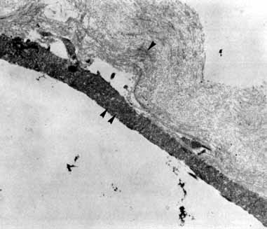
| Fig. 4 Transmission electron micrograph shows folded collagen (arrowhead) conshstent with posterior hyaloid (morphologically type 2 collagen). Adherent to the denser posterior surface is a cellular pseudopod (double arrowheads). The cell body is not present in this grid. (Margherio R, Trese MT, Masgherio A, et al: Surgical management of vitreomacular traction syndromes. Ophthalmology 96:1437, 1989) |