|
|
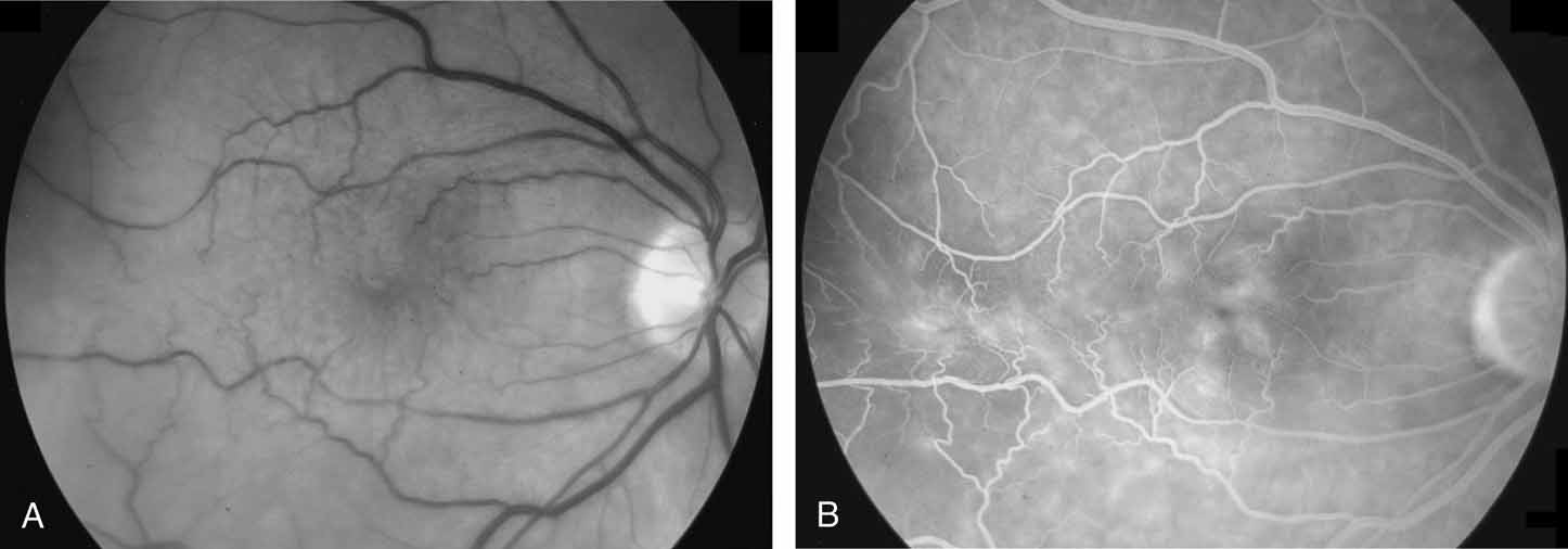
| Fig. 8 A. Black and white photograph taken with a green 540-nm filter, showing dragging and distortion of the macular vessels. B. Fluorescein angiogram. Note distortion of macula and vessels, with associated vascular leakage and cystoid macular edema. |