Chapter 26
Penetrating and Lamellar Keratoplasty
Scott Blackmon, Terry Semchyshyn and Terry Kim
Main Menu   Table Of Contents

Search

PENETRATING KERATOPLASTY
POSTOPERATIVE MANAGEMENT
EARLY POSTOPERATIVE COMPLICATIONS
LATE POSTOPERATIVE COMPLICATIONS
OTHER COMPLICATIONS
LAMELLAR KERATOPLASTY
DEEP LAMELLAR KERATOPLASTY
PREHARVESTED DONOR CORNEAS
EVOLVING CONCEPTS
DEEP LAMELLAR ENDOTHELIAL KERATOPLASTY
ACKNOWLEDGEMENT
REFERENCES

PENETRATING KERATOPLASTY
Penetrating keratoplasty (PK) is one of the most common forms of tissue transplantation currently performed. It can be an extremely successful procedure, with dramatic visual improvement for the patient. However, it can also be one of the most challenging and frustrating procedures for a patient to endure, with a prolonged convalescence, delayed visual improvement, and many postoperative challenges. The technique of keratoplasty, or corneal grafting, involves removing the dysfunctional elements of the cornea and replacing those elements with healthy tissue. Full thickness keratoplasty is termed penetrating keratoplasty, and partial-thickness keratoplasty is termed lamellar keratoplasty.

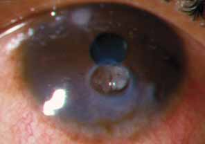
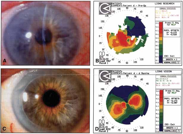
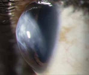
The current number of procedures performed on an annual basis is decreasing slightly; a total of 32,744 corneal transplants were performed in 2002, marking the first time since 1986 that the number of procedures performed has been less than 33,000.1 This downward trend has coincided with the decrease in incidence of pseudophakic corneal edema (PCE) (Fig. 1) and aphakic corneal edema (ACE). The percentage of PKs performed to treat PCE has dropped from 19.8% in 2001 to 18.4% in 2002, which was the lowest percent reported in this category in 15 total years of tracking by the Eye Bank Association of America (EBAA). It is possible that this decrease reflects improved cataract removal technique and technology, such as phacoemulsification and posterior chamber intraocular lens placement. The indications for PK have shifted over the past several decades, with PCE the most common indication since 1980, whereas keratoconus (Fig. 2) and ACE were the most common indications prior to 1980.2 Data is tracked for a total of 16 indications for transplant by the EBAA. Pseudophakic corneal edema, keratoconus (particularly in younger patients), Fuchs' endothelial dystrophy (particularly in older patients), and regrafting secondary to rejection or other reasons currently sit atop the list of indications for PK.

Fig. 1 Pseudophakic corneal edema is the leading indication for penetrating keratoplasty in the United States.

Fig. 2 Keratoconus is one of the most common indications for penetrating keratoplasty. Descemet's tears may occur spontaneously in this condition, leading to acute hydrops.

Although the overall number of penetrating keratoplasties has decreased, there are certain indications that have increased. Patel3 examined the incidence and indications at a major university setting in the northeastern United States and noted that the incidence of regraft was increasing. The number of regrafts increased from 9% to 16%, comparing the years 1983 to 1988 with 1989 to 1995. Within the regraft category, the number of multiple regrafts had also increased (11% from 1989–1991, jumping to 29% from 1992–1995). Regrafting and multiple regrafting were associated with an increased failure rate during the study period. A subsequent study at the same institution by Cosar4 showed that the incidence of regraft had increased to 18.1% from 1996 to 2000. The leading indications in their study population also included PCE (27.2%), keratoconus (15.4%), and Fuchs' endothelial dystrophy (15.2%). Differing incidences of regraft have been noted in other reports.5–7 One analysis noted only an 8.7% incidence of regraft.8 In this population, keratoconus (rather than PCE) was the leading indication for transplantation (45.6%).

With regards to availability of tissue, the transplant surgeon enjoys a relative abundance of donor corneal tissue, so the waiting-list experience endured by other transplant candidates (e.g., kidney or heart transplant) does not occur for the corneal transplant patient, with few exceptions. A total of 42,143 donations were made in 2002. The pool of suitable donor corneas will likely decrease in the future given the increasing incidence of refractive procedures. Donor screening (in the form of potential donor questionnaires and eye tissue analyses to detect evidence of prior refractive surgery) will likely become more important as the popularity of these corneal refractive procedures continues to increase.9–11

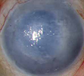
The prognosis for graft clarity can be divided into four groups (Table 1). This grouping system takes into account wound healing ability, presence of vascularity at the graft-host junction, and underlying infectious processes. Keratoconus, in group 1, has an excellent prognosis for graft clarity, and may account for the lower total number of regrafts reported by Edwards.8 The majority of PKs are performed for optical indications, so as to improve vision by removing an optically opaque or dysfunctional cornea. Tectonic (to preserve globe integrity), therapeutic (to remove progressive conditions, such as an expanding fungal infiltrate) palliative, (to improve comfort in a painful eye with bullous corneal edema), and cosmetic (to improve appearance in a blind eye) indications are less frequently encountered reasons for transplantation.


TABLE 1. Prognosis for Graft Clarity*


  Diagnosis Morphology Prognosis
Group 1KeratoconusAvascular central corneal thinning, scarring, or edema surrounded by healthy corneal tissueExcellent, 90% or more
 Central or paracentral inactive scarsLattice or granular Dystrophies  
 Central Fuchs' dystrophy (early)  
 Rotating grafts or autografts  
Group 2Advanced Fuchs' dystrophyLesion that extends partially or totally toVery good, 80%–90%
 Pseudophakic corneal edema periphery with an adequate surface and 
 Aphakic corneal edema mild-to-moderate vascularity 
 Inactive herpes simplex keratitis  
 Iridocorneal endothelial syndromes  
 Interstitial keratitis  
 Macular dystrophy  
Group 3Active bacterial keratitisExtremes of corneal thickness, perforations,Fair, 50%–80%
 Active herpes simplex keratitis  peripheral descemetoceles, active disease 
 Congenital hereditary endothelial dystrophy  
 Active fungal keratitis  
 Mild chemical burns  
 Moderate keratitis sicca  
Group 4Severe chemical burnsSevere fibrovascular replacement of cornea,Poor, 0–50%
 Radiation burns conjunctival ischemia, anterior chamber 
 Ocular cicatricial pemphigoid obliteration, limbal stem cell deficiency 
 Stevens-Johnson syndrome  
 Neuroparalytic disease  
 Congenital glaucoma  
 Epithelial downgrowth  
 Anterior chamber cleavage syndromes  
 Limbal stem cell deficient states  
 Multiple graft failures  

*This table is meant to be a guideline and is not absolute. The prognosis for each group is worsened by the presence of elevated intraocular pressure, intraocular inflammation, and lid and conjunctival defects.
†Failed grafts generally have the prognosis for the group of their primary diagnosis or slightly less. (From Buxton JN, Norden RA: Indications and contraindications. In Brightbill FS (ed): Corneal Surgery: Theory, Technique, and Tissue. St Louis: CV Mosby, 1986)


PREOPERATIVE EVALUATION

A complete patient and family history must be taken, including vision prior to onset of symptoms, presence of amblyopia, timing of prior surgery, medical treatments of the underlying condition, current ocular and systemic medications, presence of glaucoma, presence of systemic illness, and vision in the contralateral eye. The patient's desire and ability to adhere to a prolonged postoperative course must be ascertained. A social work consultation can be extremely helpful to this end. Family and/or supportive caregiver situation, transport access, ability to receive help with administration of medications if necessary, and patient motivation can help determine likelihood of patient compliance. Previous medical records from referring physicians detailing prior ocular heath, phakic status, prior medical treatments, and prior ocular surgery can be helpful.

A complete ocular examination must be performed by the surgeon to determine prognosis and to identify factors that may need to be addressed at the time of transplantation. Preoperative visual acuity, as well as rigid gas permeable contact lens refraction for selected patients, such as those with keratoconus or central scarring, are measured. Intraocular pressure (IOP) and gonioscopy, if possible, are performed, with the knowledge of current medications and potential need for concomitant glaucoma tube shunt surgery. Given the potential exposure to long-term corticosteroid use and possible steroid sensitivity, the possibility and status of glaucoma must be addressed. The presence or absence of a relative afferent pupillary defect in the operative eye is also documented, which is especially helpful in the glaucomatous or traumatized eye.

The patient's underlying corneal disorder or disorders are identified during slit-lamp examination. The status of the limbus and presence of corneal neovascularization or conjunctivalization is noted. This may aid in the evaluation of stem cell function and re-epithelialization potential after transplantation. Ocular surface disease, as seen in patients with limbal stem cell deficient processes (e.g., alkali burns, Stevens-Johnson syndrome, and aniridia), can be associated with impaired healing and may require adjunctive therapy at the time of corneal transplantation. In addition to limbal stem cell deficiency, abnormalities of the tear film and decreased mucin production (as seen in ocular cicatricial pemphigoid, Stevens-Johnson syndrome, and chemical burns) present a relative contraindication to PK. Adequate ocular lubrication is essential to the proper healing and functioning of a corneal graft. In these select patients, it has beenfound that limbal stem cell transplantation and systemic immunosuppression combined with penetrating keratoplasty has been a successful approach to ocular surface rehabilitation. Keratoprostheses, which replace diseased corneal tissue with an integrated clear plastic device, have also been used to improve visual function in patients with severe limbal stem cell deficiency and are addressed fully in a separate chapter.

The centration and extent of corneal pathology are noted, and the graft size needed to treat the condition is estimated. The Haag-Streit slit lamp is equipped with a variable height beam, and this beam is helpful in estimating the size of corneal lesions.

The patient's lenticular status is also very important in the preoperative evaluation. The presence of an anterior chamber lens and its role in possible contribution to the need for transplantation should be determined, with possible lens removal or exchange at the time of transplantation. If the patient is phakic, lenticular opacity is assessed, being aware that corticosteroid use in the postoperative period may hasten the progression of a cataract. Therefore, combined corneal transplantation, lens removal, and intraocular lens placement or “triple procedure” may be considered. If lens removal and exchange are indicated, axial length measurement is performed.

Ocular inflammation must be controlled prior to consideration of surgery. The presence of anterior chamber cell and flare, keratic precipitates, peripheral anterior synechiae (PAS), and posterior synechiae are noted. In addition to controlling inflammation, inciting agents must also be identified and addressed. Herpetic disease, including stromal inflammation, must be adequately controlled, and we recommend a time interval of at least 6 months prior to consideration of keratoplasty in these eyes.

Eyelid function and presence of scarring are noted, especially in the patient with concomitant ocular and facial burns. Inadequate lid closure and lagophthalmos can result in corneal drying and exposure, thereby placing the graft at increased risk. Patients with advanced corneal scarring and cicatrization of the fornix are also at risk for exposure keratopathy. Excessive ectropion or entropion with trichiasis can oftentimes be adequately treated by oculoplastic consultation and surgical repair. A prerequisite to oculoplastic surgical assistance, though, is control of ocular and conjunctival inflammation. Lid margin telangiectasia, meibomian gland dysfunction, lid margin hypertrophy, and other signs of rosacea are noted and treated to decrease inflammation and potential stimuli for limbal neovascularization.

Retinal status must be evaluated but may be limited by the view to the posterior pole through a diseased cornea. Contralateral retinal examination may yield information regarding glaucoma, macular degeneration, and overall retinal status. If the view is unsatisfactory, B-scan ultrasonography is indicated.

Informed consent to the procedure is obtained only after a frank discussion with the patient (and his or her family members or caregivers, if the patient so desires). The transplant surgeon should take time to discuss the diagnosis, risks, and potential complications, benefits, alternative treatment options, and details of the procedure and postoperative care. Given the prolonged convalescence, realistic expectations regarding visual recovery, and the timetable for possible recovery, are key points in the discussion. It is important to mention the potential need for rigid gas permeable contact lens wear to treat postoperative irregular astigmatism and to identify the patient's (and/or caregiver's) ability to handle such a lens. Dedication and commitment to follow-up examination must also be stressed. Again, social work evaluation can be helpful in gauging both the patient's and the family's understanding of the procedure and of the follow-up care needed for keratoplasty patients.

PREOPERATIVE MEDICATIONS

Penetrating keratoplasty has been found to be a safe procedure based on the patient's overall medical health and can often be performed utilizing regional anesthesia. At our institution, topical fluoroquinolone antibiotic drops are given every 5 minutes for three drops total preoperatively as a prophylactic measure. In phakic patients undergoing PK only, pilocarpine 2% is given every 5 minutes for three drops total to produce a miotic pupil, thus protecting the lens. Posterior chamber pseudophakic eyes also receive pilocarpine to maintain lenticular position.

In older patients, or in those with higher intrathoracic pressures (such as individuals with stout necks), intravenous mannitol 20% is given preoperatively. Prior to administration, it is essential for the anesthesia staff to assess the patient's overall medical status. Mannitol acts as an osmotic diuretic to help deturgesce the vitreous and thereby decrease posterior vitreous pressure. In combined (keratoplasty and cataract extraction) cases, it may be prudent to place a condom-style catheter on male patients and to have a bedpan ready due to the added length of the case. A Honan balloon can be placed on the eye and inflated to a pressure of 30 mm Hg for 10 minutes, or digital massage can be performed to lower IOP. In combined procedures, we also use the cycloplegic-mydriatic combination of cyclopentolate hydrochloride 1%, phenylephrine 5%, and tropicamide 0.5% one drop every 5 minutes for three doses total for pupil dilation.

SURGICAL TECHNIQUE

There are multiple accepted ways of performing penetrating keratoplasty, and it continues to be an evolving procedure. Surgeons may utilize various techniques in achieving the same goal of a successful transplant and improved visual acuity for the patient. We will present one such approach to the procedure, and we recognize that there are many other acceptable and successful approaches. We will also discuss penetrating keratoplasty in the aphakic and pseudophakic patient, as well as concurrent PK, lens removal, and intraocular lens placement.

Patient positioning is a vital step in ensuring patient comfort for a relatively lengthy procedure while maximizing surgeon access. A tense, uncomfortable patient is apt to move unpredictably during the procedure, and even the most cooperative patient may experience fatigue and strain, resulting in a Valsalva maneuver. This can lead to a rise in posterior pressure, increasing the likelihood of anterior chamber shallowing, vitreous loss, and even suprachoroidal hemorrhage. By placing the head of the bed slightly higher than the foot of the bed in a reverse-Trendelenburg position, posterior pressure can be minimized, especially in obese patients with increased intrathoracic pressure. Also essential is surgeon comfort; the surgeon should also have direct access to both a preparation table and the patient. Placing the microscope height at a level to keep the surgeon's back and shoulders erect can help minimize surgeon fatigue. A wrist rest can be used to aid in surgeon comfort, by providing a place to rest one's hands. It can also aid in patient comfort, by minimizing pressure placed by the surgeon on the forehead and neck. A wrist rest also improves stability and helps minimize the possibility of inadvertently applying pressure on the globe or orbit during the procedure. At some surgical facilities, an ergonomist is available to examine ideal heights for microscope, bed, and wrist rest placement. A good general rule to follow is that a more comfortable position can be maintained for longer periods of time, thereby lessening fatigue.

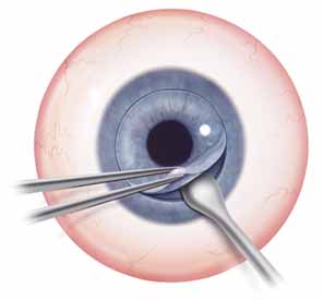
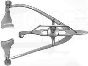
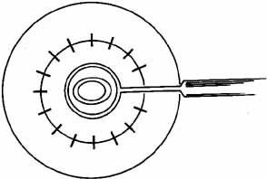
Adequate anesthesia is also important in maintaining patient comfort. Both general and local regional anesthetics can be used in penetrating keratoplasty. Pediatric or uncooperative patients as well as those with tremors, claustrophobia, high level of anxiety, or a history of ocular trauma with possible penetrating injury, are all candidates for general anesthesia. The majority of patients with nontraumatic indications for PK, however, can receive adequate anesthesia and akinesia with local regional approach. After the administration of mild intravenous sedation by the anesthesia team while in the operating room, a local lid block and retrobulbar block can be performed. Local anesthesia consists of both retrobulbar block and lid block, such as an O'Brien or Van Lint block, to minimize orbicularis function and to decrease the risk of elevated intraorbital pressure while the globe is open. An O'Brien lid block, which consists of a 3 to 5 cc injection of an equal mix of lidocaine 2% and bupivacaine 0.75% into the orbicularis muscle along the inferior and superior orbital rim, can provide adequate lid akinesia, minimizing the posterior pressure that can occur with a vigorous lid squeeze. Retrobulbar anesthesia, consisting of 3 to 5 cc of the same mix of lidocaine and bupivacaine, can provide excellent pain control and minimize globe movement during the procedure. It is important to withdraw the plunger on the syringe immediately prior to administering the block to identify inadvertent vascular canalization. Also, monitoring the globe position by retracting the upper lid (often done by an assistant) helps to minimize the risk of optic nerve trauma. It is important to not inject too large an amount of retrobulbar anesthetic, as this increase in orbital volume can result in increased posterior pressure. After the retrobulbar block, ocular massage, if the globe is intact, can be performed. Alternatively, a Honan balloon, which consists of an inflatable chamber applied over the eye and secured with an adjustable headband, can be placed for several minutes. The eyelids and skin around the eye are then cleansed with betadine antimicrobial solution. In the setting of betadine allergy, benzalkonium chloride can be used as an alternative. A fenestrated drape is then placed over the operative eye. A variety of lid specula have been devised for penetrating keratoplasty; a common goal in all of these devices is maximal exposure with minimal pressure on the globe. A locking lid speculum, such as a Maumenee-Park speculum (Fig. 3), with adjustable fissure width and blade angulations, is suitable for PK because it minimizes globe pressure. Positioning the head and neck so as to make the plane of the visual axis perpendicular to the floor and to the operating microscope is helpful in establishing a symmetric trephination. Also, tilting the head slightly away from the operative eye aids exposure.

Fig. 3 The Maumenee-Park lid speculum width adjustable fissure with and variable locking blade angulation, can minimize the pressure applied to the globe. (Courtesy of Katena Products, Inc.)

In preparing the eye for trephination, the use of a scleral fixation ring, such as a Flieringa's ring, sized to a diameter slightly larger than the corneoscleral limbal junction, can be helpful in maintaining the form of the globe. Although we do not routinely use these rings in adults, we have found scleral fixation rings to be helpful in the pediatric population, when, oftentimes, the globe and sclera are soft and easily distensible. This ring is secured with a series of 7-0 vicryl sutures and is removed at the completion of the procedure. A caliper, such as a Castroviejo caliper, is used to measure the corneal diameter and the diameter of trephination needed to clear the visual axis. Paton considered the ideal graft size to be between 6.5 and 9.0 mm.12 The smaller the trephination, the greater the distance from limbal vascularity and from the drainage structures in the angle. Therefore, the risk of rejection of a smaller graft may be lessened, in theory, given the greater distance from the peripheral limbal vasculature and the resultant immunologic response to the donor tissue. Also, by increasing the distance from the angle structures, the risks of surgeon-induced secondary angle closure, PAS, and iris incarceration are theoretically lessened. However, a smaller diameter graft is more susceptible to decentration and higher amounts of surgically-induced astigmatism. We have found that, for the majority of patients, the visual axis may be cleared with host trephinations of 7.5 to 8.0 mm. There are instances, such as infectious keratitis and peripheral corneal melting, in which an oversized therapeutic graft is indicated. Keratoconus patients with significant peripheral and inferior thinning may also benefit from a larger trephination. We often mark the visual axis with a methylene blue marking pen to help determine centration and trephine placement. Some surgeons prefer to slightly decenter the graft inferonasally so as to approximate the usual functional visual axis. The corneal periphery is marked with an eight-blade radial keratotomy marker, and these marks can be extended manually with a methylene blue marking pen to maintain orientation and identification of the corneoscleral limbal junction. These marks should extend beyond the trephination diameter, so as to help guide the radial placement of the transplant sutures. In maximizing the radial orientation, postoperative wound torque can be minimized, with a possible reduction in postoperative astigmatism. The operative microscope light is then turned off, the light source obturator is engaged, or a corneal light shield is placed on the cornea to minimize potential photic injury to the macula. The trephination of the host cornea is performed only after the preparation of the donor corneal tissue. Therefore, at this point in the procedure, attention is turned to the donor tissue.

The donor corneal tissue is now commonly available in Optisol preservative media. The tissue endothelial cell count, infectious disease status (including human immunodeficiency virus, hepatitis B and C virus, and syphilis), and overall epithelial status are reviewed prior to trephination. This information is typically shared by the supplying eye bank with the transplant surgeon 24 hours prior to the procedure, with acceptance or rejection of the tissue based on the individual patient's needs. A higher endothelial cell count is often desirable in a younger patient, whereas a lower endothelial cell count may be acceptable for a therapeutic tectonic graft (in which the need for repeat transplantation for optical improvement is more likely). Also, the donor tissue is visually inspected by the surgeon at the slit lamp for confirmation of tissue quality prior to the procedure, noting epithelial and endothelial quality and overall graft appearance. The donor tissue identification, patient identification, and operative site are confirmed with the operating room staff, and the donor tissue container is opened. The lid is removed, and the surgeon grasps the edge of the sclera with 0.3 mm Castroviejo forceps. An assistant then pours the Optisol solution into a sterile stainless steel specimen cup. Excess Optisol is removed by touching a methylcellulose sponge perpendicularly to the edge of the scleral rim, being careful to avoid touching the endothelial donor surface. By removing the excess fluid, the potential for graft slippage during trephination is minimized. The donor tissue is then centered on a Teflon trephination block. We use the Weck corneal donor trephine, and at this point, the trephination chamber guide is engaged atop the block. There are several other trephination systems available, including Hannah, Katena, Hessburg-Barron, Iowa PK Press, in addition to free-hand manual trephination, which can also be used to prepare the corneal button. The donor tissue is routinely oversized by 0.25 mm compared to the host trephination for the majority of transplants. Donor tissue is sometimes oversized by 0.50 mm in patients with shallower anterior chambers, anterior chamber intraocular lenses, and aqueous tube shunts. The donor trephine diameter is confirmed and then the donor trephine cylinder is engaged and pressed firmly against the Teflon cutting block. This trephination is done in a careful but brisk fashion to minimize endothelial crush artifact. A distinctive, firm end point is felt (“crunch” sensation), and the trephine is removed while the donor rim is grasped using 0.3 mm Castroviejo forceps. By oversizing the donor tissue by 0.25 to 0.50 mm, tissue apposition is maximized. In patients with keratoconus, in an effort to minimize postoperative myopia, the host and donor trephination may be of equal size, depending on the trephination system. The surgeon then grasps both the donor trephine and the trephination chamber guide. This stabilization minimizes the risk of the trephine dropping a second time and engaging the Teflon block. Often, the donor button remains in place on the Teflon block; however, at times, the button may become lodged in the trephine itself. If this situation is encountered, a medicine dropper filled with Optisol can be used to gently remove the button by applying soft suction to the epithelial surface. The graft is then gently placed onto the Teflon block while slowly releasing the suction created by the stopper. A drop of filtered Optisol solution is placed on the endothelial surface, the Teflon block (with donor tissue in place) is covered with a protective lid, and the donor rim and Optisol solution are cultured for bacteria and fungi. The donor tissue is then set aside on a table in a safe location. Care is taken to ensure that the sterile drapes of the table do not reach the level of the floor. If this should happen, the drapes may become caught on the wheels of the table and potentially dislodge and contaminate the prepared donor corneal tissue.

After the donor corneal tissue is safely secured, attention is then returned to the host cornea. Multiple host tissue trephines are available, and we have found success using the Hessburg-Barron vacuum trephine system. This trephine includes a central crosshair to help align trephination. The position of the trephine is stabilized using a manual vacuum system, which is activated by a handheld syringe. The diameter is confirmed by reading the printed mark on the top edge of the trephine. The blade is advanced completely by turning the trephine in a clockwise direction. A moistened fluorescein strip is then gently touched to the bottom of the trephine. The trephine is then centered on the host tissue by aligning the central crosshair with the previously placed central mark on the host cornea. The trephine is then gently touched to the cornea in a perpendicular fashion so as to leave an impression of fluorescein. The trephine is removed, and the centration of this marking is evaluated. Adjustments in the orientation of the trephine can then be made. The blade is then retreated in the trephine housing until it becomes flush. This step is done under direct visualization. Three turns of one-quarter rotation are then performed to draw the blade up into the housing. The vacuum syringe plunger is depressed, and the trephine placed onto the host cornea, making any adjustment of centration as is warranted. The plunger is then released rapidly, and the vacuum level is assessed. A satisfactory vacuum level can be noted by spontaneous trephine adherence to the cornea and host globe movement with gentle lateral pressure on the trephine. The centration is examined by viewing the crosshair and by estimating the distance from the corneoscleral limbal junction to the trephine. This estimate is done by viewing the trephine from the sides and without aid from the operating microscope. Upon confirmation of centration, the edge of the trephine is grasped and a total of 10 turns of one-quarter rotation are performed. The trephine chamber is closely monitored, and, if any aqueous egress is noted (signifying full-thickness trephination and entrance into the anterior chamber), the trephination is halted, vacuum is released, and the trephine is removed. Early entrance into the anterior chamber may be more common in patients with ectatic disorders such as keratoconus, or in patients with prior inflammation and corneal thinning.

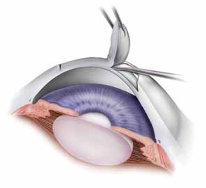
Once the trephine is removed, it is helpful to verify the depth of trephination by tracing the edge of the incision with smooth forceps. If any corneal vascularity is present, phenylephrine 2.5% can be touched to the areas affected, using a moistened methylcellulose sponge. Limbal cautery should be used sparingly as thermal injury may distort the corneal tissue. The trephined host tissue is grasped by 0.12 mm Maumenee-Park forceps (Fig. 4), and tension is gently placed toward the center of the cornea to expose the deepest extent of the trephination. A sharp 15-degree straight blade is used to enter perpendicularly into the anterior chamber, making the motion of the incision away from the surgeon. Care is also taken to not enter too deeply to avoid touching the iris or lens. The incision is extended 1 to 2 clock hours. Viscoelastic material is placed into the eye to deepen the chamber to provide space into which the blades of corneal scissors can be safely placed. Corneal scissors curved to the right and corneal scissors curved to the left are used to complete the excision of the host tissue. The blades are held parallel with the trephination to minimize asymmetric tissue overlap. The scissors are gently elevated as the blades are engaged, and the iris structure is monitored to avoid iris incarceration and trauma. Also, to help maintain tension in the area cut by the scissors, the host button is grasped 180 degrees from the site of the cut. This tension is especially helpful at the end of the tissue removal, when only 1 to 2 clock hours remain. Peripheral anterior synechiae can be lysed with a cyclodialysis spatula after the removal of host tissue.

Fig. 4 The 0.12-mm Maumenee-Park forceps allow for precision manipulation of corneal tissue. (Courtesy of Katena Products, Inc.)

The host tissue is set aside and, upon completion of the procedure, will be sent to pathology for histologic examination and special staining as warranted by the indication for corneal grafting. Until the donor tissue is securely sutured into place, however, the original host tissue should be available, in case it is needed to close the eye. Also, a temporary keratoprosthesis, such as the Cobo keratoprosthesis, should be available to help maintain the integrity of the globe. At this point of the procedure (i.e., when the host cornea is being removed and the globe pressure is equal to atmospheric pressure), the risk of suprachoroidal hemorrhage is greatest. This potentially devastating complication is caused by the rupture of a choroidal vessel, which results in rapid and uncontrolled bleeding in the suprachoroidal space. The hemorrhage may force the iris, lens, vitreous, retina, and other intraocular contents to prolapse out through the corneal trephination wound. Risk factors for surprachoroidal hemorrhage include previous intraocular surgery, elevated IOP, myopia, advanced age, and arteriosclerotic disease. Patients with large body habitus may be prone to increased intrathoracic pressure and may generate additional posterior pressure. These patients may be best approached with general anesthesia. Intraoperative risk factors include rapid decompression of IOP upon entry into the anterior chamber, Valsalva maneuver performed by the patient at the time of trephination, or compression and occlusion of the endotracheal tube resulting in a mechanical Valsalva maneuver. Management of a suprachoroidal hemorrhage is based on when it occurs during the procedure.13 Rapid recognition and response afford the best chance of salvaging the eye. If the hemorrhage occurs upon initial entry into the anterior chamber, the host cornea is sutured back into place, using 8-0 silk or 9-0 nylon sutures, which not only are easier to visualize and tie in rapid sequence, but also have greater tensile strength than 10-0 nylon sutures. Drainage of the hemorrhage can be accomplished by entering into the choroidal space by making a sclerotomy. If the hemorrhage occurs after the host cornea has been removed, the temporary keratoprosthesis is quickly placed into the wound to compress the hemorrhage and to keep intraocular contents in their correct anatomic positions. Our routine during host tissue removal is to have the assistant hold the temporary keratoprosthesis for rapid placement, and the assistant only returns it to the instrument table when all four cardinal sutures have been placed and tied. In the absence of a temporary keratoprosthesis, a surgeon may use his or her finger to tamponade the hemorrhage. Pressure is maintained until the hemorrhage stabilizes. Sclerotomy can be performed to drain the surprachoroidal hemorrhage, and then the donor or host tissue is sutured into place. Alternatively, sclerotomy and drainage of the suprachoroidal hemorrhage can be done at a later date if the intraocular contents can be successfully reposited at the the time of surgery. If possible, irrigating fluid can be placed into the anterior chamber via keratoprosthesis attachment or anterior chamber maintenance port. This fluid can help deepen the anterior chamber, resulting in more anatomic orientation of intraocular contents and may also facilitate evacuation of the hemorrhage.

Upon successful entry into the anterior chamber and removal of the host tissue, the donor tissue can then be secured to the patient. We will describe standard phakic keratoplasty and will then discuss specific situations, such as combined keratoplasty and cataract removal and also keratoplasty in the pseudophakic patient. Up to this point in the description of the procedure, the steps taken for combined procedures are similar to those in standard phakic keratoplasty.

Simple Penetrating Keratoplasty

In a simple PK, such as in patients with keratoconus, the donor cornea is transferred from the Teflon block to the surface of the eye using a Paton spatula. This transfer is done immediately adjacent to the patient's eye in the operating field to minimize the likelihood of inadvertent tissue loss onto a nonsterile field. The surgeon's dominant hand is used to lift the donor graft. Viscoelastic substance is placed on both the endothelial side of the donor button and on the host corneal tissue rim. The donor tissue is brought to the host rim, and it is tipped slightly by touching the cut edge of the graft to the 6 o'clock position on the viscoelastic-coated host rim.

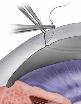
The tissue is tipped by rotating the wrist and the Paton spatula so that the graft is positioned almost perpendicularly to the host cornea. This position facilitates a precise grasping of the top edge of the donor tissue by split-tined Hofmann-Polack forceps (Fig. 5). These speciallydesigned forceps allow for the uninterrupted passage of a needle between the two toothed tines. After the elevated edge of the graft is grasped by the Hofmann-Polack forceps, the surgeon quickly exchanges the Paton spatula for a preloaded needle driver containing a 10-0 nylon suture. The suture is first placed onto the donor cornea at the 12 o'clock position. The action of suture placement involves not only the passage of the needle onto the graft tissue, but also the placement of the graft tissue onto the needle. In this sense, one can think of “placing the graft onto the needle” in addition to passing the needle through the graft. The optimal exit depth of the suture is approximately 90% thickness, with the exit path extending radially through the stromal side cut of the donor graft tissue (Fig. 6). The donor tissue is released by the Hofmann-Polack forceps, and then the host rim is grasped.

Fig. 5 Split-tined Hofmann-Polack forceps can be effective in stabilizing corneal tissue and allowing precise visualization of the passage of the suture needle. (Courtesy of Katena Products, Inc.)

Fig. 6 Suturing technique for penetrating keratoplasty. Forceps are used to approximate the edges of the host and donor cornea, while the suture is placed at approximately 90% depth.

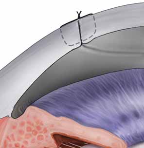
The previously placed methylene blue radial keratotomy marks are used to guide the positioning of the 12 o'clock suture pass. The suture is passed though the two tines of the forceps, making sure that the pass is completed in as radial an orientation as possible. The ratio of the distance from the entry of the needle on the donor to the exit on the host cornea is ideally one-third donor to two-thirds host. The Hofmann-Polack forceps are then exchanged for 0.12 mm Maumenee forceps. The suture is tied in place using a slipknot (two-one throw) and then cut using Vannas corneal scissors (Fig. 7). Both tags can be left long so as to be able to adjust suture tension based on intraoperative keratoscopy after the placement of the first eight sutures. The cut sutures are gently draped away from the visual axis, and the donor tissue is grasped at the 6 o'clock position, 180 degrees away from the first suture. A similar technique is used in passing the suture at this position. Symmetry of the pass can be estimated by noting the tissue alignment as well as the symmetry at 3 o'clock and 9 o'clock. Also, as gentle tension is applied in tying the suture, a straight tension line will be noted in the graft tissue. This line should be oriented from 6 o'clock to 12 o'clock. Sutures are then passed at the 3 o'clock and 9 o'clock meridians, while taking into account the amount of tissue on either side of the pass. Upon completion of the four cardinal sutures, a symmetric diamond shape should be noted on the donor tissue. A total of eight interrupted sutures are placed, the ends of which are temporarily left long. The graft is then wetted with balanced salt solution, and a manual keratoscope, such as a circular metal loop or a von Luhnen keratoscope, is used to estimate astigmatism based on the compression or expansion of the reflected rings or ovoid reflex from the corneal surface. If suture adjustment is needed, the suture is grasped proximal to the knot. The suture is cut and rotated by pulling the grasped suture. The tension of the suture can then be adjusted by retying the knot based on keratoscopy. For the slipknots not needing adjustment, a third locking throw is placed. The details of suture adjustment to minimize postoperative astigmatism are discussed more thoroughly in the suturing techniques section.

Fig. 7 Suturing technique for penetrating keratoplasty. Schematic illustration showing the 10-0 nylon suture at proper depth after the suture is tied in a slipknot fashion. The knot will then be rotated and buried in the cornea.

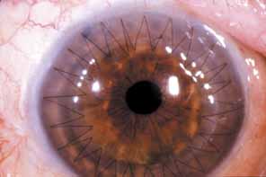
In patients undergoing simple keratoplasty, a running suture can be used (Fig. 8). A half section of 10-0 nylon suture is used, placing two suture bites between each pair of previously placed interrupted sutures. The ratio of the radial orientation is one-fourth the distance to either interrupted suture. On the final pass of the suture, the needle is left in place and the edge of the needle track is slightly expanded, using a 15-degree steel blade. This slight expansion facilitates the passage of a tied knot through corneal tissue. The suture tension is adjusted using two smooth tying forceps, grasping the suture in an “end over end” fashion to maintain tension throughout the adjustment. The suture tightening begins 180 degrees from where the knot will be placed, and two passes in both directions are made. The cornea is again wetted, and intraoperative keratoscopy is performed with appropriate suture adjustment. A knot is then tied and the suture is gently rotated to bury the knot. In patients with peripheral corneal neovascularization, multiple interrupted sutures can be placed, thereby facilitating suture removal based on local suture vascularity. A total of 16 interrupted sutures are adequate to approximate the graft to the host tissue in the majority of patients.

Fig. 8 Clear graft after penetrating keratoplasty utilizing and showing a continuous (running) 24-bite suture. (Courtesy of Alan Carlson, MD)

Combined Penetrating Keratoplastyand Cataract Extraction

When a cataract extraction is to be performed along with the PK, the patient's pupil is dilated preoperatively with cyclopentolate hydrochloride 1%, phenylephrine 5%, and tropicamide 0.5%, one drop of each every 5 minutes for three total doses. We favor an extracapsular technique in removing the lens nucleus. There are instances, such as in Fuchs' endothelial dystrophy, where corneal clarity is sufficient to facilitate the performance of a continuous curvilinear capsulorrhexis through a paracentesis incision prior to removal of the host corneal tissue. The anterior chamber is filled with viscoelastic substance during this procedure, which allows for additional control of the capsulorrhexis by flattening the anterior curvature of the crystalline lens. By stabilizing the anterior chamber with viscoelastic material, a capsulorrhexis is less likely to proceed radially toward the zonules, causing radial tears and potentially destabilizing the capsular support necessary for adequate intraocular lens positioning. We also favor this approach in the appropriate patient because it minimizes the duration of time spent between removal of the host cornea and placement of the donor tissue.

If, however, the cornea is too opaque to confidently visualize the anterior capsular surface, we perform a capsulorrhexis with an open anterior chamber and use an extracapsular technique for removal of the cataract unless obvious lens dislocation or significant traumatic zonular rupture necessitates an intracapsular cataract extraction. A small stab incision is made in the central capsule with a bent needle cystotome, and the edge is grasped with fine Utrata capsulorrhexis forceps. The capsule is gently torn circumferentially to create a smooth-edged, round opening. To facilitate removal of the entire lens nucleus, the opening should be 5 to 6 mm in diameter. If the tear extends toward the anterior zonules it will begin to turn outwardly and may result in a tear extending to the equator of the lens. If this occurs, the capsulotomy should be restarted in the opposite direction with fine scissors and completed using the Utrata forceps. The anterior capsule is then removed with forceps. The lens is loosened from the capsular bag by introducing a cannula just under the capsule and irrigating fluid around the periphery of the lens cortex (i.e., hydrodissection). Visualizing a wave of fluid traversing the red reflex can be helpful in confirming adequate hydrodissection. The lens is left in place until the cornea is trephined. The chamber is reformed and the paracentesis wound is secured by placing a 10-0 nylon suture. After the successful trephination of host tissue, attention can then be turned back to lens removal. The lens can be gently hydrodissected once again and rotated out of the capsular bag using a 25-gauge hydrodissection cannula. If adequate hydrodissection has been performed earlier, the lens is usually easily removed. If required, slow, steady, and gentle pressure at the 6 o'clock and 12 o'clock positions just posterior to the limbus with a muscle hook or lens loop and forceps can help express the lens nucleus.

Residual cortex is removed with either a manual irrigation-aspiration cannula, such as the Simcoe cannula, or with an automated irrigation-aspiration setup. The former allows for a central aspirating port and a peripheral irrigation port to minimize the amount of tension placed on the capsular bag. Cortical removal is more tedious and time-consuming in an open-sky technique than in a closed system because no fluid pressure can be built up and air frequently enters the aspiration port. Caution must be exercised to not tear the posterior capsule or rupture the zonules by rushing. Capsular stability provided by a successful capsulorrhexis makes cortical cleanup much easier.

The most commonly accepted method for visual rehabilitation after cataract extraction is implantation of an intraocular lens. This is also true after corneal transplantation; the combination of penetrating keratoplasty, cataract extraction, and intraocular lens implantation has become known as the triple procedure. We prefer posterior placement of the intraocular lens within the capsular bag. Placement within the capsular bag reduces contact of the optic and haptics to the pigmented, vascularized uveal tissue, reducing the risks of haptic erosion into vascular structures and the constant chafing on the uvea that may lead to chronic, low-grade uveitis, pigment dispersion, and glaucoma.

In the absence of adequate capsular support, the surgeon can place an intraocular lens in the anterior chamber. Alternatively, one can place a fixated posterior chamber intraocular lens with either iris or scleral fixation. There are anterior chamber intraocular lenses currently available that are relatively well tolerated by the corneal endothelium. These lenses are relatively easy to place and, when accompanied by a surgical peripheral iridectomy, can be a viable option in older transplant patients. However, because an anterior chamber lens is positioned anterior to the pupil, the iris cannot protect the graft endothelium from the optic. Even with the use of viscoelastic substances, significant endothelial cell loss and possible graft decompensation may result from intraocular lens touch. In younger patients (under 50) in whom longer term graft viability and endothelial function needed, implantation of a lens anterior to the capsule (in the ciliary sulcus) or a suture fixated lens may be possible and preferable to an anterior chamber lens. Intraocular lens suturing techniques are discussed in the section on aphakic and pseudophakic keratoplasty.

The technique involved in placing the posterior chamber intraocular lens into the capsular bag is relatively simple and is aided by having an 8-mm opening in the cornea through which to work. A small amount of viscoelastic is injected through a 30-gauge blunt-tipped cannula between the anterior and posterior capsule at the 12 o'clock to 6 o'clock position. The inferior haptic is then slipped into this space. The superior haptic is grasped with angled tying forceps about 1 mm from the end and swung centrally over the optic while gently depressing the intraocular lens to bring the haptic below the level of the anterior capsule. The haptic is then slowly allowed to recoil while observing it slip into position within the capsular bag. Usually, centration is excellent and rotation of the intraocular lens is unnecessary. A topical miotic agent is injected onto the surface of the iris to constrict the pupil while gently depressing the optic to minimize lens capture by the pupillary edge. We do not routinely perform an iridectomy in an otherwise quiet eye undergoing a triple procedure with a posterior chamber lens. In patients with anterior chamber intraocular lenses, a peripheral iridectomy is performed. Once the lens is adequately positioned, the donor tissue is transferred and sutured in place.

Aphakic and Pseudophakic Keratoplasty

Aphakic corneal edema and pseudophakic corneal edema are common indications for PK. Because of this, any surgeon who wishes to perform corneal transplants should be completely familiar and comfortable with the technique and instrumentation required for anterior vitrectomy, iris surgery, and intraocular lens manipulations.

As mentioned earlier, a thorough preoperative evaluation must be completed before the patient enters the surgical suite. Particular attention should be paid to the status of the opposite eye. Is it phakic, aphakic, or pseudophakic? This will often dictate which surgical procedure would be most appropriate for the second eye with regard to intraocular lens placement, removal, or exchange. The vitreous should be evaluated carefully. An intact hyaloid face that is well behind the pupil may allow the surgeon to avoid the necessity of proceeding with an anterior vitrectomy and thus to avoid the increased risk of retinal detachment or cystoid macular edema that may accompany vitreous manipulation. A broken hyaloid face or loose vitreous in the anterior chamber will require an anterior vitrectomy, as discussed below, to complete the transplant safely and reduce the potential postoperative complications associated with vitreous incarceration. Iris and pupil abnormalities, such as synechiae, corectopia, iridodialysis, or pupillary membranes, may require anterior segment reconstruction to create a more favorable anatomic or physiologic ocular status. Poorly controlled glaucoma is generally an indication for removal of an anterior chamber lens and argues against primary placement or replacement of this type of lens.

The host cornea is removed as described earlier. If the eye is to remain aphakic and the vitreous face is intact, a topical miotic agent is injected to constrict the pupil, and viscoelastic is placed onto the iris and hyaloid face before transferring the donor cornea. Based on the surgeon's preference, a peripheral iridectomy can be performed as prophylaxis against postoperative pupillary block glaucoma. When loose vitreous is present in the anterior chamber, an anterior vitrectomy must be performed. Mechanical suction-cutting units are superior to the simple cellulose sponge vitrectomy technique. An Ocutome mechanical vitrectomy unit that has a guillotine cutting action works well for our patients. The cutting rate should be set at 300 to 400 cuts per minute, and suction should be low to moderate. Care should be taken to avoid traction on the vitreous so as not to create a retinal tear. The vitrectomy is adequate when all vitreous has been removed from the anterior chamber and the iris bows backward into the vitreous cavity. In cases of preexisting cystoid macular edema or when a suture-fixated posterior chamber intraocular lens is to be placed, a deeper and more complete vitrectomy is possible and may be advantageous. Once the mechanical vitrectomy is completed, a dry cellulose sponge is touched to the anterior surface of the iris and pupillary margin for 360 degrees to ensure that no vitreous strands remain. Scissors are used to cut residual vitreous that adheres to the sponge. Balanced salt solution is gently irrigated over the iris into the posterior chamber to refill the globe.

There are a great variety of ways in which a pseudophakic eye may present. The intraocular lens may be in the anterior or posterior chamber or may be supported by the iris. The lens may be in a good, stable position; or it may be loose and dislocated; or it may be entrapped in PAS, a pupillary membrane, or even through the cataract wound. Often, a well-positioned, stable posterior chamber lens can be left in place during penetrating keratoplasty. It is unlikely that this lens will compromise graft integrity or function. However, one report indicates a slightly decreased graft survival rate in eyes requiring a corneal transplant in which a posterior chamber lens was left in place rather than exchanged at the time of the keratoplasty.14 Decreased graft survival rate has not been reported by other authors, though. Miotic agents and viscoelastic substance are injected onto the surface of the iris and lens before transferring the donor button.

We remove iris-supported lenses and closed-loop haptic anterior chamber intraocular lenses at the time of corneal transplantation. Fortunately, the current anterior chamber lens designs are often better tolerated than older anterior chamber lens designs, and fewer older lenses are encountered today. Many of the older designs of anterior chamber lenses associated with pseudophakic corneal edema can be considered a significant contributing factor in corneal endothelial decompensation. Although it is well noted that other factors, such as initial cataract surgical technique and complications, glaucoma, uveitis, and corneal dystrophy, may all influence corneal decompensation, a significant variable that may now be modified is the removal of an ill-positioned or otherwise inadequate intraocular lens. General principles will be outlined and can be applied and modified to address a given anterior chamber lens type. The reader is referred to the literature for a more detailed discussion of removal of intraocular lenses at the time of PK.15,16

Iris-supported lenses are relatively easy to remove with gentle manipulations of the pupil and lens. Often a suture is present securing the superior haptic to the iris. This should be identified and removed. The intraocular lens should be manipulated cautiously because synechiae or vitreous adhesions to the lens may be present. Blunt or sharp dissection is used to free the intraocular lens, which is then removed from the eye.

Anterior chamber intraocular lenses may be flexible, rigid with solid footplates, or have open- or closed-loop haptics. Initially, the intraocular lens may be grasped with angled tying forceps testing whether or not it may be gently removed from the eye. Some solid lenses may be removed this way, but it may sometimes be necessary to cut this type of intraocular lens in two pieces for removal. Closed-loop haptics present perhaps the most difficult challenge for a safe removal, and the reader is referred to the literature for a discussion regarding the removal of this particular type of anterior chamber lens.16 PAS are lysed with a cyclodialysis spatula by gently sweeping the angle. Large iris defects, such as an old sector iridectomy, may be repaired to bring the iris and pupil into a more anatomically correct configuration and to prevent the floppy edges of the iris from becoming incarcerated into the corneal wound. One10-0 Prolene suture on a blunt-tapered cardiovascular needle passed at the sphincter muscle is used to just approximate the tissue edges. An additional suture in the mid-portion of the iris may be required to close large defects. Care should be taken to not tie the suture too tightly because it may lead to localized iris necrosis.

Replacement of an intraocular lens is considered, if the opposite eye is phakic or pseudophakic, or if the patient has demonstrated intolerance to contact lenses or aphakic spectacle correction. In the past, the only option for intraocular lens fixation in an aphakic eye or after removal of another intraocular lens during penetrating keratoplasty was to implant an anterior chamber lens. The Multiflex-style anterior chamber intraocular lens has four-point fixation with minimal angle touch and good compression characteristics to lessen the chance of angle erosion or chronic pain but may tend to vault forward toward the corneal endothelium. The lens is relatively easy to implant through a keratoplasty opening, and the overall surgical results have been good. Hassan17 showed that 63% of patients in which this type of intraocular lens was implanted at the time of keratoplasty had a visual acuity of 20/40 or better at 2 and 3 years. Acetylcholine is used to create miosis, and viscoelastic may be used to open the angle, after which the lens should be positioned with the footplates away from preexisting iridectomies or other areas of iris defects.

The development of alternative intraocular lens fixation techniques has increased the surgeon's options regarding style and position of the pseudophakic lens after keratoplasty. Because not all eyes are good candidates for an anterior chamber intraocular lens due to PAS, iris defects, or glaucoma, suture fixation of posterior chamber intraocular lenses has become an increasingly popular technique in our surgical armamentarium.

The posterior chamber intraocular lens may be suture-fixated to the posterior iris or to the sclera18; both techniques have potential advantages and disadvantages. The surgeon who chooses to suture-fixate a posterior chamber intraocular lens should select one of the techniques and concentrate on becoming adept primarily in that technique. Contrary to what some of the literature would indicate, these are not necessarily simple procedures; they add time and complexity to the operation and require skill and forethought to successfully complete.

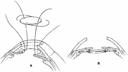
One of the techniques used for posterior chamber intraocular lens implantation during penetrating keratoplasty is iris fixation.19 A 6-mm, three-piece modified J-loop, 0-degree or 10-degree angulated Prolene haptic, four-hole intraocular lens can be used. It is suture-fixated to the posterior aspect of the iris with 9-0 Prolene on a blunt-tapered needle (Fig. 9). The lens is held securely in place with the haptics positioned within or posterior to the ciliary sulcus. As opposed to the old style of iris-fixated intraocular lenses, pseudophacodonesis is nearly nonexistent, and torsion of the intraocular lens along the fixational point axis does not seem to occur, probably because of the stability provided by the haptics posterior to the iris.

Fig. 9 Visual results of suture-fixated posterior chamber lenses during penetrating keratoplasty. Using a BVIOO-4 Ethicon needle, two prolene sutures are placed through the midperipheral iris, one at approximately 3 o'clock and one at 9 o'clock. Each suture also passes through one hole in the optic of the intraocular lens with the knots being tied between the iris and the anterior surface of the optic. The haptics of the lens are positioned at 3 o'clock and 9 o'clock within the ciliary sulcus. (From Price FW Jr, Whitson WE: Visual results of suture-fixated posterior chamber lenses during penetrating keratotomy. Ophthalmology 96:1234, 1989, with permission)

Suture fixation of a posterior chamber intraocular lens to the sclera requires passage of two Prolene sutures 180 degrees apart posterior to the iris, theoretically through the ciliary sulcus and out of the sclera. Either single-armed suture tied under preplaced partial-thickness scleral flaps or double-armed suture tied and rotated into the sclera can be used to avoid erosion of the Prolene knots through the conjunctiva. The sutures are tied to the tangential point of each haptic, and the intraocular lens is then implanted through the pupil. Again, there is no pseudophacodonesis, and potential lens tilt is minimized by haptic/optic approximation to the posterior iris. Both posterior lens implants with preplaced eyelets integrated to the haptics and one-piece acrylic lenses can be adequately fixated to the sclera. The interested reader is referred to the literature for more detailed descriptions of the various intraocular lens suture-fixation techniques.19–26

SUTURING TECHNIQUES

If the host cornea is not vascularized, a running suture technique can be used (Fig. 8). We often utilize an 8-bite interrupted and 12-bite running combination, but a variety of techniques can be used effectively. One alternative is a 12-bite interrupted and 12-bite running combination, and another is a 20- to 24-bite single running suture. Each provides the opportunity for modification of astigmatism in the postoperative period; the 12 and 12 combination provides selective interrupted suture removal while the running suture allows adjustment of local suture tension. We will first describe the 12 and 12 technique in which we use an Alcon CU-5 4-mil needle with 10-0 nylon for the interrupted sutures. In this technique, a 12-blade radial keratotomy marker is used to mark the peripheral cornea prior to trephination. The first suture is placed at the 12 o'clock position. The sutures are placed using the method described in the section on simple penetrating keratoplasty, proceeding to the 6 o'clock, 9 o'clock, and 3 o'clock positions, and then to all 12 clock-hour positions, alternating suture placement at 180 degrees to the previously placed suture.

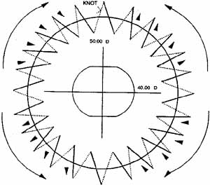
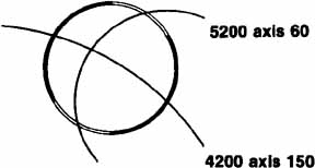
It is at this point that we take what is the last of a series of intraoperative steps designed to minimize astigmatism. Before placing the running suture, we perform qualitative keratoscopy using a von Luhnen keratoscope. Other methods may be employed, such as the use of circular metal loop on a handle (Fig. 10), or an operating microscope-mounted qualitative keratometer. The Flieringa's ring, if present, is removed. Intraocular pressure is adjusted by filling the anterior chamber. The keratoscope or wire loop is held about 2 cm directly above and parallel to the plane of the cornea. Irrigation of the graft will smooth the surface enough to see a reflection of the loop on the cornea. Most commonly, the reflection is ovoid, which indicates astigmatism of the graft; the short axis represents the steepest curvature of the cornea. Suture tension is adjusted by loosening or tightening the slip knots. Keratoscopy is repeated with additional suture adjustment as needed to minimize the corneal astigmatism. When the adjustments are completed, a final throw is placed on each suture, and the knots cut short with Vannas scissors. Burying the knots in any type of corneal surgery is essential. The knots are buried into the donor stroma in an attempt to keep them as far away from any vascularization as possible. Anteroposterior graft–host disparity in alignment can also induce astigmatism and should be identified. Sometimes a meridian is flat because the donor is anteriorly displaced or steep secondary to posterior displacement. Resuturing is then necessary to correct the problem. It is not uncommon to inadvertently take superficial bites on the recipient side when suturing away from oneself.

Fig. 10 Wire-loop keratoscope is held between the cornea and microscope. An oval image narrower at the 90-degree axis indicates corneal astigmatism with the steeper curvature at 90 degrees.

The running suture is then placed, starting on the graft side at the 11:30 position. We use 10-0 nylon on an Ethicon Ultima CS 160 6-mil needle. The needle is passed radially through the graft and host, and about 10 cm of length is pulled up to allow for completion of the running suture. Each successive pass is taken halfway between the interrupted sutures, moving in a clockwise direction, taking care to create radial, deep, and equal-length bites. After the 10:30 pass, the needle is left in place and the exit track is expanded using a 15-degree steel blade as noted in the simple PK section. The two ends are cut about 3 cm long and a single two-loop throw is placed. Slack is then taken up from the running suture with microtying forceps by starting at one side of the knot and working back around the graft. Excessive tightening should be avoided. The suture is then tied with two square knots, and the ends are cut short. The knot is rotated to lie in the graft stroma, trying not to incarcerate it within the graft–host interface. While the anterior chamber is slowly refilled with balanced salt solution, the iris is observed to drop back away from the cornea. Any areas of iris incarceration or synechiae are released by introducing a cyclodialysis spatula through the wound 2 clock hours away from the area of concern. It is then passed over the iris peripheral to the incarceration and gently swept centrally to pull the iris from the graft–host interface. The chamber is again re-formed and the eye is dried with cellulose sponges. High magnification is used to look for the presence of any wound leaks, which, if present, are to be closed with single superficial 10-0 nylon sutures.

Another suture technique that allows earlier and possibly more significant adjustment of postkeratoplasty astigmatism is the use of a single running 10-0 nylon suture, as described by McNeill and Wessels27 and by Van Meter.28 Other surgeons have had considerable success with this technique and use it to reduce and initially stabilize astigmatism within 4 to 10 weeks after keratoplasty in the majority of patients. Spectacle correction can be prescribed much earlier than with other techniques, thus providing visual rehabilitation to patients, many of whom are elderly. The advantanges of astigmatism adjustment are balanced by the risk of reliance on a single suture for graft apposition and the flexibility of suture removal in interrupted techniques in the setting of peripheral neovascularization.

The suturing technique begins similarly to that just described with the placement of four interrupted cardinal sutures at the 3 o'clock, 6 o'clock, 9 o'clock, and 12 o'clock positions. A 10-0 nylon is then passed in a running fashion beginning at the 11:30 position and proceeding in a clockwise direction. Usually, 20 bites are taken at and between each of the methylene blue marks placed by the 10-blade marker. Initial experience with this technique should be gained using a 12-blade marker and 24 suture bites. The cardinal sutures are then cut and removed, and the single running suture is securely tied. Excessive tightening tends to induce a circular row of corneal compression that can resemble a “barrel top.” Sutures that are too tight induce flattening of the graft. This results in an initial hyperopic shift from the intended result followed by a (sometimes significant) myopic shift once the sutures are removed and the graft steepens to its resting curvature. The anterior chamber is refilled with balanced salt solution, and the surface of the graft is moistened. A keratoscopy ring is used to determine graft asphericity, and the running suture is manipulated to adjust the astigmatism that is present. The suture is moved toward the steep meridians by grasping the suture initially at the flat meridian with microtying forceps held parallel to the suture to avoid breaking or cutting the suture on the edge of the forceps and gently, but firmly, sliding each successive suture pass in a direction away from the flat meridian toward the steep meridian. This will redistribute suture tension more evenly, and by a series of successive adjustments and rechecking keratoscopy readings, the graft will be brought into a more spherical shape. This same type of adjustment can be performed on subsequent postoperative visits at the slit lamp to fine-tune the refractive astigmatism.

Cycloplegic agents are not routinely administered. We provide subconjunctival injections of tobramycin, 20 mg; cefazolin, 50 mg; and dexamethasone sodium phosphate, 12 mg. Subconjunctival corticosteroids are not given in patients with herpetic corneal disease. A double eye patch is taped firmly in place, and a metal Fox shield is taped over the double eye patch to protect the eye.

Back to Top
POSTOPERATIVE MANAGEMENT
Corneal transplant surgery can often be performed on an outpatient basis, depending on medical status and requirements. The first postoperative day examination involves assessing pain or discomfort, visual acuity testing, IOP measurements, slit-lamp examination, and examination of the fundus. Initial topical medications include prednisolone sodium acetate 1% and a fluoroquinolone antibiotic, both of which are given four times daily. A combination antibiotic/corticosteroid such as tobramycin 0.3% and dexamethasone 0.1% can be used instead. The use of cycloplegia depends on surgeon preference and patient need. Patients with herpes simplex keratitis continue their oral acyclovir, 200 mg three times daily. Continued patching can sometimes be helpful in healing large epithelial defects. Bland ointments can also be used for patient comfort.

Full-time eye protection for the first 2 months is essential after penetrating keratoplasty. Eyeglasses or sunglasses are worn during waking hours, and an eye shield is taped in place for naps and at bedtime. Activity is restricted within the constraints of the patient's lifestyle. We strongly suggest not straining or lifting objects heavy enough to induce a Valsalva maneuver for the first 2 months. No swimming is allowed for at least 1 month, but a full shower may be taken after the epithelium has healed completely, often after the first week.

The patient is routinely seen 1 week postoperatively. At this time the cycloplegic agent (if used) may be discontinued. Topical antibiotic drops are continued until all epithelial defects are healed. Topical corticosteroid drops are continued four times daily for the first month. As the postoperative inflammation subsides, the corticosteroid drops may be slowly decreased to two times daily, then once daily, and finally every Monday, Wednesday, and Friday. The tapering schedule varies and is based on the preoperative diagnosis, the surgical procedure, and the condition of the eye at each postoperative visit. We attempt to taper the dosage and to stop the corticosteroids over the course of the first 12 months in patients with routine phakic keratoplasties. In aphakic eyes that have undergone a vitrectomy, the corticosteroids are continued for at least 6 months. We taper corticosteroid dosage over a very prolonged period in any patient with a history of prior graft rejection. Many of these patients require low-dose corticosteroids indefinitely. If the IOP becomes elevated, the prednisolone sodium acetate may be substituted with topical fluorometholone phosphate or acetate, or with loteprednol etabonate. When the transplant is performed for herpes simplex corneal disease, concomitant antiviral coverage with trifluorothymidine is used on a drop-for-drop basis until once-daily dosing of corticosteroid is reached, at which time the antiviral agent is stopped.

The greatest advantage of the 20- or 24-bite single running suture is the ability to adjust corneal astigmatism in the office during the early postoperative period. Suture adjustment can be performed as soon as the graft epithelium is healed and smooth enough to provide a good reflecting surface to evaluate the mires of a keratometer, a photokeratoscope, a handheld keratoscope, such as the Klein, or a computer-analyzed digital keratoscope. This is usually 1 week after the epithelium is healed and topical antibiotics, which frequently cause mild to moderate punctate keratopathy, have been stopped. The suture adjustment is typically at the 2- to 4-week postoperative visit.

Manifest refraction is performed and followed by visual analysis of the keratoscopic images to show the areas of steep and flat corneal curvatures. The long axis of the ovoid denotes the flat meridian, while the short axis denotes the steep meridian. The computer-assisted corneal topography units commercially available provide color-coded maps in which the “cool” colors (blues and greens) indicate relative flat curvatures, while the “warm” colors (yellows, oranges, reds) indicate the relatively steep areas of corneal curvature. The goal of suture adjustment is to achieve a perfectly symmetric graft surface without any suture-induced astigmatism.

Multiple topical anesthetic drops and antibiotic drops are applied, and the patient is instructed to hold the eye very still during the adjustment to avoid the risk of breaking the single running suture. Comfortable positioning at the slit lamp is essential. Blunt-tipped microtying forceps are used to break the epithelium and anterior stromal adhesions in the graft–host interface for 360 degrees. After the relatively tight (steep) meridians are determined, the forceps are used to gently pull individual loops, sequentially moving the suture away from the flat (loose) meridian toward the steep meridian, thereby redistributing suture tension (Fig. 11). A drop of artificial tears is placed on the cornea, and a handheld keratoscope is used to reassess the corneal curvature. The adjustment process is repeated until circular mires are noted on keratoscopy. Antibiotic drops are again placed on the eye; a patch is optional but not usually necessary. We use a broad-spectrum topical antibiotic drop, such as a fluoroquinolone, in addition to the usual topical corticosteroid drops four times daily for 1 week after suture adjustment. The patient returns in 1 to 3 weeks when the process can be repeated if greater than 2 to 3 diopters (D) of corneal astigmatism persist. Spectacle correction can be provided approximately 4 weeks after suture adjustment as the cornea appears to be relatively stable at that time. The remainder of the postoperative care is per regular routine.

Fig. 11 Schematic example of the single continuous suture. Ten diopters (50.00 D-40.00 D) of postkeratoplasty astigmatism are present at axis 90 degrees. By using straight tying forceps at the slit lamp, loose suture from the horizontal axis is gently pulled into the vertical axis where suture tension is high. Arrowhead represents the direction of individual suture pull to reduce suture tension in tight meridian. Bowed arrow represents general direction of suture adjustment. (From Van Meter WS, Gussler JR, Soloman KD, Wood TO: Postkeratoplasty astigmatism control. Ophthalmology 98:179, 1991, with permission)

The timing of suture removal in the 12-bite interrupted, 12-bite running suture technique is based on several factors. In an otherwise uneventful postoperative course, we begin to remove individual interrupted sutures 3 to 4 months after surgery to modify suture-induced corneal astigmatism. Manifest refraction, keratometry, and keratoscopy are used to determine the axis of steep cylinder, indicating the tight suture(s) that need(s) to be removed. The suture is cut at the midportion with a 25-gauge needle that is bent at the tip or with any sharp sterile blade. The central end is then grasped with microtying forceps, pulling the knot out from where it is buried in the graft. The knot should not be pulled through the graft–host interface, which could cause the newly formed scar to separate. Monthly follow-up with additional suture removal as necessary will minimize residual postkeratoplasty astigmatism. Any suture that becomes eroded or loose, or that attracts a clump of mucus, acts as a nidus for infection and should be removed. Vascularization stimulated by loose sutures may grow into the graft–host interface, increasing the risk of rejection. Sutures remaining in an otherwise quiet eye with minimal astigmatism may be left for several years, although over time they will hydrolyze and may loosen or break, at which time they will need to be removed. Eventually, nearly all patients will need to have the sutures removed; it is usually easier to remove these after 1 to 2 years, before they have significantly hydrolyzed. To remove the running suture, we use a 25-gauge needle bent at the tip or a razor-blade fragment to cut every other loop and to pull up the intervening loops, which are then removed with tying forceps. After any suture removal, we use topical fluoroquinolone four times daily for 7 days.

Although some patients may have excellent uncorrected visual acuity, most patients require spectacle correction after PK. Contact lenses are required for monocular aphakes but may also be necessary for those patients with moderate regular astigmatism or with irregular astigmatism. Rigid gas-permeable lenses should be fitted in these eyes. Even though we have successfully done so on occasion, we are less comfortable fitting soft contact lenses over a transplant because of the tendency of the lens to induce corneal vascularization; close monitoring of these patients is necessary.

None of the currently available suturing techniques has yet proven to be better than any other with regard to postkeratoplasty astigmatism once all the sutures have been removed. Significant residual astigmatism after removal of all sutures may require corneal relaxing incisions or astigmatic keratotomy for visual rehabilitation.

Back to Top
EARLY POSTOPERATIVE COMPLICATIONS
Postoperative complications can arbitrarily be divided into those that occur in the immediate postoperative period and those that occur weeks to months after surgery. Early complications primarily involve the corneal wound, epithelial healing, and maintenance of normal anterior segment relationships. Anterior displacement of the wound is caused by sutures placed at unequal depths on the donor and recipient sides and will frequently cause high postkeratoplasty astigmatism and poor wound healing. Anterior wound gape results from sutures tied too loosely and may be associated with wound leaks, chronic epithelial defects, and poor wound healing. Loose sutures will collect mucus, which rubs over the graft with each blink, often resulting in epithelial defects and discomfort. The mucus attracts many bacterial organisms that can invade the corneal stroma along the loose suture tract, setting up an infectious keratitis. Any loose suture is nonfunctional and should be removed. A shallow anterior chamber and low IOP in the immediate postoperative period are likely caused by a wound leak. If the site of the leak is not obvious, a Seidel test should be performed. We use a fluorescein strip (barely wetted with sterile saline or nonpreserved artificial tear drops) to touch the graft–host interface for 360 degrees. With the cobalt blue light at the slit lamp, the concentrated fluorescein appears black. Dilution of the fluorescein by aqueous flowing through a wound leak will create a fluorescent green stream that is easily seen. Treatment of the wound leak may consist of patching or placing a bandage soft contact lens with concomitant aqueous suppression to help minimize flow across the leak. Persistent or significant wound leaks will need to be sutured with a superficial bite of10-0 nylon. With the advent of tissue adhesives, it may eventually be possible to treat focal wound leakage with strategically placed glue. Currently, however, the most satisfactory treatment for persistent wound leakage is approximation of the tissue wound edges with 10-0 nylon suture.

Epithelial problems after penetrating keratoplasty include defects secondary to cell loss during or after surgery, epithelial irregularity, and superficial punctate keratopathy secondary to an inadequate tear film, localized trauma from aberrant eyelashes or eyelid scarring, and delayed or poor healing secondary to toxic keratitis medicamentosa from topical drops. Stem cell deficient states may result in poor epithelialization and potential conjunctivalization of the graft tissue. Persistent epithelial defects increase the chances of graft infection, scarring, and failure. Prompt initiation of therapy directed at the specific cause is required to promote epithelial healing.

A transplanted cornea that never becomes clear is considered a primary graft failure and is a result of nonviable graft endothelial cells. Multiple factors may contribute to this endothelial decompensation. Surgical trauma or improper handling and storage of the donor cornea may lead to decreased endothelial cell function. Usually, the cause of endothelial loss is not known. A repeat transplant should be performed.

Iris incarceration in the wound may be caused by mild or severe trauma, which creates a wound leak that is spontaneously closed as the iris moves into the posterior defect. An undetected capture of the iris by a full-thickness suture pass may also occur. A small incarceration may seem insignificant but serves to bring vascularized tissue into the corneal wound, increasing the chances of graft vascularization and rejection. In addition, progressive formation of PAS may occur, leading to severe glaucoma.

Most postoperative hyphemas are small and resolve without sequelae. Large hyphemas may obstruct the trabecular meshwork, resulting in increased IOP and possible corneal blood staining. Blood that enters the vitreous cavity in aphakic eyes may take months to clear.

Pupillary block glaucoma secondary to an intact hyaloid or intraocular lens pushing against the posterior iris manifests as a shallow anterior chamber with increased IOP. Another common cause is an air bubble in the vitreous, introduced at the time of surgery, blocking the pupil and iridectomy. Simple repositioning of the patient may move the air bubble and break the attack. Endophthalmitis may rarely occur in the early postoperative period.

Back to Top
LATE POSTOPERATIVE COMPLICATIONS

ASTIGMATISM

Astigmatism remains the single most common factor limiting visual acuity after PK.29–31 Each individual step in the operative technique potentially plays an important role in the amount of eventual postoperative astigmatism. We have outlined an approach that pays meticulous attention to those particular steps over which the surgeon may exert control to minimize the cylindrical error. Many surgeons believe that the final astigmatism is greatly influenced by the two trephination steps (i.e., trephining the recipient bed and punching the donor corneal button).12,31,32 Graft–host size disparity, wound apposition and alignment, inequality of suture tightness, nonradial or irregularly spaced sutures, wound healing, vascularization, and the timing of suture removal or adjustment all come to bear on the final refraction. When poor visual acuity secondary to high postkeratoplasty astigmatism occurs in spite of all good intentions, several options exist to maximize the chances for clear, useful vision.

Some patients tolerate surprisingly large amounts of cylinder in their spectacle correction. This is especially true of patients with keratoconus, perhaps because of years spent dealing with large amounts of irregular astigmatism. In patients who cannot adapt to glasses, rigid gas-permeable contact lenses may be fit over the graft to minimize regular or irregular astigmatism; however, difficulty with contact lens fitting and wear is not uncommon with large amounts of corneal cylinder.

Surgical options must be considered for those patients not able to tolerate spectacles or contact lenses.33–37 Relaxing incisions can be performed for postoperative corneal astigmatism up to 8.5 D (Fig. 12). If the eye has greater than 8.5 D of astigmatism, one can perform relaxing incisions with augmentation sutures 90 degrees away. The Ruiz procedure, or trapezoidal keratotomy, can be used on patients with greater than 6.0 D of astigmatism and a spherical equivalent at least 3.0 D more myopic than the fellow eye. The procedure has been found to be very powerful, with vector-corrected cylindrical changes of as much as 16.5 D, but also extremely unpredictable, with overcorrections, undercorrections, significant axis changes, and protracted instability of refractive change. Thus, it is not recommended as a preferred surgical approach for the correction of postkeratoplasty astigmatism. A wedge resection (Fig. 13) is reserved for eyes with very large amounts of cylinder (up to 20 D) because of the prolonged healing time and intervening irregular astigmatism and poor vision. We perform a repeat keratoplasty on an otherwise clear graft with large amounts of residual astigmatism only after all other methods have been exhausted and have failed to provide an adequate visual result.

Fig. 12 Relaxing incisions are placed in the graft–host interface along the steepest meridian as indicated here by a keratometry reading of 52.00 × 60 degrees. (From Krachmer JH, Fenzl RE: Surgical correction of high postkeratoplasty astigmatism. Arch Ophthalmol 98:1400, 1980. Copyright © 1980, American Medical Association)

Fig. 13 The wedge resection is designed to steepen the flattest corneal meridian (keratometry reading of 42.00 × 150 degrees) by removing a wedge of corneal tissue and suturing the edges together. (From Krachmer JH, Fenzl RE: Surgical correction of high postkeratoplasty astigmatism. Arch Ophthalmol 98:1400, 1980. Copyright © 1980, American Medical Association)

GRAFT REJECTION

Corneal allograft rejection is one of the most common causes of graft failure after an initially successful penetrating keratoplasty (Fig. 14).38–41 It results from a complex series of immunologically mediated events that may be viewed in their most simple form as an arc with afferent and efferent arms. The afferent arm consists of the presentation and recognition of antigenic stimuli (donor corneal tissue), whereas the efferent arm is the host's cellular response to the foreign antigen. Corneal immunogenicity is likely controlled by a multiplicity of factors, perhaps the most important of which is the human leukocyte antigen (HLA) system, which is the major histocompatibility complex in humans.42–44 Four major loci on chromosome 6 code for the various tissue antigens. These loci have been designated HLA-A, B, and C (class I antigens) and HLA-DR (class II antigens). Class I antigens have been found on corneal epithelial, stromal, and endothelial cells,42–44 whereas Langerhans cells, which are dendritic cells of mesenchymal origin located within the corneal epithelium, appear to be the primary cell type expressing class II antigens.42–45 Transient corneal cells of hematopoietic origin (primarily B lymphocytes and macrophages) may also express class II antigens. These two classes of antigens seem to affect different steps in the process of antigenic recognition by the recipient's immune surveillance system. Differences in graft and host antigen expression controlled by the HLA system play a significant role in initiating the immunologic response, which results in an allograft rejection reaction. HLA crossmatching of donor and recipient in high-risk patients (heavily vascularized corneas or a history of prior graft rejections) has been proven to decrease somewhat the incidence of corneal allograft rejection.46 Crossmatching has not been shown to improve the already favorable rate of rejection in low-risk cases and is therefore generally not performed. The use of topical cyclosporin A, a potent suppressor of lymphocyte activity, has been suggested as beneficial in reducing the incidence of graft rejections in high-risk patients.47

Fig. 14 Graft failure after penetrating keratoplasty.

Graft rejection is diagnosed in a transplant that has remained clear for at least 10 days after PK. The three distinct clinical forms of corneal allograft rejection that occur are epithelial rejection, subepithelial infiltrates, and endothelial rejection.39–46,48 Epithelial rejection is characterized by an epithelial rejection line (Fig. 15). This is recognized as a fine, raised, white line that stains in a punctate pattern with fluorescein or rose bengal. It begins in the periphery of the graft, marching centrally in a circumferential pattern over several days to weeks. Usually, the patient is asymptomatic and the eye may otherwise appear quiet. Histologically, the graft epithelial cells are destroyed by an advancing line of lymphocytes and are replaced by epithelial cells originating from the recipient. Although an epithelial rejection reaction in itself is not a threat to graft clarity or survival, it is frequently associated with more severe forms of graft rejection.38 It should be considered a sign of heightened immunologic activity in the transplanted eye; therefore, aggressive treatment with topical corticosteroids should be instituted and will usually prevent progression to an endothelial rejection reaction.

Fig. 15 An epithelial rejection line (arrow) begins in the graft periphery and marches centrally as a raised white line that stains with fluorescein or rose bengal.

Subepithelial infiltrates are small, discrete opacities located immediately beneath the epithelium that may be seen diffusely scattered across the graft (Fig. 16). They are very similar in appearance to the subepithelial lesions present in epidemic keratoconjunctivitis. The lesions can be subtle and are often missed with a narrow slit-lamp beam; they are best seen with a broad beam casting diffuse side illumination. Subepithelial infiltrates clear with topical corticosteroids but may leave faint scars. They may be found at one time or another in about 15% of transplants and, like epithelial rejections, may be a sign of a generalized, low-grade, chronic immunologic reaction. We treat both epithelial rejection lines and subepithelial infiltrates with topical prednisolone sodium phosphate or acetate 1% every 3 hours while awake. The patient should return in 1 week, and, if improved, the drops are tapered by reducing the frequency of instillation by half every 3 days.

Fig. 16 Multiple subepithelial infiltrates (arrow points to one such lesion) are present on this graft in an otherwise quiet eye.

As opposed to the relatively mild effects of epithelial rejections and subepithelial infiltrates, an endothelial rejection reaction can result in rapid failure of a graft. The average time of onset in one series was 8 months,39 but the first episode of endothelial rejection may be seen as early as 2 weeks or as late as 35 years postoperatively. Symptoms may be mild or severe and patients may present with any combination of what we call the three danger signs of rejection: decreased vision, pain, and redness. We have found decreased visual acuity to be the most sensitive indicator of endothelial rejection and therefore counsel our patients to test their vision daily by viewing a household object under consistent lighting conditions. The patient is instructed to contact us immediately if there is redness lasting longer than 24 hours, decreased vision, or pain.

The signs of endothelial rejection (Fig. 17) include circumlimbal injection, an anterior chamber reaction, keratic precipitates present on the graft, an endothelial rejection line, and graft edema. The anterior chamber reaction is usually mild, frequently with no more than 1+ cell or flare. The keratic precipitates may be diffusely scattered or grouped or may form an irregular line of precipitates (Khodadoust line) that begins at the graft periphery, often near an area of vascularization or synechial formation, and then progresses over the endothelial surface of the donor tissue, leaving in its wake a decompensated, edematous graft. Histologically, lymphocytes are seen adherent to the endothelial surface, often separating destroyed endothelium from normal appearing endothelial cells.

Fig. 17 Corneal allograft rejection may be accompanied by an endothelial rejection line (black arrow), epithelial edema, Descemet's striae, and keratic precipitates (white arrow).

Prompt and aggressive treatment may prevent graft failure and allow a seemingly decompensated graft to regain borderline endothelial function with a return of corneal clarity. We divide our patients with endothelial graft rejection into two groups and vary the treatment accordingly: (1) in a patient who presents with a mild endothelial rejection reaction without graft edema 6 months or longer after their PK, we begin topical treatment only, consisting of prednisolone sodium phosphate or acetate 1% every 1 hour while awake, and dexamethasone ointment at bedtime; (2) endothelial rejection reactions that present with any evidence of graft edema or that occur within 6 months after penetrating keratoplasty receive the same intensive topical medication regimen, but, in addition, we give a subconjunctival injection of dexamethasone sodium phosphate, 12 mg to 24 mg, or a sub-Tenon's injection of methylprednisolone, and begin oral prednisone, 80 mg, in the average-sized adult. In particularly severe rejections, we also use methylprednisolone sodium succinate, 125 mg slow intravenous push, at the time of presentation. We warn our patients about the possible side effects of systemic corticosteroids and question them about a history of diabetes mellitus, hypertension, peptic ulcer disease, tuberculosis, or other chronic infections or contraindications to systemic corticosteroid use such as pregnancy.

Patients with severe endothelial rejections will fit into one of three groups after about 7 days of treatment:

  1. The rejection reaction has improved significantly with less inflammation, keratic precipitates, and edema, in which case we discontinue the oral prednisone but continue intensive topical medications, beginning a slow taper in 3 to 7 days.
  2. No improvement in graft edema and little or no change in the amount of anterior chamber reaction have occurred. Oral prednisone is stopped because continued use is not likely to provide any therapeutic benefit and dosages of intensive topical medications are tapered. Regraft may be necessary.
  3. Partial improvement in the amount of graft edema, keratic precipitates, and anterior chamber reaction has occurred; we taper the oral prednisone over several weeks while continuing intensive topical medication. Repeat subconjunctival corticosteroid injections are used infrequently but may be helpful.

Back to Top
OTHER COMPLICATIONS
Many other complications can occur in the late postoperative period, some of which are peculiar to corneal transplant surgery and others of which may be seen after any intraocular surgery. Chronic progressive nonspecific endothelial decompensation manifests as a gradual onset of graft edema secondary to endothelial dysfunction not associated with prior rejection, uveitis, or glaucoma. Recurrence of host disease in the graft may be seen in several situations. Herpes simplex keratitis can frequently recur in the graft, whereas bacterial keratitis is far less common. Several of the corneal dystrophies may recur after PK, with Reis-Bückler dystrophy being the most common.49,50 Among the classic stromal dystrophies, lattice dystrophy recurs more frequently than either granular or macular dystrophy.51,52

Transmission of donor disease to the host may occur and can be serious or even life threatening. Reports in the literature on transplantation of infected donor tissue into previously healthy recipients reveal cases of bacterial endophthalmitis,53–58 fungal endophthalmitis,59–61 fungal keratitis,53 and bacterial keratitis.53 Transmission of rabies to four patients62–64 and Creutzfeldt-Jakob disease to one patient65 have led to the deaths of each of these individuals. Corneal grafts from 5 donors who were not previously known to be harboring HIV have been transplanted into 10 healthy recipients.67–69 Serial antibody testing for HIV has not indicated seroconversion over a period of at least 130 days. All corneal donors are currently tested for hepatitis B and C, syphilis, and HIV infection. Positive tests preclude the use of this donor tissue for transplantation.

Infectious crystalline keratopathy is a serious, nonsuppurative, bacterial infection of the graft that may occur in patients maintained on chronic topical corticosteroids. It is caused by several different organisms, with Streptococcus viridans and anaerobic bacteria being the most frequent isolates. Cultures are often negative; therefore, corneal biopsy for histologic examination and microbiologic evaluation is recommended for accurate diagnosis.

Epithelial irregularity or haziness secondary to chronic ocular surface disease, such as keratoconjunctivitis sicca or blepharitis, can cause decreased vision and may put the eye at risk for other problems, such as infectious keratitis, corneal melting, and stromal scarring.

Fibrous ingrowth, also called retrocorneal membrane, results from the growth of fibrocytes across the posterior aspect of the graft. Epithelial downgrowth resulting from poor wound apposition, iris or vitreous incarceration, or possibly from introduction of epithelial cells into the eye at the time of surgery may also occur and will often present as a posterior corneal line similar in appearance to an endothelial rejection line (Fig. 18).70 Other findings include a fine translucent membrane on the surface of the iris that turns white with argon laser applications, large clumps of cells floating in the anterior chamber that do not resolve with the use of topical corticosteroids, and the rapid onset of severe glaucoma that is refractory to medical treatment. Other causes of increased IOP after penetrating keratoplasty include inflammatory anterior and posterior synechiae, iris incarceration leading to angle closure, mechanical compression of the trabecular meshwork by sutures, malignant glaucoma, corticosteroid-induced glaucoma, and preexisting glaucoma.71,72 Cataract formation or progression results from the surgical insult on normal lenticular physiology as well as from significant intraocular inflammation and chronic use of topical corticosteroids. Vitreous adhesions to the wound in aphakic or pseudophakic eyes may be associated with chronic uveitis and cystoid macular edema. Vitreous traction also increases the risk of a retinal tear or detachment.

Fig. 18 Epithelial downgrowth can be seen as a fine translucent line on the corneal endothelium (arrow) in this eye after penetrating keratoplasty.

Back to Top
LAMELLAR KERATOPLASTY
Lamellar keratoplasty (LK) is a technique whereby a partial-thickness corneal transplant is performed for various indications. The traditional method of performing LK is relatively difficult compared with penetrating keratoplasty (PK). New technology has emerged which may increase the utilization of donor corneas by increasing the numbers of lamellar keratoplasties performed.

Although the number of PKs performed far outweigh the number of LKs performed, there has been a resurgence of interest in lamellar procedures, in part, because of the popularity of refractive procedures. Laser in situ keratomileusis (LASIK) and automated lamellar keratoplasty (ALK), both performed to decrease reliance on spectacle correction, have borrowed principles from traditional lamellar keratoplasty. We should certainly see a sharp rise in the numbers of lamellar keratoplasties performed over the next several years.

HISTORICAL BACKGROUND

The first successful human corneal transplant was performed by von Hippel73 in 1886 and was a lamellar procedure. Lamellar keratoplasty was performed more often than penetrating keratoplasty for the treatment of corneal opacities in the first half of the 20th century. In the 1950s, Eascott and Barraquer74–76 developed the technique of “cryolathing,” which enabled donor corneas to be frozen, carved on a modified jeweler's lathe, and then transplanted into a recipient's eye. There were only a few advances in LK procedure and technology until recent years, when interest in lamellar procedures once again rose, primarily for refractive indications.

The use of antibiotics and corticosteroids in ocular surgery as well as improvements in microsurgical techniques led to a shift in the proportion of LKs relative to the number of PKs performed in the last half of the 20th century. As reviewed by Rich,77 lamellar keratoplasties constituted less than 1% of all corneal transplants by 1997.78 This contrasts sharply with the 30% proportion performed 50 years prior.79,80 Surgeons have become better equipped over the years to perform penetrating keratoplasties as well as to handle postoperative complications.

STATISTICS

According to the Eye Bank Association of America's 2002 Statistical Report1, the number of corneal transplants performed in the United States during the 2002 year was 32,744. Of these, 334 were lamellar, an increase from 251 the year before. The most common indication for LK was ulcerative keratitis or perforation (32.3%), followed by unspecified anterior stromal scarring (27.8%), keratoconus (20.7%), corneal degenerations (12.0%) and others (7.2%). Of the many reasons why tissue intended for surgery was not suitable for transplantation, corneal defects topped the list (41.5%). At the end of 2002, there were 1,116 persons on waiting lists for corneal tissue in the US, with 2,378 persons scheduled for corneal transplant surgery.

INDICATIONS

Indications for lamellar keratoplasty are similar in many regards to those for PK. These can be broken down into five main categories, as described by Rich77: optical, tectonic, therapeutic, refractive, and cosmetic. Although there are areas of overlap within these categories, it does provide a useful framework. For example, while LK performed in a patient with a large corneal scar over the visual axis is mainly for optical purposes, it may also provide improved cosmesis.

Optical lamellar keratoplasty is the most common indication for LK. This procedure is often performed for anterior stromal scars resulting from nonperforating trauma and resolved infectious keratitis, including bacterial, viral, fungal, and acanthamoeba keratitis. Various forms of corneal ectasia, including keratoconus, can be successfully managed with LK. Another indication includes stromal dystrophies that spare Descemet's membrane and the corneal endothelium. In addition, as the numbers of refractive surgeries have increased, the numbers of complications arising from these procedures have also increased. Patients whose irregular astigmatism or other surgical abnormalities (e.g., from refractive keratotomy or keratomileusis) are not correctable with contact lens wear may be good candidates for LK.

Tectonic lamellar keratoplasties are performed to provide support for a structurally weak cornea, oftentimes to prevent corneal perforation. Tectonic LKs may be performed for descemetoceles (Fig. 19), corneal thinning disorders/melts, and autoimmune or noninflammatory peripheral ulcerative processes. Specifically, Mooren's ulcer, rheumatoid arthritis-associated thinning, Terrien marginal degeneration, pellucid marginal degeneration, and others have been treated successfully.

Fig. 19 A descemetocele (shown here) may be repaired with a tectonic lamellar graft.

In addition, tectonic LK may be performed to provide structural integrity and to restore normal corneal thickness after the excision of corneal lesions, such as limbal dermoids or pterygia. Although tectonic LKs are performed primarily for structural integrity, the procedure frequently improves vision by reducing large amounts of astigmatism, as seen in Terrien marginal degeneration81 and in pellucid marginal degeneration. In the latter disease, a single tectonic LK may not be enough to fully support the integrity of the globe. In these instances in which the area of thinning is too extensive, total LK for tectonic purposes combined with PK at a later time for optical indications may be performed.82 The use of LK in pterygium excisions, due to the advent of conjunctival autografts and the use of antimetabolites, is usually limited to cases in which there are significantly thinned recipient beds. A case series of 11 eyes with limbal dermoids removed via lamellar keratoplasty reported good results with respect to cosmesis, graft survival, and stability of vision.83

Therapeutic lamellar keratoplasty is most often performed to arrest progression of disease (as in removal of neoplasms or of areas of the cornea involved in inflammatory or degenerative processes, if limited to the anterior stroma). LK may also be used to decrease pain, as in advanced pseudophakic corneal edema.

Refractive lamellar keratoplasty modifies the anterior curvature of the cornea to decrease refractive error and, thus, reliance on corrective lenses. This indication was described in the 1950s by Barraquer.76 In addition, LASIK and related refractive surgery techniques, including epikeratophakia, use principles of lamellar approaches to corneal surgery to help reduce refractive error.

Cosmetic grafts are performed to improve the appearance of the eye, often related to severe opacification in eyes with little visual potential.

ADVANTAGES OF LAMELLAR KERATOPLASTY OVER PENETRATING KERATOPLASTY

Lamellar keratoplasty has several advantages over full thickness procedures for corneal transplantation. Patients who are good candidates for LK should certainly be considered for this procedure after weighing the risks and benefits of this procedure and of PK.

Importantly, the anterior chamber is not entered, thus avoiding the risks of intraocular surgery. Major complications, including suprachoroidal hemorrhage and endophthalmitis, are thus avoided. Other less serious complications associated with intraocular surgery, including iris prolapse, flat anterior chamber, serous choroidal detachment, hypotony, glaucoma, cataract formation or progression, and synechial formation, are also avoided. Intraoperative and postoperative complications are indeed less common and less severe with partial-thickness grafting.

In monocular patients or in patients who have had complications arising from penetrating keratoplasty in the fellow eye, a lamellar procedure may be a more viable option because of the inherent lower risks of severe vision loss. Visual rehabilitation is usually faster with LK, which would be more important for monocular patients and in many other patient demographics. In addition, because of the increased contact area in lamellar transplants (involving both the intrastromal lamellar interface and peripheral graft-host interface), the tensile strength is greater than in full-thickness grafts. Therefore, this procedure may represent a better surgical option for young, physically active patients or for patients subject to trauma and graft dehiscence, including elderly patients, handicapped patients, and patients with mental deficiencies.84 It may also be the procedure of choice for patients with anterior pathology who have a history of poor compliance.

Since the endothelium remains intact in LK, there is no risk of endothelial rejection. In full-thickness grafts, the endothelium comes from the donor cornea. Therefore, there is a substantial risk of endothelial rejection. Topical corticosteroids, with their inherent risks of infection, cataract formation, and increased IOP, are used frequently and for extended periods of time with PK. On the other hand, topical corticosteroids can be tapered much more quickly in LK, thus minimizing these risks. Additionally, the incidence of graft rejection is much lower with LK than with PK, reported to be as high as 20% in penetrating keratoplasty in some studies.85,86 The decreased incidence of allograft rejection is felt to be related to the decreased antigenicity of lamellar grafts, possibly because of lack of donor endothelium or because of the extraocular nature of LK.79

Lamellar tissue can be fashioned into various shapes, including annular, crescentic, doughnut, horseshoe, or hemicircular. Such tissue can be used to treat corneal pathology usually outside of the visual axis, for both tectonic and therapeutic purposes. For example, the surgeon may want to use these shaped grafts to treat Mooren's ulcers,87 recurrent pterygia,88 or dermoids,89 among others.

In unstable or inflamed eyes, LK may be preferred over PK, for example, in cases of paracentral or peripheral corneal melts. In fact, a technique of intralamellar autopatching with lamellar keratoplasty has been reported to be effective for both the tectonic and optical needs of eyes with paracentral corneal perforations, thus avoiding two surgeries.90

Furthermore, the timing of lamellar surgery is more flexible than with penetrating keratoplasty. This is because donor endothelium and even donor keratocytes do not require viability and can thus be frozen and stored for up to 6 to 12 months, a much longer time period than grafts used for PK, which do require cellular viability. Two studies from the 1950s showed that the viability of keratocytes after freezing is essentially zero, but that clarity of the graft after thawing remains quite good.74–76

Eyes with keratoconus and many other forms of anterior corneal pathology contain healthy endothelium with normal cell counts. Donor tissue used for PK in these situations must have high initial endothelial cell counts to withstand the pronounced endothelial cell loss in the first few months after the procedure91 and also the slow, progressive loss of cells that occurs long-term.92 LK, on the other hand, is associated with minimal loss of endothelial cells.93

DISADVANTAGES OF LAMELLAR KERATOPLASTY VERSUS PENETRATING KERATOPLASTY

There are certainly instances when performing a penetrating keratoplasty would be preferred. There are also instances in which a lamellar keratoplasty would be contraindicated. In all circumstances, the surgeon and patient must weigh the risks and benefits of both procedures so that an informed and appropriate decision can be made.

Penetrating keratoplasty, since it involves full-thickness grafting, removes endothelial and Descemet's pathology along with the remaining thickness of the host cornea. In this way, disease processes that involve the posterior layers of the cornea are excised. Fuchs' dystrophy causes the buildup of excrescences along Descemet's membrane, leading to endothelial dysfunction with resultant corneal edema and decreased visual acuity. Conservative measures, including hypertonic saline preparations, may be used initially, but many of these patients will ultimately require PK to restore visual acuity. LK would be contraindicated in Fuchs' dystrophy since the partial-thickness grafting procedure leaves host endothelium and Descemet's membrane intact.

The most common indications for performing corneal transplantation in the United States are those to restore vision (i.e., optical indications). Penetrating keratoplasty has shown better results than standard lamellar keratoplasty with regards to visual acuity in several studies.79,94–102

In one large series, patients undergoing penetrating or lamellar keratoplasty for keratoconus were evaluated. Eighty-eight percent of patients undergoing PK achieved a best-corrected visual acuity of at least 20/30, whereas only 62% of LK patients achieved this level of vision.100 Patients undergoing LK using standard techniques can expect visual acuities no better than 20/30 to 20/40,79,95,96,99 with the typical visual acuity being somewhat lower than this. Traditionally, LK has resulted in visual outcomes inferior by one line of Snellen visual acuity, on average, than PK.

There are four main factors that have traditionally limited the final visual outcome in patients undergoing lamellar keratoplasty. These include (1) imperfections in the free-hand dissection of donor corneal tissue, (2) imperfections in the free-hand dissection of host corneal tissue, (3) debris in the graft-host interface, and (4) mechanical folds in the posterior layers over the visual axis. This latter phenomenon is felt to result from flattening of the posterior layers by the overlying donor tissue and occurs most commonly in cases of advanced keratoconus. The four factors above may contribute to interface haze and irregularity, with subsequent scarring.

Traditional LK is technically more difficult and time-consuming than PK, and many surgeons who have done hundreds of PKs may not have performed a single LK. As discussed below, this trend is changing and is likely to continue to change as factors that limit visual outcomes and technical difficulties are being addressed.

TECHNIQUE

As with any surgical procedure, the risks, benefits, and alternatives to performing a lamellar keratoplasty must be discussed with the patient, and informed consent must be obtained.

Preoperative evaluation is vital to obtaining a successful outcome. Particular attention is paid to the thickness of the corneal pathology to be excised. A thorough evaluation of the endothelial cell layer and of the remainder of the cornea to detect any associated corneal thinning or any evidence of a preexisting ocular disease must be performed. If endothelial cell function is inadequate, reduced graft clarity secondary to inability to deturgesce the stroma as well as delayed epithelialization of the donor graft may occur postoperatively. Relative contraindications to performing an LK include previous breaks in Descemet's membrane, poor endothelial cell function, and preexisting ocular disease that may potentially delay graft epithelialization or reduce graft clarity if the pathology is not eliminated preoperatively or in cases of recurrence.

The technique of performing lamellar keratoplasty for optical indications involves excising the diseased host cornea to a depth chosen during the preoperative evaluation at the slit lamp and replacing it with a donor cornea whose endothelium, Descemet's membrane, and varying amounts of posterior stroma have been removed. The procedure may vary significantly if the LK is being performed for other indications. Typically, patients receive retrobulbar anesthesia along with monitored anesthesia care. Some circumstances require the use of general anesthesia, usually in young or uncooperative patients.

Donor corneas are harvested from the donor eye intraoperatively, either from a corneoscleral rim or from a whole globe (less commonly). The donor cornea should always be constructed before an incision is made in the host cornea. A Weck (manual) trephine is used to cut the donor cornea to the desired depth in the anterior-posterior axis. The size of the trephine should be chosen such that the diameter of the donor cornea will be 0.25 to 0.75 mm larger than the diameter of the excised cornea. This is to allow adequate wound apposition between the donor cornea and stromal bed. This size is also chosen to maximize the chances for normal postoperative corneal curvature. The endothelium, Descemet's membrane, and posterior stromal layers are removed from the donor tissue using lamellar dissection instruments. This technically difficult and time-consuming step may be skipped entirely by using a preharvested donor cornea, cut to a specified thickness and diameter, as will be discussed later.

A radial keratotomy marker is used to create eight radial marks on the host cornea. The center of the cornea is also marked. A corneal trephine (e.g., Hessburg-Barron vacuum trephine) is placed on the cornea with special attention to the centration of the trephine. The diameter of the trephine depends on the extent of pathology in the eye; usually an 8.0 to 9.0 mm trephine is used. Vacuum suction is then used to create a tight adhesion between the trephine and the surface of the cornea. The trephine is used to create an anterior-posterior incision into the cornea. The thickness of the incision depends on the number of revolutions of the trephine. With standard trephines, each one-quarter turn incises 60 microns of host cornea.

The host cornea is then dissected along the posterior stromal lamellae using 0.12 mm forceps to hold the edge of the cornea, applying gentle traction as it is being separated from the recipient bed with a microsurgical blade and/or a cyclodialysis spatula (Figs. 20 and 21). A microsurgical blade is preferred since blunt dissection with a cyclodialysis spatula typically results in a less even dissection plane. Since irregularities in the stromal bed can limit final visual acuity, all attempts are made to create a smooth and regular host dissection in the lamellar planes. The parallel arrangement of the stromal fibers (lamellae) in the posterior two-thirds of the cornea helps to facilitate this.

Fig. 20 Lamellar keratoplasty technique. After trephination of the host cornea to the desired depth, the cornea is dissected along the posterior stromal lamellae using 0.12 mm forceps to hold the edge of the cornea, applying gentle traction as it is being separated from the recipient bed with a microsurgical blade and/or a cyclodialysis spatula.

Fig. 21 Lamellar keratoplasty technique. Cross-sectional view of the lamellar (partial-thickness) dissection of the host cornea.

After irrigating the recipient bed with balanced saline solution and making sure that the recipient bed is visibly free of debris, the donor cornea is placed stromal side down in the recipient bed, making certain that it is well-centered and without any folds. Four cardinal 10-0 nylon sutures are then placed at the 12 o'clock, 6 o'clock, 3 o'clock, and then 9 o'clock positions. These are tied in a slipknot fashion and, after ensuring equal and appropriate tension, tied down permanently and rotated to bury the knots. The use of qualitative intraoperative keratometry may be used to ensure adequate tension. Four or 12 additional 10-0 nylon sutures are then placed. Tension is again assessed, and then the sutures are tied and rotated. Some surgeons routinely place a running 10-0 nylon suture in addition to the interrupted sutures, although this is generally not required.

A bandage soft contact lens is applied to the eye and a drop of ciprofloxacin (or other broad spectrum antibiotic) and a drop of prednisolone acetate 1% are placed in the eye. Bacitracin or erythromycin ointment is then applied, and the eye is then patched and shielded until the first postoperative visit.

POSTOPERATIVE MANAGEMENT

Patients are seen on postoperative day 1, at which time the patch is removed. Visual acuity assessment, IOP measurement, and slit-lamp examination are performed. The patient is usually started on topical corticosteroids (e.g., prednisolone acetate 1%) four times daily, topical antibiotic drops (e.g., fluoroquinolone) four times daily, and topical antibiotic ointment at bedtime (e.g., erythromycin or bacitracin). If there is significant graft edema, the frequency of corticosteroid use can be increased accordingly.

Patients are routinely seen on postoperative day 1, postoperative day 7, and once between postoperative days 21 through 28. Subsequent visits are then spaced out over longer time intervals and vary on a case-by-case basis. At all postoperative visits, particular attention should be paid to suture integrity, status of the graft, and IOP. If increased IOP results (which is much less common in lamellar than in penetrating keratoplasty), topical pressure-lowering drops or systemic agents should be administered to prevent rejection, to clear corneal edema, and to prevent damage to the optic nerve. Topical carbonic anhydrase inhibitors are relatively contraindicated in both LK and PK because they may compromise the corneal endothelium.

Sutures in the steep axis are selectively cut based on corneal topographic map readings to reduce postoperative astigmatism starting at 3 to 4 months postoperatively. All sutures may be removed by 6 to 9 months after the procedure. A drop of topical antibiotic (e.g., fluoroquinolone) should be instilled in the eye immediately after a suture is cut and then used four times daily for 7 days.

In lamellar keratoplasty, topical corticosteroids are generally tapered over 2 to 3 weeks after the surgery. If there are signs of graft rejection, the frequency of topical corticosteroid use should be increased to every 1 to 2 hours while awake with frequent follow-up. Signs or symptoms of graft rejection or failure may include decreased vision, photophobia, foreign body sensation, conjunctival injection, epithelial defects or bullae, stromal edema, and neovascularization. Pachymetry is a useful tool to objectively monitor changes in the amount of graft edema, if present.

Major complications are relatively rare, given the extraocular nature of the procedure. The most common complication incurred in LK is delayed epithelialization of the donor surface, which may result from damage to limbal stem cells or from prolonged surgery with exposure of the ocular surface. Persistent epithelial defects may be a stimulus for corneal neovascularization and melting. Corneal perforation intraoperatively is one of the most important complications because the risks of all intraocular procedures then become applicable. If Descemet's membrane is violated at the time of trephination, the keratotomy incision should be immediately closed and the procedure delayed for several weeks to allow healing of the disrupted Descemet's membrane. Other complications include allograft rejection, sterile corneal melting, microbial keratitis, epithelial downgrowth, recurrence of pathology, failure to improve vision or even decreased vision, and the need for subsequent surgery (including regraft). Complications specific to LK include interface haze and interface irregularity, which, as mentioned earlier, may limit final visual outcomes.

Back to Top
DEEP LAMELLAR KERATOPLASTY
Deep lamellar keratoplasty (DLKP), is being performed increasingly for pathology that involves the posterior stroma. However, it carries with it a higher incidence of perforation into the anterior chamber. This usually necessitates conversion to PK with its inherent intraocular risks. The goal of DLKP is to remove all stromal tissue overlying the recipient pupillary zone, while leaving Descement's membrane and endothelium intact. This would theoretically create a much smoother recipient bed than traditional LK. Deep dissections have been theorized to lead to improved visual acuity versus traditional LK, regardless of the depth of corneal pathology.

One concern that researchers have is the concern over the potential loss of corneal endothelium immediately after and several years after DLKP—an issue not yet fully resolved. One report indicates, however, that the cell loss after DLKP is minimal. The postoperative endothelial cell density in this study93 was 2417 cells/mm2, compared to the normal adult density of approximately 2400.103

Another complication that should be mentioned is the formation of a double anterior chamber after DLKP. This can occur after an uncomplicated procedure, although it occurs more frequently after perforation of Descemet's membrane. The double anterior chamber will usually resolve spontaneously. Introduction of air or of expandable gas (such as SF6) may sometimes be indicated, however.

Perforation is indeed more common in DLKP versus traditional LK. One of the reasons for this stems from the poor visualization of the posterior layers of the cornea as well as poor visualization of the cornea-anterior chamber interface. This adds to the technical difficulty of the procedure, making it more tedious and time-consuming than PK and traditional LK. Many substances have been tried, with varying success, to facilitate dissection down to Descemet's membrane, while avoiding perforation. For example, trypan blue injection into the stromal fibers has been shown to help facilitate visualization and removal of the posterior stromal layers.104

The introduction of air into the anterior chamber during DLKP has also been used to facilitate visualization of the endothelial-anterior chamber interface, thus making the dissection technically easier.105 This procedure, unlike traditional LK, could theoretically promote entry of microorganisms into the anterior chamber, carrying with it the risk of severe intraocular infection.

Lamellar dissection with the injection of intrastromal air just above Descemet's membrane to separate it from the overlying stroma was first reported in 1985 by Archila.106 In one study using this technique, 3 out of 10 eyes experienced perforation with only 1 having complete dissection down to Descemet's membrane.107 Another report on four eyes using the air LK technique also found that the dissection was incomplete, leaving the area with most of the pathology undissected.108

“Hydrodelamination” with saline has been employed to help facilitate removal of posterior stromal fibers. The largest series using this technique involved 120 diseased eyes operated on in Japan.109 The technique involves the use of intrastromal injection of saline to induce whitening of the posterior stromal fibers after trephination and removal of three-quarters of the host cornea has been performed. This technique reportedly assisted in the visualization of the posterior layers. Perforation still occurred 39% of the time, although all self-sealed after injection of air into the anterior chamber. The mean visual acuity was 20/30 with no significant difference between eyes experiencing perforation and those that did not. The visual acuities reported here are similar to those obtained with PK, although this was not a prospective study.

Recently, a prospective study of 48 eyes (24 undergoing DLKP, 24 undergoing PK) was performed. The report indicated that better visual acuity, less astigmatism, higher rate of graft clarity, and more endothelial density was obtained in the DLKP group versus the PK group. Importantly, good results were obtained in donor corneas that were deemed not suitable for PK.110

The air LK technique, combined with intrastromal viscoelastic injection and limbal paracentesis to prevent bulging of Descemet's membrane during the final resection of posterior stromal fibers, has also been reported, with good outcomes.93

Deep LK has potential advantages over traditional LK and may eventually be used, along with some of the newer technologies, including the excimer laser, cryolathed donor tissue, or the microkeratome to consistently attain visual acuities better than those seen with PK and with a risk of perforation much lower than the current rate of up to 25%, even in skilled hands. There is controversy regarding whether DLKP can be performed for keratoconus, because the risk of perforation in the thinned corneas may be higher. However, in one report published in 2000, patients undergoing this procedure in cases of advanced keratoconus enjoyed improved visual acuity in 24 of 26 eyes without perforation.111

Back to Top
PREHARVESTED DONOR CORNEAS
As stated earlier, one of the factors that may limit the final visual outcome in patients undergoing lamellar keratoplasty is the free-hand dissection of donor corneal tissue. The technical difficulty in this step—removing the posterior layers of the donor cornea—has been perhaps the most significant disadvantage of LK versus PK and certainly has led many surgeons to favor the latter method.

One way to overcome the limitations in this step is to perform lamellar keratoplasty using preharvested cryolathed donor tissue (Fig. 22). This tissue is commercially available (Cryo-Optics, Houston, and Casey Eye Institute, Portland, OR) and can be fashioned into the surgeon's specifications with respect to diameter (Cryo-Optics) and thickness. The current technique of cryolathing donor tissue is a modification of Barraquer's112 method from the mid-20th century and of Rich's113 technique described in 1980. A contact lens lathe has a precision on the order of 10 microns, far exceeding the precision and the uniformity of free-hand dissections.113

Fig. 22 A preharvested cryolathed donor cornea was used in this eye that underwent lamellar keratoplasty. Preoperatively, there was a large stromal scar secondary to a Pseudomonas ulcer, limiting visual acuity to 20/200. At 8 months postoperatively, the graft is clear, and the best-corrected visual acuity is 20/30 (A). A small tangential slit beam shows the graft-host interface (B). Haze at the graft-host interface is common, as illustrated here.

In a study using animal corneas to compare the free-hand technique versus cryolathing to prepare donor corneas, the tissue constructed from the latter technique was smoother upon histologic examination, adhering to a single lamellar plane.77 In addition, the thickness of the cryolathed tissue in this animal model could be accurately predicted.

The same author described surgical outcomes on 164 eyes undergoing lamellar keratoplasty using preharvested cryolathed donor tissue for various indications.77 Of the 72 patients in the study undergoing LK for optical indications, 60% experienced increased visual acuity, with 31% achieving 20/40 or better. One patient achieved a visual acuity of 20/15. Many of the patients who did not experience an increase in visual acuity had maculopathy, amblyopia, or similar preexisting conditions. The most common complication in all of the eyes, as with traditional LK, was persistent epithelial defects (17%). Seven of the 164 grafts required subsequent surgery, either regrafting or enucleation.

Benson and colleagues114 have reported results of LK using preharvested cryolathed donor corneas performed on 28 eyes with keratoconus. Follow-up ranged from 6 to 24 months. Ninety-one percent of the eyes saw 20/30 or better at 2 years, with an average reduction of 13 diopters between preoperative and postoperative keratometry measurements. The resolution of mechanical folds (which often limits visual acuity in LK) and more rapid visual rehabilitation was found to be associated with younger age of patients, independent of the degree of preoperative ectasia.

In a follow-up study, diathermy applied to the steep meridian of the peripheral recipient bed allowed accelerated visual rehabilitation, with 93% of the keratoconus eyes achieving visual acuity of greater than or equal to 20/30 at 6 months. The mechanism behind this faster visual rehabilitation is postulated to be the resolution of recipient bed folds via diathermy.115

Importantly, a large study of LK using cryolathed tissue found that out of 280 eyes, none experienced allograft rejection over a 10-year period.116 This lack of rejection, in contrast to traditional LK, may be due to the absence of active keratocytes in cryolathed donor tissue.

Utilization of donor corneas may increase as a result of expanded application of lamellar procedures.77 In one study, up to 48% of donor corneas were rejected for PK, primarily because of insufficient endothelial cell counts.117 Many of these corneas, especially those rejected for insufficient endothelial cell counts, could certainly be used for lamellar procedures, while others could be used for research and training purposes. Although the supply of donor corneas meets the demand on a national level in the United States, there are regional as well as international shortages. The United States is by far the largest exporter of donor corneas in the world, with 13,881 exported to other countries in 2002.1

Back to Top
EVOLVING CONCEPTS
Excimer laser technology has been applied to lamellar keratoplasty in the preparation of donor, and also recipient, corneas. The advantage of using the excimer laser is that a smooth-surfaced cornea of desired thickness can be obtained with much less surface irregularity versus free-hand dissection,118 and also at a fraction of the time. Initial studies were performed on pig119 and rabbit corneas120 and later on human eyes.121 Buratto et al.121 also studied the effects of excimer laser ablation to the posterior surface of the donor corneas prior to LK to improve refractive error. The current problem with the excimer laser is its expense, lack of availability, and the uncertainty regarding possible decline in endothelial cell count.

Microkeratome technology has progressed over the last couple of decades due, in large part, to LASIK and other refractive surgery techniques. Automated microkeratomes could theoretically be used to achieve smoother interfaces within both the recipient and the donor corneas versus free-hand dissections. This should theoretically translate into improved visual acuity versus traditional LK. In reality, the mean visual acuities have been only slightly better than free-hand dissections.122,123 In corneas with irregular surface contours, microkeratome dissection may lead to irregularities in the stromal bed. Tissue cut with a manual microkeratome (the Barraquer microkeratome) has tapered edges, which translates into difficulty obtaining identical diameters in the donor cornea and recipient cornea. It is also user-dependent such that cut regularity and uniformity may be less than ideal. Newer automated microkeratomes may produce more parallel (rather than tapered) edges, such that the donor and recipient corneas prepared with this instrument more closely approximate with respect to diameter and with more parallel faces.124 In addition, the cut obtained with the automated microkeratome is regular and homogenous, in contrast to the manual microkeratome.

With continued improvements and availability of microkeratomes, excimer lasers, and prefashioned cryolathed donor tissue, consistently improved visual acuities should become a reality, perhaps significantly better than PK for certain indications.

Sutureless LK may also become a reality as technology for fashioning donor and recipient corneas continues to improve. Advantages of sutureless LK would beless astigmatism, ease of postoperative management, and faster visual rehabilitation.124 This technique has been tried in animal models and in humans with mixed results.77,124 Graft displacement or folds in the graft may occur, requiring the need for suture placement postoperatively or the need for regraft.

Research is also underway to determine the feasibility of using biologic tissue glues instead of sutures in LK. Tisseel fibrin sealant (Baxter Healthcare Corporation, Glendale, CA) and Hemaseel APR fibrin sealant (Haemacure Corporation, Sarasota, FL) are such adhesives and have already been applied in surgical procedures involving cardiopulmonary bypass and repair of the spleen.

Back to Top
DEEP LAMELLAR ENDOTHELIAL KERATOPLASTY
Lamellar transplantation has also been utilized to transplant the deeper corneal tissues as well. Although penetrating keratoplasty can yield excellent results, there still exist multiple inherent problems, such as risk of dehiscence, corneal suture infection and neovascularization, and irregular postoperative astigmatism. Therefore, a posterior lamellar procedure, which replaces only dysfunctional endothelium and a small layer of posterior corneal stroma, may benefit select patients with endothelial pathology but with healthy anterior stroma.

Posterior lamellar transplantation has only recently become feasible. This surgical approach to replacing the corneal endothelium was first reported by Ko125 in 1993. There are two main techniques currently used in posterior transplantation. One approach utilizes a microkeratome to cut a partial-thickness stromal flap, similar to a LASIK flap. This flap is retracted, and then the posterior corneal layer is trephined and removed. A previously prepared posterior lamellar corneal button is then sutured into place using absorbable sutures, or alternatively, laid into place on the underside of the lamellar flap without suture placement. The overlying flap (consisting of host cornea) is then sutured into place. This technique has been called endothelial lamellar keratoplasty (ELK) by Jones and Culbertson,126 endokeratoplasty by Busin, Arffa, and Sebastiani,127 and microkeratome-assisted posterior keratoplasty by Azar.128

The other technique of posterior lamellar transplantation, referred to as deep lamellar endothelial keratoplasty (DLEK), uses a scleral pocket incision,129 with the creation of a complete corneal stromal lamellar pocket (Fig. 23). A modified thin trephine130 is passed into this pocket, and the posterior layer of stroma and endothelium are trephined and replaced with a similar donor lamellar section. An air bubble is used to allow positioning of the lamellar flap, and upon the removal of air, the grafted tissue adheres to the donor stroma by the endothelial pump mechanism.

Fig. 23 The deep lamellar endothelial keratoplasty technique. (From Terry MA, Ousley PJ: Corneal Endothelial Transplantation: Advances in the Surgical Management of Endothelial Dysfunction. Contemporary Ophthalmology 1:26, 2002. Published courtesy of Lippincott Williams & Wilkins, Inc.)

Terry and Ousley130 studied topographic changes after DLEK in cadaveric eyes and noted 0.40 +/- 0.50 D astigmatic changes with the procedure. Corneal power varied only -0.20+/-0.40 D after the procedure. Melles131 reported the first successful procedure in a patient with pseudophakic corneal edema in 1998. In the first series of seven patients, Melles noted average postoperative astigmatism of 1.54 +/- 0.81 D and excellent endothelial cell counts of 2520 +/- 340 cells/mm2.132

Terry and Ousley reported the first series of patients in the United States who received DLEK (Fig. 24). Average refractive astigmatism change after the procedure was 1.13 +/- 1.50 D, with total average refractive astigmatism of 2.28 +/- 1.03 D. At 6 months, no significant change in astigmatism was noted, and average astigmatism was reported as 2.31 +/- 0.38 D. A total of 35 procedures using a lamellar pocket with a 9 mm scleral incision has been performed by Terry133 as of November 2003. Mean postoperative astigmatism averaged 1.63 +/- 0.97 D, (+0.39 +/- 1.12 D net change compared to preoperative astigmatism). Corneal power averaged 43.9 +/- 1.50 D (only -0.1 +/- 0.68 D difference from preoperative power). Also, endothelial cell count averaged 2189 +/- 440 cells/mm2. Patients averaged only 23% endothelial cell loss at 6 months compared to preoperative cell counts, compared with approximately 30% cell loss for conventional penetrating keratoplasty. Mean postoperative visual acuity averaged 20/50, with a range from 20/25 to 20/200.

Fig. 24 All images are taken from the same patient who underwent deep lamellar endothelial keratoplasty (DLEK). A. Preoperative corneal edema. B. Preoperative corneal topography reveals highly irregular astigmatism due to epithelial edema secondary to Fuchs' endothelial dystrophy. C. Six months' postoperative photo after DLEK. Note the corneal clarity of the central cornea. D. Six months' postoperative topography after DLEK. Note the smooth, regular corneal topography. The regular astigmatic pattern closely resembles the patient's preoperative refractive astigmatism prior to the onset of epithelial edema. (From Terry MA, Ousley PJ: Corneal Endothelial Transplantation: Advances in the Surgical Management of Endothelial Dysfunction. Contemporary Ophthalmology 1:26, 2002. Published courtesy of Lippincott Williams & Wilkins, Inc.)

This technique offers the advantage of preserving the patient's corneal surface and topography and does not require corneal sutures, thereby eliminating a full thickness corneal wound incision. By retaining a patient's preoperative corneal curvature, the possibility of a high refractive error in a pseudophakic patient is reduced. The introduction of immunogenic tissue with an allograft, however, remains, and the possibility of endothelial rejection with graft failure is still present. Three patients in this series experienced endothelial graft rejection, and all were successfully managed with topical corticosteroids. Also, as with all lamellar surgery, interface haze between the donor button and host tissue can result.

This procedure is evolving and has also been performed though a smaller 5 mm scleral opening. Two sets of long curved corneal scissors in place of a modified trephine to remove the host deep lamellar button has been employed. Terry133 recently reported a total of 11 patients who had undergone this smaller incision procedure with 6-month follow-up data. In order to insert the endothelial button, the tissue must be folded. However, endothelial counts averaged 2135 +/- 614 cells/mm2, with an average 24% cell loss compared to preoperative cell counts. Postoperative astigmatism averaged 1.43 +/- 0.76 D. Variation from preoperative astigmatism averaged 0.70 +/- 1.40 D.

These procedures and their modifications are currently being evaluated by the Endothelial Keratoplasty Group (EKG), an international organization that studies the management of patients with endothelial disorders requiring transplantation. This group includes multiple international sites, and the surgical approach to endothelial disease remains a dynamic one. With regards to the future of this procedure, the possibility of utilizing the femtosecond laser to create a smooth lamellar plane could potentially improve the precision of this procedure to help establish a smooth tissue interface. This technology is currently being studied and the future of endothelial replacement, like the entire spectrum of the future management of corneal disorders, appears promising.

Back to Top
ACKNOWLEDGEMENT
The authors wish to gratefully acknowledge the contributions made by William E. Whitson, Robert W. Weisenthal, and Jay H. Krachmer, authors of the previous chapter. Some of their material is incorporated into this chapter.
Back to Top
REFERENCES

1. Eye Bank Association of America 2002 Eye Banking Statistical Report. Washington, DC: Eye Bank Association of America, 2002

2. McNeill J: Indications and outcomes in penetrating keratoplasty. In Krachmer JH (ed): The Cornea. St. Louis: Mosby, 1997:1151–1562

3. Patel NP, Kim T, Rapuano CJ, et al: Indications and outcomes of repeat penetrating keratoplasty, 1989–1995. Ophthalmology 107:719, 2000

4. Cosar CB, Sridhar MS, Cohen EJ, et al: Indications for penetrating keratoplasty and associated features, 1996–2000. Cornea 21:148, 2002

5. Liu E, Slomovic AR: Indications for penetrating keratoplasty in Canada, 1986–1995. Cornea 16:414, 1997

6. Chen WL, Hu FR, Wang IJ: Changing indications for penetrating keratoplasty in Taiwan from 1987 to 1999. Cornea 20:141, 2001

7. Sharif KW, Casey TA: Changing indications for penetrating keratoplasty, 1971–1990. Eye 7:485, 1993

8. Edwards M, Clover GM, Brookes N, et al: Indications for corneal transplantation in New Zealand: 1991–1999. Cornea 21:152, 2002

9. Priglinger SG, Neubauer AS, May CA, et al: Optical coherence tomography for the detection of laser in situ keratomileusis in donor corneas. Cornea 22:46, 2003

10. Terry MA, Ousley PJ, Rich LF, et al: Evaluation of prior photorefractive keratectomy in donor tissue. Cornea 18:353, 1999

11. Terry MA, Ousley PJ: New screening methods for donor eye-bank eyes. Cornea 18:430, 1999

12. Paton D: The principal problems of penetrating keratoplasty: Graft failure and graft astigmatism. In: Symposium on Medical and Surgical Diseases of the Cornea. Transactions of the New Orleans Academy of Ophthalmology. St. Louis: CV Mosby, 1980:248

13. Purcell JJ, Krachmer JH, Doughman DJ, Bourne WM: Expulsive hemorrhage in penetrating keratoplasty. Ophthalmology 89:41, 1982

14. Price FWJr , Whitson WE, Marks RG: Graft survival in four common groups of patients undergoing penetrating keratoplasty. Ophthalmology 98:322, 1991

15. Waring GO: Management of pseudophakic corneal edema with reconstruction of the anterior ocular segment. Arch Ophthalmol 105:709, 1987

16. Price FWJr : A controlled method of removing the Stableflex lens. J Cataract Refract Surg 14:81, 1988

17. Hassan TS, Soong HK, Sugar A, Meyer RF: Implantation of Kelman-style, open-loop anterior chamber lenses during keratoplasty for aphakic and pseudophakic bullous keratopathy: A comparison with iris-sutured posterior chamber lenses. Ophthalmology 98:875, 1991

18. Trimarchi F, Stringa M, Vellani G, Iato MS: Scleral fixation of an intraocular lens in the absence of capsular support. J Cataract Refract Surg 23:795, 1997

19. Price FWJr , Whitson WE: Visual results of suture-fixated posterior chamber lenses during penetrating keratoplasty. Ophthalmology 96:1234, 1989

20. Hall JR, Muenzler WS: Intraocular lens replacement in pseudophakic bullous keratopathy. Trans Ophthalmol Soc U K 104:541, 1985

21. Soong HK, Meyer RF, Sugar A: Posterior chamber IOL implantation during keratoplasty for aphakic or pseudophakic corneal edema. Cornea 6:306, 1987

22. Wong SK, Stark WJ, Gottsch JD, et al: Use of posterior chamber lenses in pseudophakic bullous keratopathy. Arch Ophthalmol 105:856, 1987

23. Drews RC: Posterior chamber lens implantation during keratoplasty without posterior lens capsule support. Cornea 6:38, 1987

24. Stark WJ, Goodman G, Goodman D, Gottsch J: Posterior chamber intraocular lens implantation in the absence of posterior capsular support. Ophthalmic Surg 19:240, 1988

25. Spigelman AV, Lindstrom RL, Nichols BD, et al: Implantation of a posterior chamber lens without capsular support during penetrating keratoplasty or as a secondary lens implant. Ophthalmic Surg 19:396, 1988

26. Cowden JW, Hu BV: A new surgical technique for posterior chamber lens fixation during penetrating keratoplasty in the absence of capsular or zonular support. Cornea 7:231, 1988

27. McNeill JI, Wessels IF: Adjustment of single continuous suture to control astigmatism after penetrating keratoplasty. Refract Corneal Surg 5:216, 1989

28. Van Meter WS, Gussler JR, Solomon KD, Wood TO: Post keratoplasty astigmatism control, single continuous suture adjustment vs. selective interrupted suture removal. Ophthalmology 98:177, 1991

29. Olson RF, Mattingly TP, Waltman SR, Kaufman HE: Aphakic keratoplasty: Visual acuity and optical errors. Ophthalmology 87:680, 1980

30. Perl T, Charlton KH, Binder PS: Disparate diameter corneal grafting: Astigmatism, intraocular pressure and visual acuity. Ophthalmology 88:774, 1981

31. Troutman RC: Microsurgery of the Anterior Segment of the Eye. In The Cornea: Optics and Surgery, vol. 2. St Louis: CV Mosby, 1977:200

32. Olson RD: Modulation of postkeratoplasty astigmatism by surgical and suturing techniques. Int Ophthalmol Clin 23:137, 1983

33. Krachmer JH, Fenzl RE: Surgical correction of high postkeratoplasty astigmatism. Arch Ophthalmol 98:1400, 1980

34. Mandel MR, Shapiro MB, Krachmer JH: Relaxing incisions with augmentation sutures for the correction of postkeratoplasty astigmatism. Am J Ophthalmol 103:441, 1987

35. Troutman RC: Corneal wedge resections and relaxing incisions for postkeratoplasty astigmatism. Int Ophthalmol Clin 23:161, 1983

36. Lavery GW, Lindstrom RL: Clinical results of the trapezoidal astigmatic keratotomy. J Refract Surg 1:70, 1985

37. Price FWJr , Whitson WE: The art of surgical correction for post keratoplasty astigmatism. Int Ophthalmol Clin 31(3):59, 1991

38. Khoudadoust AA: The allograft rejection: The leading cause of late failure of clinical corneal grafts. In Porter R, Knight J (eds): Corneal Graft Failure. Ciba Found Symp 15:151, 1973

39. Alldredge OC, Krachmer JH: Clinical types of corneal transplant rejection: Their manifestations, frequency, preoperative correlates, and treatment. Arch Ophthalmol 99:599, 1981

40. Chandler JW, Kaufman HE: Graft reaction after keratoplasty for keratoconus. Am J Ophthalmol 77:543, 1974

41. Price FWJr , Whitson WE, Marks RG: Five-year corneal graft survival. Arch Ophthalmol 111:799, 1993

42. Mayer DJ, Daar AS, Casey TA, Fabre JW: Localization of HLA-A,B,C and HLA-DR antigens in the human cornea: Practical significance for grafting technique and HLA typing. Transplant Proc 15:126, 1983

43. Treseler PA, Foulks GN, Sanfilippo F: The expression of HLA antigens by cells in the human cornea. Am J Ophthalmol 98:763, 1984

44. Whitsett CF, Stulting RD: The distribution of HLA antigens on human corneal tissue. Invest Ophthalmol Vis Sci 25:519, 1984

45. Gillette TE, Chandler JW, Greiner JV: Langerhans cells of the ocular surface. Ophthalmology 89:700, 1982

46. Foulks GN, Sanfilippo F: Beneficial effects of histocompatibility in high-risk corneal transplantation. Am J Ophthalmol 94:622, 1982

47. Belin MW, Bouchard DCS, et al: Topical cyclosporin in high risk corneal transplants. Ophthalmology 96:1144, 1989

48. Shapiro MB, Mandel MR, Krachmer JH: Rejection: Clinical forms, diagnosis, and treatment. In Brightbill FS (ed): Corneal Surgery: Theory, Technique, and Tissue. St. Louis: CV Mosby, 1986:310

49. Olson RF, Kaufman HE: Recurrences of Reis-Bücklers corneal dystrophy in a graft. Am J Ophthalmol 85:349, 1978

50. Caldwell DR: Postoperative recurrence of Reis-Bücklers corneal dystrophy. Am J Ophthalmol 85:567, 1978

51. Herman S1, Hughes WF: Recurrence of hereditary corneal dystrophy following keratoplasty. Am J Ophthalmol 75:689, 1973

52. Brownstein S, Fine BS, Sherman ME, Zimmerman LE: Granular dystrophy of the cornea: Light and electron microscopic confirmation of recurrence in a graft. Am J Ophthalmol 77:701, 1974

53. Ghandi SS, Lamberts DW, Perry HD: Donor to host transmission of disease via corneal transplantation. Surv Ophthalmol 25:306, 1981

54. Shaw EL, Aquavella JV: Pneumococcal endophthalmitis following grafting of corneal tissue from a (cadaver) kidney donor. Ann Ophthalmol 9:435, 1977

55. Leveille AS, McMullan FD, Cavanagh HD: Endophthalmitis following penetrating keratoplasty. Ophthalmology 90:38, 1983

56. Rotkis WM: Infections. In Brightbill FS (ed): Corneal Surgery: Theory, Technique, and Tissue. St. Louis: CV Mosby, 1980:322

57. Khodadoust AA, Franklin RM: Transfer of bacterial infection by donor cornea in penetrating keratoplasty. Am J Ophthalmol 87:130, 1979

58. Matoba A, Moore MB, Merten JL, McCulley JP: Donor to host transmission of streptococcal infection by cornea stored in McCarey-Kaufman medium. Cornea 3:105, 1984

59. Beyt BE, Waltman ST: Cryptococcal endophthalmitis after corneal transplantation. N Engl J Med 298:825, 1978

60. Larsen PA, Lindstrom RL, Doughman DJ: Torulopsis glabrata endophthalmitis after keratoplasty with an organ-cultured cornea. Arch Ophthalmol 96:1019, 1978

61. Insler MS, Urso LF: Candida albicans endophthalmitis after penetrating keratoplasty. Am J Ophthalmol 104:57, 1987

62. Houff SA, Burton RC, Wilson RW, et al: Human-to-human transmission of rabies by a corneal transplant—Idaho. MMWR 28(10):109, 1979

63. Galian A et al: Human to human transmission of rabies via a corneal transplant—France. MMWR 29(3):25, 1980

64. Thongchareon P et al: Human to human transmission of rabies via corneal transplant—Thailand. MMWR 30(37):473, 1981

65. Duffy P, Wolf J, Collins G, et al: Possible person to person transmission of Creutzfeldt-Jakob disease. N Engl J Med 290:692, 1974

66. L'age-Stehr J, Schwarz A, Offerman G, et al: HTLV-III infection in kidney transplant recipients. Lancet 2:1361, 1985

67. Pepose JS, MacRae S, Quinn TC, Ward JW: Serologic markers after the transplantation of corneas from donors infected with the human immunodeficiency virus. Am J Ophthalmol 103:798, 1987

68. Schwarz A, Hoffmann J, L'age-Stehr J, et al: Human immunodeficiency virus transmission by organ donation. Transplantation 44:21, 1987

69. Simonds RJ, Holmberg SD, Hurwitz RL, et al: Transmission of human immunodeficiency virus type 1 from a seronegative organ and tissue donor. N Engl J Med 326:11, 726, 1992

70. Feder RS, Krachmer JH: The diagnosis of epithelial down-growth after keratoplasty. Am J Ophthalmol 99:697, 1985

71. Thoft RA, Gordon JM, Dohlman CH: Glaucoma following keratoplasty. Trans Am Acad Ophthalmol Otolaryngol 78:OP–352, 1974

72. Karesh JW, Nirankari VS: Factors associated with glaucoma after penetrating keratoplasty. Am J Ophthalmol 96:160, 1983

73. von Hippel A: Methode der Hornhauttransplantation. Graefes Arch Ophthalmol 34:108–130, 1888

74. Eascott HH, Cross AG, Leigh AG, et al: Preservation of grafts by freezing. Lancet 1:237, 1954

75. Eascott HHG, Cross AG, Leigh AG, et al: Preservation of grafts by freezing. Lancet 1:237, 1954

76. Barraquer JI: Method for cutting lamellar grafts in frozen cornea: New orientation for refractive surgery. Arch Soc Am Oftalmol Optom 1:271–286, 1958

77. Rich LF: Expanding the scope of lamellar keratoplasty. Tr Am Ophth Soc 97:771–814, 1999

78. Eye Bank Association of America 1997 Eye Banking Statistical Report. Washington, DC: Eye Bank Association of America, 1997

79. Fine M: Lamellar corneal transplants. In Dabezies OH, Gitter KA, Samson CLM (eds): Symposium on the Cornea. Transactions of New Orleans Academy of Ophthalmology. St. Louis: Mosby, 1972:204–213

80. Arentsen JJ: Lamellar graftin. In Brightbill FS (ed): Corneal Surgery Theory, Technique and Tissue. St. Louis: Mosby, 1993:360–368

81. Ehrlich M, Phinney R, Mondino B, Pettit T: Techniques of lamellar keratoplasty. Int Ophthalmol Clin 28:24–29, 1988

82. Kremer I, Sperber L, Laibson P: Pellucid marginal degeneration treated by lamellar and penetrating keratoplasty. Arch Ophthalmol 111:169–170, 1993

83. Scott JA, Tan DT: Therapeutic lamellar keratoplasty for limbal dermoids. Ophthalmology 108:1858–1867, 2001

84. Haugen OH, Hovding G, Eide GE, Bertelsen T: Corneal grafting for keratoconus in mentally retarded patients. Acta Ophthalmologica Scandinavica 79:609–615, 2001

85. Kirkness C, Ficker L, Steele A, Rice N: The success of penetrating keratoplasty for keratoconus. Eye 4:673–688, 1990

86. Wood T: Lamellar transplants in keratoconus. Am J Ophthalmol 83:543–545, 1977

87. Christiansen JM, Arentsen JJ: Surgical therapy of Mooren's ulcer. Ophthalmology 7:1507–1509, 1975

88. Laughera PA, Arentsen JJ: Lamellar keratoplasty in the management of recurrent pterygium. Ophthalmic Surg 17:106, 1986

89. Zaidman GW, Johnson B, Brown SI: Corneal transplantation in an infant with corneal dermoid. Am J Ophthalmol 93:78, 1982

90. Titiyal JS, Ray M, Sharma N, et al: Intralamellar autopatch with lamellar keratoplasty for paracentral corneal perforations. Cornea 21:615–618, 2002

91. Olson T: Postop changes in endothelial cell density of corneal grafts. Acta Ophthal 59:863–870, 1985

92. Ing JJ, Ing HH, Nelson LR, et al: Ten-year postoperative results of penetrating keratoplasty. Ophthalmology 105:1855–1865, 1998

93. Morris E, Kirwon J, Sutatha S, Rostran C: Corneal endothelial specular microscopy following deep lamellar keratoplasty with lyophilised tissue. Eye 12:612–622, 1998

94. Hamilton W, Wood TO: Inlay lamellar keratoplasty. In Kaufman HE, McDonald MB, Barron BA, Waltman SR (eds): The Cornea. New York: Churchill Livingstone, 1988:683–695

95. Casey TA, Mayer DJ: The history of corneal grafting. In Casey TA, Mayer DJ (eds): Corneal Grafting. London: WB Saunders, 1984:9–16

96. Paton D: Lamellar keratoplasty. In: Symposium on Medical and Surgical Diseases of the Cornea. Transactions of New Orleans Academy of Ophthalmology. St Louis: Mosby, 1980:406–427

97. Polack FM: Corneal Transplantation. New York: Grune & Stratton, 1977:123–133

98. Moore TE: Keratoplasty. In Leibotitz HM (ed): Corneal Disorders Clinical Diagnosis and Management. Philadelphia: WB Saunders, 1984:530–532

99. Barraquer JI: Lamellar keratoplasty (special techniques). Ann Ophthalmol 4:437–469, 1972

100. Richard JM, Paton D, Gasset AR: A comparison of penetrating keratoplasty and lamellar keratoplasty in the surgical management of keratoconus. Am J Ophthalmol 86:807–811, 1978

101. Anseth A: Keratoplasty for keratoconus: A report of fifty operated. Acta Ophthalmol 45:684, 1967

102. Boruchoff SA, Jenson AD, Dohlman CH: Comparison of suturing techniques in keratoplasty for keratoconus. Ann Ophthalmol 7:433–436, 1975

103. Laing R: Specular microscopy. In Krachmer JH (ed): The Cornea. St. Louis: Mosby, 1997:313–334

104. Balestrazzi E, Balestrazzi A, Mosca L, Balestrazzi A: Deep lamellar keratoplasty with trypan blue intrastromal staining. J Cat Refr Surg 28:929–931, 2002

105. Melles GRJ, Lander F, Tietveld FJR, et al: A new surgical technique for deep stromal, anterior lamellar keratoplasty. Br J Ophthalmol 83:327–333, 1999

106. Archila E: Deep lamellar keratoplasty dissection of host tissue with intrastromal air injection. Cornea 3:217–218, 1985

107. Price F: Air lamellar keratoplasty. Refract Corneal Surg 5:240–243, 1989

108. Chau G, Dilly S, Sheard C, Rostron C: Deep lamellar keratoplasty on air with lyophilized tissue. Br J Ophthalmol 76:646–650, 1992

109. Sugita J, Kondo J: Lamellar keratoplasty and deep lamellar keratoplasty. Folia Ophthalmol Jpn 45:1–3, 1994

110. Panda A, Bageshwar LMS, Ray M, et al: Deep lamellar keratoplasty versus penetrating keratoplasty for corneal lesions. Cornea 18:172–175, 1999

111. Amayem AF, Anwar M: Fluid lamellar keratoplasty in keratoconus. Ophthalmology 104:76–80, 1997

112. Barraquer JI: Queratomileusis para lar correccion de la miopia. Arch Soc Am Oftal Optom 5:27–48, 1964

113. Rich LF: A technique for preparing corneal lamellar donor tissue using simplified keratomileusis. Ophthalmic Surg 11:606–608, 1980

114. Benson WH, Goosey JD: Lamellar keratoplasty. In Krachmer JH (ed): The Cornea. St. Louis: Mosby, 1997:1833–1842

115. Bansal J, Wang Y, Kiang E, et al: Accelerated visual rehabilitation with diathermy after lamellar keratoplasty for keratoconus. Invest Ophthalmol Vis Sci 35:1476, 1994

116. Cottle F, Benson W, Goosey J: Incidence of graft rejection following lamellar keratoplasty. Invest Ophthalmol Vis Sci 39:S1069, 1998

117. Moyes AL, Holland EJ, Palmon FE, et al: Tissue utilization at the Minnesota Lions Eye Bank. Cornea 14:571–577, 1995

118. Gabay S, Slomovic A, Jares T: Excimer laser-processed donor corneal lenticules for lamellar keratoplasty. Am J Ophthalmol 107:47–51, 1989

119. Kubota K, Seitz B, Tetsumoto K, Naumann GO: Lamellar excimer laser keratoplasty: Reproducible photoablation of corneal tissue. Doc Ophthalmol 82:193–200, 1992

120. Cano D, Downie N, Young I, et al: Excimer laser lamellar keratoplasty. Aust N Z J Ophthalmol 23:189–194, 1995

121. Buratto L, Belloni S, Valeri R: Excimer laser lamellar keratoplasty of augmented thickness for keratoconus. J Refract Surg 14:517–525, 1998

122. Haimovici R, Culbertson W: Optical lamellar keratoplasty using the Barraquer microkeratome. Refract Corneal Surg 7:42–45, 1991

123. Hanna K, David T, Besson J, Pouliquen Y: Lamellar keratoplasty with the Barraquer microkeratome. Refract Corneal Surg 7:177–178, 1991

124. Jimenez-Alfaro I, Perez-Santonja JJ, Telleria GG, et al: Therapeutic lamellar keratoplasty with an automated microkeratome. J Cataract Refract Surg 27:1161–1165, 2001

125. Ko WW, Feldman ST, Freuh BE, et al: Experimental posterior lamellar transplantation of the rabbit cornea. Invest Ophthalmol Vis Sci 34:1102, 1993

126. Jones DT, Culbertson WW: Endothelial lamellar keratoplasty (ELK). Abstract 342. Invest Ophthalmol Vis Sci 39(4):S76, 1998

127. Busin M, Arffa RC, Sebastiani A: Endokeratoplasty as an alternative to penetrating keratoplasty for the surgical treatment of diseased endothelium: Initial results. Ophthalmology 107:2077, 2000

128. Azar DT, Jain S, Sambursky R, Strauss L: Microkeratome-assisted posterior keratoplasty. J Cataract Refract Surg 27:353, 2001

129. Melles GRJ, Eggink FAGJ, Lander F, et al: A surgical technique for posterior lamellar keratoplasty. Cornea 17:618, 1998

130. Terry MA, Ousley PJ: Endothelial replacement without surface corneal incisions or sutures: Topography of the deep lamellar endothelial keratoplasty procedure. Cornea 20:14, 2001

131. Melles GRJ, Lander F, Beekhuis WH, et al: Posterior lamellar keratoplasty for a case of pseudophakic bullous keratopathy. Am J Ophthalmol 127:340, 1999

132. Melles GRJ, Lander F, van Dooren BT, et al: Preliminary results of posterior lamellar keratoplasty through a sclerocorneal pocket incision. Ophthalmology 107:1850, 2000

133. Terry MA: The Endothelial Keratoplasty Group: American Academy of Ophthalmology Annual Meeting presentation, November 4, 2003

Back to Top