|
|
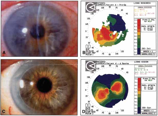
| Fig. 24 All images are taken from the same patient who underwent deep lamellar endothelial keratoplasty (DLEK). A. Preoperative corneal edema. B. Preoperative corneal topography reveals highly irregular astigmatism due to epithelial edema secondary to Fuchs' endothelial dystrophy. C. Six months' postoperative photo after DLEK. Note the corneal clarity of the central cornea. D. Six months' postoperative topography after DLEK. Note the smooth, regular corneal topography. The regular astigmatic pattern closely resembles the patient's preoperative refractive astigmatism prior to the onset of epithelial edema. (From Terry MA, Ousley PJ: Corneal Endothelial Transplantation: Advances in the Surgical Management of Endothelial Dysfunction. Contemporary Ophthalmology 1:26, 2002. Published courtesy of Lippincott Williams Wilkins, Inc.) |