Chapter 62
Onchocerciasis
DANIEL ETYA'ALE and HUGH R. TAYLOR
Main Menu   Table Of Contents

Search

DEFINITION
LIFE CYCLE OF O. VOLVULUS
PATHOGENESIS OF HOST DISEASE
PATHOLOGY
CLINICAL MANIFESTATIONS
DIAGNOSIS
CONTROL AND MANAGEMENT
PREVENTION OF ONCHOCERCIASIS
REFERENCES

DEFINITION
Onchocerciasis, also known as “river blindness,” is an insect-borne disease caused by a nematode worm, Onchorcerca volvulus, and transmitted by black flies of the genus Simulium. It is endemic across much of sub-Saharan Africa, in circumscribed areas of Central and South America, and in the Arabic Peninsula. Recent estimates suggest that worldwide, at least 120 million persons are exposed to the disease and 18 million persons are infected.1 More than 99% of these live in sub-Saharan Africa, and at least 1 million persons are either blind or severely disabled by the disease.

Onchocerciasis has a devastating impact in hyperendemic areas, where almost every person will be infected, and half of the population will be blinded by the disease before they die (Fig. 1). Once blind, they have a life expectancy of only one third that of the sighted, and most die within 10 years.2

Fig. 1. A common sight in hyperendemic villages. A father blinded by onchocerciasis is led to his field by his child. Blindness has a devastating impact in these subsistence farming communities.

Back to Top
LIFE CYCLE OF O. VOLVULUS

THE VECTOR

Although the species of blackfly vary in different geographic areas, they are all members of the family Simuliidae. In Africa, the chief vector of O. volvulus is Simulium damnosum, a species complex made up of 30 or more closely related cytospecies (Fig. 2). Other important vectors include Simulium naevi, which occurs mainly in East Africa, and Simulium albivirgulatum, considered an important vector in the “Cuvette Centrale” of the Democratic Republic of the Congo. In the Americas, the main vectors are the Simulium ochraceum complex in Guatemala and Mexico, Simulium exiguum in Ecuador, Simulium metallicum and Simulium exiguum in Venezuela, and Simulium guianense in the “Amazonia” focus.1,3

Fig. 2. Simulium damnosum, the biting black fly that transmits onchocerciasis. (Photo courtesy of Charles D. Mackenzie, PhD.)

The blackfly larvae require well-oxygenated water to mature, and eggs are laid in rapids in fast flowing rivers and streams. Because of seasonal variation in volume and rate of flow, a particular stretch of river may be either a permanent breeding site or a temporary breeding site only at certain times of the year. Blackflies usually do not travel more than 1 or 2 km from their breeding sites, although at times they may be carried hundreds of kilometers by monsoonal winds. Female black flies require a blood meal to initiate ovulation, and it is during this meal that they may transmit or receive the onchocercal infection.

THE PARASITE

O. volvulus is one of a group of filarial nematodes. Other members include Wuchereria bancrofti and Brugia malayi, which produce lymphatic filariasis and elephantiasis and Loa loa, which causes loiasis. The adult worms live encysted in fibrous nodules, which may be single or conglomerate. Each nodule contains between two to three female worms lying in a twisted, tangled mass (hence the term volvulus). The adult female O. volvulus organism is a white threadlike worm, 30 to 70 cm long and only 0.3 mm across (Fig. 3). Adult males have a similar appearance but are thinner and much shorter (about 2.5 to 5 cm long).

Fig. 3. Adult worms of Onchocerca volvulus can be freed from excised nodules by collagenase digestion, which removes all of the host tissue. This nodule contained two adult females, which are much longer and thicker than the one male seen emerging at the upper right. These three worms were obtained from the same nodule.

Adult female worms have a life span of 8 to 10 years but may live up to 15 years. During their lifetime, each releases millions of first-stage larvae, also known as microfilariae. A microfilaria is 320 to 360 μm long and 5 to 10 μm in diameter (Fig. 4) and lives for 6 to 30 months.1,2 In hyperendemic areas, the total microfilaria load in the body of affected individuals may be as high as 150 million.4

Fig. 4. Microfilaria seen by phase-contrast microscopy. The rounded cephalic end is clearly distinguishable from the pointed caudal end. This microfilaria is straightening as it dies.

CYCLE OF INFECTION

Microfilariae enter a female blackfly when she bites an infected person. A small percentage of these pass to the fly's midgut, penetrate the gut wall, and enter its thoracic muscles. After several molts, they become third-stage infective larvae, which then migrate to the fly's proboscis and are ready to be transferred during the next blood meal. Flies infected during their first blood meal carry developing larvae during their second blood meal, but they are not infectious until their third blood meal. Most flies die after their third reproductive cycle.

After entering the skin of the human host through the bite of an infected blackfly, the infective larvae (usually two to six) migrate through the subcutaneous tissues. They rapidly molt to become fourth-stage larvae and then molt again 4 to 6 weeks later. Over a period of 12 months, each larva develops into a mature adult male or female worm. Adult worms become encapsulated by host fibrous tissue to form characteristic nodules (onchocercomas). The adult worms reproduce sexually, and new microfilariae escape through the nodule wall and migrate throughout the host's body, particularly the skin. If they are then taken up by a biting blackfly, microfilariae develop into larvae and the cycle continues. There is no multiplication of the parasite during its development in the vector nor during its early development in the human host. One microfilaria can, therefore, only develop into one adult worm. Repeated infections, often accumulated over a period of many years of exposure are required before a heavy load of adult worms and pathogenic microfilariae can build up in the human host.3

NATURAL HISTORY AND EPIDEMIOLOGY

Longitudinal studies carried out in areas of ongoing transmission5,6,7 have shown that onchocerciasis is a progressive disease, and the intensity of infection in a person is the cumulative result of many years of exposure. In these areas, most persons are infected with O. volvulus during the first years of life (Fig. 5). The intensity of infection and the number of microfilariae per milligram of skin snip increase with host age. Men are often more heavily infected than women, but this is usually related to different occupational exposures. As a general rule, the closer a person is to the breeding sites and the longer the time spent nearby, the higher the exposure.

Fig. 5. Age-specific prevalence of infections, blindness, and impaired vision in three villages in an area of hyperendemic onchocerciasis in the valley of the Sissili River, Ghana. (Report of WHO Expert Committee. Epidemiology of Onchocerciasis. Technical Report Series No. 597. Geneva, World Health Organization, 1976.)

About one third of persons older than 15 years have microfilariae in the anterior chamber of the eye. Intraocular microfilariae are more common in those who have high skin-snip counts. In the worst affected areas, 10% of the population and half of those older than 40 years may be blind.3 Severe eye lesions and blindness are more common in heavily infected persons.

The geographic distribution of onchocerciasis is characterized by local foci of disease. Foci may vary in intensity but are always located in the vicinity of rivers or streams that form the breeding sites of the blackflies. One measure of the risk of infection is the annual transmission potential (ATP), which is the calculated number of infective larvae transmitted to a person who is continuously exposed to blackflies for 1 year. In the worst areas, the ATP may be 90,000 infective larvae per person per year. ATPs of greater than 1500 are associated with a high prevalence of blindness, and in West Africa, before current control programs, values over 2500 were often associated with the subsequent desertion of the involved villages by the populace.1

Regional geographic differences also occur, and these are explained by variation in transmission and by local variation in the parasite-vector complex, although host factors may also be involved8 (Table 1 and Fig. 6). There are a number of different strains of O. volvulus, each of which is associated with, and transmitted by, a particular species of Simulium, which also vary from region to region. Most of these can now be identified and characterized using deoxyribonucleic acid (DNA) detection techniques.9


Table 1. Comparison of General Characteristics of Onchocerciasis in Savanna and Rain Forest Areas of West Africa and Central America


CharacteristicsWest Africa SavannaWest Africa Rain ForestCentral America
Skin countsVery highModerateLow to moderate
NodulesLess common, usually below waistMore common, usually below waistLess common, usually above waist
Annual transmission potentialModerateHighLow
PrevalenceHighVery highLow
BlindnessCommon (5%–10%)Less common (1%–2%)Uncommon (% unknown)
Major cause of blindnessSclerosing keratitisChorioretinitisAnterior uveitis


Fig. 6. The distribution of onchocerciasis in Africa. Regional variation is seen in its severity and manifestations. The countries in the Onchocerciasis Control Program (OCP) and the African Program for Onchocerciasis Control (APOC) are indicated.

Back to Top
PATHOGENESIS OF HOST DISEASE

DEATH OF MICROFILARIAE

Nearly all the lesions of onchocerciasis and their resulting pathologic changes, including those in the eye, are directly or indirectly related to the local death of microfilariae. Access of microfilariae into the eye may occur by a number of different routes.10 It seems likely that they enter the cornea from the skin by way of the conjunctiva. Those in the uveal tract and aqueous may well be blood borne, although direct invasion along the vessels and nerves also occurs.

Generally, live microfilariae stimulate very little inflammatory response. They appear to pass undetected by the host's immune system despite the presence of a demonstrable host antibody and cell-mediated immune response.11,12 Inflammation is associated with dead or dying microfilariae and is probably initiated by the release of, or exposure to, microfilarial antigens. The mechanisms that protect live microfilariae are unclear. More heavily infected persons show evidence of immunosuppression, which can be reversed with treatment.

In the untreated disease, a relatively small number of microfilariae are dying at any one time, and each initiates a limited inflammatory response. With time, many microfilariae die, and, cumulatively, this produces considerable local inflammation and scarring.

THE MAZZOTTI REACTION

Many microfilaricidal drugs used in the past, such as diethylcarbamazine (DEC), killed most microfilariae almost immediately. The synchronous death of millions of microfilariae was believed to release a massive quantity of antigenic or toxic material. This was associated with a marked inflammatory response, which was recognized clinically as the Mazzotti reaction.13 In those with onchocerciasis, DEC almost always caused side effects that, although sometimes mild, could be very severe2 and even precipitate irreversible vision loss. A few deaths have also been reported with its use.

The administration of even a single 50-mg dose of DEC (Mazzotti test) can initiate the previously mentioned response. This was a dramatic and dangerous method of diagnosing onchocerciasis in the past,14 and its use can no longer recommended.

Back to Top
PATHOLOGY

SKIN

The earliest changes in the skin in onchocerciasis are mild and are often limited to a perivasculitis.14 In more heavily infected persons, microfilariae can be found, especially at the epidermal-dermal junction (Fig. 7). Live microfilariae are usually not surrounded by inflammatory cells.

Fig. 7. Microfilaria lying in the dermis. Although it has been cut into a number of sections, it is clearly recognizable, because the nuclei of the microfilaria are much smaller than those of the host cells. The absence of an inflammatory infiltrate around the microfilaria is characteristic (hematoxylin-eosin, ×700).

Later changes include hyperkeratosis, acanthosis, and parakeratosis, with an increase in inflammation and fibrosis. Dermal collagen is disrupted by the deposition of increasing amounts of mucin ground substance and by fibrous scar tissue. With more advanced fibrosis and inflammation, microfilariae become less common in the superficial dermis and are found mainly in the deeper layers. Ultimately, the dermis becomes fibrotic and is covered by a thinned and atrophic epithelium. There are areas of depigmentation and hyperpigmentation (Fig. 8). Some patients have a much more severe, reactive onchodermatitis, often confined to one limb. This was first described in Yemen and is called “sowda,” or black limb. Here, the most prominent histologic change is an extensive infiltrate of plasma cells.14 Microfilariae are seen occasionally in the deep dermis or in skin snips. These patients have a high cellular immune response to onchocercal antigens, which is suppressed if their disease becomes generalized.

Fig. 8. An example of severe atrophic onchocerciasis skin changes, leopard skin (A). Dermal atrophy has led to islands of residual pigment around hair follicles (B). (Photos courtesy of Charles D. Mackenzie, PhD.)

NODULES

Adult worms may at times be found free in fascial planes, although they are far more commonly encapsulated in nodules. Some nodules are palpable and visible, lying subcutaneously over bony prominences, especially the iliac crests, greater trochanters, ribs, knees, ankles, coccyx, and skull. Others, probably more numerous, lie deep and impalpable, attached to the fibrous capsules of joints (especially the hip joint) or close to the periosteum of bones or in intramuscular fasciae. Occasionally, nodules are found in even deeper structures such as intracranially or in the wall of the aorta.

When examined microscopically, dense scar tissue surrounds and encases the adult worms, which are coiled up like a ball of string. Developing microfilariae can be found in the uteri of most female worms. Degenerative females often become calcified. Nodules may show an inflammatory infiltrate of varying intensity and occasionally areas of necrosis and liquefaction.

THE EYE

As in the skin, many of the changes in the front of the eye at least are related to invasion by, and local death of, microfilariae. The pathophysiology of chorioretinal disease is far less clear and may also be related to immune complex disease,12,15 the presence of antiretinal antibodies,16 or other mechanisms.17 Much of the chorioretinal disease seems to start at the level of the retinal pigment epithelium.18 Few eyes have been examined histopathologically, and most of these have had end-stage disease that often obscures the earlier changes.19 Live microfilariae cause little intraocular reaction and may be found in the conjunctive, cornea, posterior sclera, anterior and posterior chambers, vitreous, uveal tract, inner retina, and optic nerve and its sheath.

OTHER TISSUES

Microfilariae have been found in many of the deeper organs, including the liver, kidney, spleen, pancreas, lung, peripheral nerves, and arteries. They have also been found in tears, blood, urine, cerebrospinal fluid, sputum, vaginal secretions, and peritoneal fluid.

Back to Top
CLINICAL MANIFESTATIONS

SKIN CHANGES

The actual bite of the blackfly is usually unnoticed, although a painful wheal develops quickly and resolves in 2 or 3 days. Pruritus is the most common early manifestation of onchocerciasis. It develops after a latent or “prepatent” interval of 1 to 2 years following initial infection. During this time, the infective larvae mature into adult worms. Symptoms develop as microfilariae appear in the skin, and the prevalence of most types of onchocercal skin disease show statistically significant correlation with onchocercal endemicity. Itching and scratching may be mild and intermittent or severe and unremitting, in which case they often lead to excoriation and secondary infection, a syndrome complex widely known as “filarial scabies” (la gâle filarienne). A standard clinical method of classifying and grading the dermal changes of onchocerciasis has been developed20 and endorsed by World Health Organization (WHO) Expert Committee on Onchocerciasis Control.1

In long-standing and heavy infections, disfiguring skin lesions develop, the most common being a mottled depigmentation of the shins, known as “leopard skin” and generalized dermal atrophy.

NODULES

Onchocercal nodules are firm, round masses more commonly found around the pelvis in infected persons but also occur around the head and shoulders, especially in persons living in Central America. They may also be found on the lateral chest wall and in the limbs, where they are often fixed to the skin. New nodules tend to develop around older nodules. Although an individual nodule will usually be 0.25 to 1 cm in diameter and contain two or three females and one or two males, large matted conglomerations of nodules can form. Nodules are usually painless and, by themselves, cause little trouble.

OCULAR CHANGES

Intraocular Microfilariae

The earliest sign of ocular involvement in onchocerciasis is the invasion of the eye by microfilariae. Microfilariae in the anterior chamber can be seen easily with a slit lamp, especially if a patient sits with his or her head between the knees for 2 to 5 minutes to allow any microfilariae in the anterior chamber to fall to the center of the cornea. When the patient sits up, the microfilariae are concentrated in one area behind the central cornea. The microfilariae are seen as small, wriggling white “worms.” They often can be followed as they circulate in convection currents in the aqueous.

Live microfilariae can be more difficult to find in the cornea because they are far less mobile, almost transparent, and are often coiled. Dead microfilariae are more opaque and straighter and, therefore, much easier to see. Microfilariae are best seen with high magnification (×25) using retroillumination produced by an oblique beam reflected from the iris. They are most common in the peripheral cornea, especially temporally and nasally, but they may be found in the anterior vitreous or retina and, at times, attached to the lens capsule or to Descemet's membrane. Intraretinal microfilariae are best seen during contact lens examination of the fundus and appear as small, highly refractile objects with a greenish tinge.21

Punctate Keratitis

In the cornea, dead microfilariae are usually surrounded by an inflammatory infiltrate that appears as an ill-defined punctate, fluffy, or snowflake opacity about 0.5 mm in diameter. These opacities are focal collections of lymphocytes and eosinophils with local edema.14,18 Although punctate keratitis sometimes occurs in persons who have not received treatment, it is more common in those receiving DEC treatment when many microfilariae die at one time (Fig. 9). Initially, dead microfilariae can be recognized in the center of these opacities. The opacities clear without visible sequelae.

Fig. 9. Snowflake opacities (punctate keratitis) usually occur in the peripheral cornea. Although a few opacities are common in patients with microfilariae in the cornea, they are especially seen during treatment with suramin or diethylcarbamazine (DEC), as was the case here.

Sclerosing Keratitis

With time, subtle changes begin at the limbus, initially in the interpalpebral fissure and inferiorly. These changes progress until ultimately the whole cornea is opaque and vascularized. An increased limbal translucency, or haze, is the earliest change. In time, a fibrovascular pannus and inflammatory infiltrate, composed mainly of lymphocytes and eosinophils, develops at the level of Bowman's membrane.14 This opacification advances in an arc, which is most marked at each side and below (Fig. 10). Usually, the remaining areas of clear cornea show heavy infiltration of microfilariae. The opacity progresses to cover the central cornea, and eventually the whole cornea may become opaque.

Fig. 10. Sclerosing keratitis usually begins at the nasal and temporal periphery and slowly progresses centrally. This Guatemalan man also has a pear-shaped distortion of his pupil, which is characteristic of onchocercal uveitis.

Anterior Uveitis

The presence and severity of anterior uveitis is extremely variable. A mild nongranulomatous uveitis with a minimum of flare and an occasional cell commonly occurs when there are microfilariae in the anterior chamber or the cornea. At times, however, the eye may be heavily infiltrated by microfilariae and yet have no detectable anterior chamber reaction, whereas in other cases a severe anterior uveitis may develop. When this occurs, inferior-posterior synechiae often develop, leading to a characteristic pear-shaped deformity of the pupil and retropupillary fibrosis. Chronic granulomatous anterior uveitis also causes a loss of the iris pigment frill and a pumice-stone appearance of the iris. Extensive synechiae may cause seclusion and occlusion of the pupil, secondary cataract, or secondary glaucoma. Mild uveitis has been related to the microfilarial invasion of the iris; the more severe granulomatous uveitis is related to invasion of the ciliary body.14,18

Chorioretinitis

The great spectrum of fundal changes that is seen clinically seems to result from a few basic pathologic processes. Although numerous names and eponyms have been given to the different clinical pictures they present, it is simpler to consider the chorioretinal changes as these basic processes.15 Histopathologically, the retinal pigment epithelium may show areas of pigment migration atrophy or focal hyperplasia.21 Chronic nongranulomatous chorioretinitis occurs with infiltration by lymphocytes, plasma cells, and eosinophils, with secondary degenerative changes in the overlying retinal pigment epithelium, neuroretina, and choriocapillaris. Eventually, there may be apposition of the internal limiting membrane of the retina and the larger choroidal vessels.

Clinically, the most common change is a granular atrophy of the retinal pigment epithelium often with intraretinal dispersion of pigment from the pigment epithelium22,23 (Fig. 11). Although areas of severe atrophy are easy to see, milder forms may only be revealed by fluorescein angiography. Chorioretinal atrophy is common and is usually associated with retinal pigment clumping (Fig. 12). Well-defined, white plaque-like areas of subretinal fibrosis with neovascularization may occur in these areas. Each of these three changes—retinal pigment epithelial atrophy, chorioretinal atrophy, and subretinal fibrosis—represents an end stage of, or the structural change caused by, previous acute inflammation induced either by the local death of microfilariae or possibly by other immune mechanisms.

Fig. 11. Discrete areas of retinal pigment epithelial atrophy are one of the most common fundal changes seen in onchocerciasis. Usually they occur temporal to the macula, but they may occur elsewhere. When seen through a hazy cornea, they may need to be differentiated from drusen.

Fig. 12. Large area of chorioretinal atrophy, which shows characteristic sparing of the uveal region until late in the disease.

Areas of active inflammation in the retina can sometimes be seen.23 They appear as areas of posterior retinal edema that demonstrate leakage of retinal capillaries and veins on fluorescein angiography. Pale, ill-defined areas of swelling in the choroid may also occur. These also show leakage on fluorescein angiography and are believed to be choroidal granulomas. Transitory retinal pigment epithelial abnormalities have also been seen with DEC therapy.24 A severe retinal vasculitis may rarely occur.

It is the variation in prominence and the distribution of each type of change that leads to the wide variation in the final morphologic picture. The extent of involvement is not clearly related to the severity of the infection. The macular region is often spared until late in the disease.

Optic Neuritis and Optic Atrophy

Optic neuritis followed by postneuritic optic atrophy is often seen in patients with onchocerciasis.25,26,27 This is often associated with scarring and pigment disturbance at the disc margin. Primary optic atrophy may also occur, and both have been presumed to be caused by the disease. Microfilariae have been found within the optic nerve and its sheaths,18 and it is reasonable to assume that the occasional death of these microfilariae initiates an inflammatory response and subsequent atrophy. Optic atrophy also would be expected from loss of ganglion cells in areas of retinal atrophy and from secondary glaucoma.

In many cases, active optic neuritis may last several weeks and up to a year or even longer. Prevalence of optic atrophy varies from 1% to 4% in hyperendemic, rainforest and savanna communities of Cameroon, to 6% to 9% in Sierra Leone, to 9% in the Guinea Savanna of Northern Nigeria. Optic neuritis and optic atrophy have also been found to occur more commonly in persons who have received treatment, either with DEC or suramin, than in those who have not been treated.28

Other Ocular Changes

Patients with onchocercal ocular disease may present with a variety of ocular manifestations ranging from mild (itching, redness, pain photophobia, blurring of vision) to the more severe symptoms of night blindness and or visual field loss, depending on which tissue of the eye is affected.

Blindness and Visual Field Loss

Blindness is by far the most serious consequence of onchocerciasis. The prevalence of blindness rises with increasing subject age. As mentioned earlier, males are more frequently affected than females because male-dominated occupations such as farming and fishing lead to greater exposure to infected vectors.

The true magnitude onchocerciasis-related blindness still remains largely underestimated because most available data are based on blindness defined using visual acuity alone (less than 3/60 in the better eye). As recent studies in northern Nigeria28,29 have clearly shown, onchocerciasis can cause severe reduction in peripheral vision, leaving affected individuals with only a small island of central vision, which is usually preserved until late in the disease process. These individuals may be greatly disabled long before any reduction of their visual acuity is noticeable. Thus, when visual field constriction is also taken into account, blindness prevalence rates are likely to be 25% to 30% higher.26

Back to Top
DIAGNOSIS

SKIN SNIP

Worldwide, the skin snip is still the most widely used and easiest method to demonstrate the presence of microfilariae in a person.1 The skin snip, which can be obtained using a razor blade (or better, by using a scleral punch), is placed in fluid, and the microfilariae that emerge are counted under a microscope. The technique of skin snip and its many refinements is widely described in the literature.30,31

In Central America, skin snips for microfilarial count are often taken from the deltoid or the scapular region. In Africa, they are usually taken over the iliac crest. Outer canthal skin snips have been suggested as a general indicator of potential ocular involvement and the risk of developing onchocerciasis-related blindness but probably reflect higher total counts rather than a particular local ocular risk.

Although fairly specific as a diagnostic method, the collection of skin snips is not a very sensitive way of detecting early, light, or prepatent infections. Moreover, it is becoming increasingly unacceptable, partly because of a growing awareness among local populations of the potential risk of infection with HIV and partly because the method is rarely needed today in defining endemic communities eligible for mass treatment with ivermectin (simpler, noninvasive, and more rapid methods are now available).32

NODULES

The clinical detection of a typical nodule is good presumptive evidence of onchocerciasis. Nodules need to be differentiated from lipomas, sebaceous cysts, ganglia, and lymph nodes. A definitive diagnosis can be made if adult worms can be identified in an excised nodule.

Recent operational-research in sub-Saharan Africa has demonstrated that irrespective of the epidemiologic zone and pattern, the prevalence of palpable onchocercal nodules is closely related to the prevalence of infection as determined by skin snipping and nodule palpation.33 Other studies confirmed that the prevalence of microfilariae in the skin is generally twice that of palpable nodules34 and directly related to the prevalence of nodules. This explains why over the past few years, nodule palpation has superseded skin snipping as the preferred, reasonably accurate, more rapid, and risk-free method of estimating the level of endemicity in a community.

MICROFILARIAE

The clinical recognition of intraocular microfilariae is diagnostic of onchocerciasis, as is the finding of microfilariae in the urine, blood, cerebrospinal fluid, sputum, or vaginal secretions. These fluids are not normally screened for microfilariae.

IMMUNOLOGIC TESTS

As yet, a reliable immunologic test to detect infection with O. volvulus has not been developed.1 The key to the development of a test has been the need to identify one or more antigens that provide a sensitive indicator of the early or prepatent stages of infection and yet be highly specific for O. volvulus infection.

Three new diagnostic tests for onchocerciasis are currently under development: an immunologic assay, based on a three-antigen cocktail obtained using recombinant DNA technology;35 a polymerase chain reaction (PCR)-based assay, which is based on the use of DNA probes and which may also be used for “pool screening” of blackflies36; and the DEC patch test, a much safer, virtually innocuous variant of the Mazzotti test.32 Of these the DEC patch test seems to fit best the criteria of an ideal test: highly sensitive, highly specific, easy to carry out, cheap, and acceptable to the population under investigation.

Back to Top
CONTROL AND MANAGEMENT

ONCHOCERCIASIS CONTROL PROGRAMS

At the time of writing, there are three regional onchocerciasis control programs, one in Central and Latin America, the Onchocerciasis Control Program of the Americas (OEPA); and two in Africa, the Onchocerciasis Control Program1 (OCP), and the African Program for Onchocerciasis Control (APOC). Together these three regional programs cover more than 99% of all endemic populations and all but one (South Yemen) endemic countries. Moreover the rapid expansion of ivermectin treatment over the last decade and the enthusiastic response and involvement of affected communities in these efforts makes the elimination of onchocerciasis a real possibility in the not too distant future.37

STRATEGIES FOR ONCHOCERCIASIS CONTROL

The control of onchocerciasis today is based essentially on two strategies: Simulium vector control and large-scale chemotherapy with ivermectin mainly alone or in combination with vector control.

Widespread nodulectomy campaigns have been conducted in Guatemala and Mexico since the late 1930s. Available data suggest that these have achieved some measure of success in reducing the prevalence of onchocercal blindness in those countries but have little impact on disease transmission. Elsewhere, and particularly in Africa where 99% of the disease exist and fewer than two thirds of the nodules can be palpated and identified for surgical removal, nodulectomy has never been considered as an effective control measure.

VECTOR CONTROL

This is the chief strategy used in West Africa by the OCP, which was initiated in 1974 by the WHO, with funding from the World Bank and the donor community and support from participating countries.1 Its goal is to interrupt transmission of O. volvulus by regular aerial spraying to all Simulium larval breeding sites and to maintain this for at least 14 years until the infection has died out in human population.

For the first 14 years of the program, vector control was used alone, because there was no medical treatment that could be given alongside and on a large scale to alleviate symptoms or prevent further deterioration of eye lesions among high-risk groups. Today, although still used in the greater part of the program area, vector control is complemented by ivermectin treatment, used either in combination or alone. The latter is the case in the so-called Western and Southern extensions,1 of the program, where vector control was not initiated or was found to be operationally not feasible.

The achievements of OCP have been truly remarkable. Onchocerciasis has been virtually removed from the controlled area in the original seven OCP countries. In the remaining part of the program area, control activities are so advanced that the program is likely to close down in 2002 and any residual—control and surveillance—activities transferred to participating countries.38

CHEMOTHERAPY

A truly satisfactory drug for use against onchocerciasis does not yet exist. To be of lasting benefit, such a drug would kill the adult worms, in the absence of severe side effects and without the exacerbation of existing lesions and could be used on a mass scale. Although not meeting all of these criteria, a drug developed during the 1980s, ivermectin, has revolutionized the treatment of onchocerciasis.39,40 Ivermectin has rendered obsolete the two drugs that were previously used: suramin and DEC1 (Table 2). Before the introduction of ivermectin, DEC or suramin treatment were recommended only for those with the highest risk of developing blindness, because of the high incidence of severe reactions seen with these drugs.41,42 Today, the use of suramin and DEC can no longer be justified, given the safety record and the wide availability of ivermectin for individual and mass treatments of onchocerciasis.


Table 2. Action, Dosage, and Side Effects of Drugs Used to Treat Onchocerciasis


 IvermectinSuraminDiethylcarbamazine
ActionKills microfilariae (GABA agonist)Kills adults and microfilariaeKills microfilariae
AdministrationOralIntravenousOral
DosageSingle annual oral dose (150 μg/kg)3.8 g divided in 6 weekly infusions200 mg/day for 7 to 14 days
Duration of effectProlonged: at least 2 yearsLong-lasting: may give permanent cureTemporary: 6 to 12 months
Side effectsMinimal reactions, mild MazzottiSevere rash, exfoliative dermatitis, diarrhea, prostration, renal failure, coma, deathMazzotti reaction, pruritus, rash, fever, lymphadenopathy, arthralgia,exacerbation of eye changes, optic neuritis, chorioretinitis,proteinuria
Cause of side effectsMicrofilarial deathBinds to proteins, blocks enzymes, inhibits coagulation, inhibits complementsMicrofilarial death leads to immediate hypersensitivity reactions,circulating immune complexes


Ivermectin

Ivermectin is a semisynthetic, macrocyclic, lactone antibiotic widely used in the field of veterinary medicine against a wide range of animal parasites.39 Its pharmacologic effect is as a modulator of γ-aminobutyric acid (GABA)-mediated neurotransmitter effects.

Clinical trials and subsequent field experience40,43,44 have shown that ivermectin is a rapidly effective and well-tolerated, single-dose microfilaricide. At the recommended annual dose of 150 μg/kg body weight, it causes little or no Mazzotti-type reactions and is suitable for community-wide distribution. Unlike DEC, ivermectin does not cause reactions in the eye, from which microfilariae escape by natural routes and are not immediately replaced. The drug, thus, prevents further development of onchocercal lesions in the eye and the skin.45–49

Ivermectin has no demonstrable effect on the viability of the adult worms. It has, however, a suppressive action on the production of microfilariae by the female worms, which lasts between 3 and 12 months.50 This effect on the adult female may explain the delayed repopulation of the skin after ivermectin treatment.

Following ivermectin treatment there is a marked reduction in the capability of an infected person to contribute to transmission.51,52,53 There is a reduction in the number of microfilariae taken up by blackflies and in the number of infective larvae that develop. With good coverage, ivermectin can reduce but not interrupt transmission.

CURRENT IVERMECTIN USES IN ONCHOCERCIASIS CONTROL.

As mentioned previously, ivermectin is currently the only drug recommended for use in the control of onchocerciasis. It is available for both individual and mass treatments following the decision in 1987 by Merck and Company, the manufacturer, to provide it at no charge to all those suffering from onchocerciasis.54

Passive Treatment.

This is the form of treatment available to all those seeking treatment and in whom a clinical diagnosis of onchocerciasis has been made in a hospital or health center. It is directed primarily to the individual infected patient and is the main method of treatment in hypoendemic areas where the risk of blindness or severe skin disease is virtually nonexistent.

Mass Treatment.

Also known as active, large-scale, or community-wide treatment, this is the method of choice in mesoendemic and hyperendemic areas of onchocerciasis, that is, those areas where onchocerciasis is considered a public health problem. In these areas ivermectin is given once a year for at least 14 years, at the recommended dose (Table 3) to all members of the community except for the “exclusions to treatment”1,45,49,50,55,56 (Table 4). In most communities and for any round of treatment, some 80% of the total population are eligible for treatment.


Table 3. Recommended Doses of Ivermectin in Mass Treatment


Weight (kg)Height (cm)No of Tablets (3 mg)
15–2590–1191
26–44120–1402
45–64141–1583
65 or more159 or more4


Table 4. Exclusion Criteria for Ivermectin Treatment

  • Children younger than 5 years (age), or less than 15 kg (weight), or less than 90 cm (height)
  • Pregnant women
  • Lactating mothers of infants less than 1 week old
  • Severely ill persons
    Use with extreme caution in areas co-endemic with Loa loa*

*Pacque MC, Munoz B, Poetschke G et al: Pregnancy outcome after inadvertent ivermectin treatment during community-based distribution. Lancet 336:1486, 1990 and Pacque MC, Munoz B, Greene BM et al: Community-based treatment of onchocerciasis with ivermectin: Safety, efficacy and acceptability of yearly treatment. J Infect Dis 163:381, 1991


Over the years mass treatment has evolved from mobile strategies used in the early days following ivermectin donation,5 to various forms of community-based treatment of which community directed treatment with ivermectin (CDTI) is not only the latest but also the least expensive and the most likely to be sustainable.57 This is now the preferred method used throughout Africa by both OCP and APOC. Its main strength is the extent to which affected communities are involved at all stages of treatment activities; it ensures that the highest treatment coverage is achieved at each round.

Recent reports have drawn attention to the rare occurrence of severe and sometimes life-threatening adverse reactions after ivermectin in those who are infected with O. volvulus and Loa loa.58 Care must be taken when using ivermectin in areas where Loaiasis is known to be endemic.59

Back to Top
PREVENTION OF ONCHOCERCIASIS
At present, there is no simple, effective and easily replicable way of preventing onchocerciasis. In the absence of a vaccine (which is not even a remote prospect at this time), the only certain method of prevention is to avoid being bitten by blackflies. A number of preventive measures, such as avoiding known breeding sites at the time of maximum fly activity (early morning and early evening), relocating villages and their water supplies at some distance from a breeding site, wearing protective clothing, or using insect repellents, have all been suggested. However, their implementation has proved impracticable. The future and hope of onchocerciasis control will, therefore, continue to rely primarily on the effective implementation of the strategies described previously, pending the availability through ongoing research of suitable alternatives to ivermectin or better still of a safe, effective, and cheap microfilaricide.60,61
Back to Top
REFERENCES

1. Report of WHO Expert Committee: Onchocerciasis and its control. Technical Report Series No. 852. Geneva, World Health Organization, 1995

2. Prost A: The burden of blindness in adult males in the savanna villages of West 
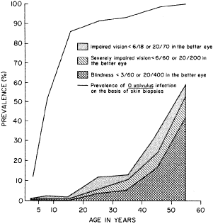
Africa exposed to onchocerciasis. Trans R Soc Trop Med Hyg 80:525, 1986

3. Report of WHO Expert Committee: Epidemiology of Onchocerciasis. Technical Report Series No. 752. Geneva, World Health Organization, 1987

4. Duke BOL: Onchocerciasis. In Johnson GJ, Minassian DC, Weale R (eds): The Epidemiology of Eye Disease. London, Chapman and Hall 1998, pp 227–247

5. Anderson J, Fuglsang H, Marshall TF deC: Studies on onchocerciasis in the United Cameroon Republic. III. A four-year follow-up of 6 rain forest and 6 Sudan-savanna villages. Trans R Soc Trop Med Hyg 70:362, 1976

6. Mackenzie CD, Williams JF, Sisley BE et al: Variations in host responses and the pathogenesis of human onchocerciasis. Rev Infect Dis 7:802, 1985

7. Budden FH: The natural history of ocular onchocerciasis over a period of 14–15 years and the effect on this of a single course of suramin therapy. Trans R Soc Trop Med Hyg 70:484, 1976

8. Duke BOL: The population dynamics of Onchocerca volvulus in the human host. Trop Med Parasitol 44:61, 1993

9. Vincent JA, Lustigman S, Zhang S et al: A comparison for the newer tests for the diagnosis of onchocerciasis. Ann Trop Med Parasitol 94:253, 2000

10. Budden FH: Route of entry of Onchocerca volvulus microfilariae into the eye [Letter]. Trans R Soc Trop Med Hyg 70:265, 1976

11. Gallin M, Edmonds K, Ellner JJ et al: Cell-mediated responses in human infection with Onchocerca volvulus. J Immunol 140:1999, 1988

12. Ottesen EA: Immune responsiveness and the pathogenesis of human onchocerciasis. J Infect Dis 171:659, 1995

13. Francis H, Awadzi K, Ottesen EA: The Mazzotti reaction following treatment of onchocerciasis with diethylcarbamazine: Clinical severity as a function of infection intensity. Am J Trop Med Hyg 34:529, 1985

14. Buck AA: Onchocerciasis: Symptomatology, Pathology, Diagnosis. Geneva, World Health Organization, 1974

15. Neumann E, Gunders AE: Pathogenesis of the posterior segment lesion of ocular onchocerciasis. Am J Ophthalmol 75:82, 1973

16. Chan CC, Nussenblatt RB, Kim MK et al: Immunopathology of ocular onchocerciasis. 11. Anti-retinal autoantibodies in serum and ocular fluids. Ophthalmology 94:439, 1987

17. Van der Lelij A, Rothova A, Stilma JS et al: Cell-mediated immunity against human retinal extract, S-antigen and interphotoreceptor retinoid binding protein in onchocercal chorioretinopathy. Invest Ophthalmol Vis Sci 31:2031, 1990

18. Garner A: Pathology of ocular onchocerciasis: Human and experimental. Trans R Soc Trop Med Hyg 70:374, 1976

19. Rodger FC: The pathogenesis and pathology of ocular onchocerciasis. IV. The pathology. Am J Ophthalmol 49:327, 1960

20. Murdoch ME, Hay RJ, Mackenzie CD et al: A clinical classification and grading system of the cutaneous changes in 
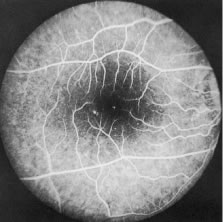
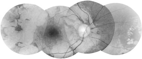
onchocerciasis. Br J Dermatol 129:260, 1993

21. Murphy RP, Taylor HR, Greene BM: Chorioretinal damage in onchocerciasis. Am J Ophthalmol 98:519, 1984

22. Newland HS, White AT, Greene BM et al: Ocular manifestations of onchocerciasis in a rain forest area of West Africa. Br J Ophthalmol 75:163, 1991

23. Bird AC, Anderson J, Fuglsang H: Morphology of posterior segment lesions of the eye in patients with onchocerciasis. Br J Ophthalmol 60:2, 1976

24. Bird AC, El-Sheikh H, Anderson J et al: Changes in visual function and in the posterior segment of the eye during treatment of onchocerciasis with diethylcarbamazine citrate. Br J Ophthalmol 64:191, 1980

25. Semba RD, Murphy RP, Newland HS et al: Longitudinal study of lesions of the posterior segments in onchocerciasis. Ophthalmology 97:1334, 1990

26. Murdoch IE, Jones BR, Cousens S et al: Visual field constriction as a cause of blindness or visual impairment. Bull World Health Organ 75:141, 1997

27. Cousens S, Cassels-Brown A, Murdoch I et al: Impact of annual ivermectin treatment on progression of onchocercal visual field loss. Bull World Health Organ 75:229, 1997

28. Abiose A, Jones BR, Cousens S et al: Reduction in incidence of optic nerve disease with annual ivermectin to control onchocerciasis. Lancet 341(8838):130, 1993

29. Abiose A: Onchocercal eye disease and the impact of Mectizan treatment. Ann Trop Med Parasitol 92:S11, 1998

30. Schulz-Key H: A simple technique to assess the total number of Onchocerca volvulus microfilariae in skin snips. Tropenmed Parasitol 29:51, 1978

31. Taylor HR, Munoz B, Keyvan-Larijani E et al: Reliability of skin snips in the diagnosis of onchocerciasis. Am J Trop Med Hyg 41:467, 1989

32. Boatin BA, Toé L, Alley ES et al: Diagnostics in onchocerciasis: Future challenges. Ann Trop Med Parasitol, 92:S41, 1998

33. Taylor HR, Duke BOL, Munoz B: The selection of communities for treatment of onchocerciasis with ivermectin. Trop Med Parasitol 43:267, 1992

34. World Health Organization: Methods for community diagnosis of onchocerciasis to guide ivermectin-based Control in Africa. Report of an informal consultation held in Ouagadougou from 19–21 November 1991. Document TDR/TDE/ONCHCERCIASIS/92.2. Geneva, WHO, 1992

35. Bradley JE, Trenholme KR, Gillepsie AJ et al: A sensitive serodiagnostic test for onchocerciasis using a cocktail of 
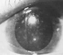
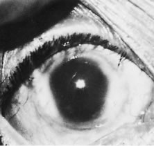
recombinant antigens. Am J Trop Med Hyg 48:198, 1993

36. Toe L, Boatin BA, Adjami A et al: Detection of Onchocerca volvulus infection by 0–150 polymerase chain reaction analysis of skin scratches. J Infect Dis 178:282, 1998

37. Etya'alé DE: Mectizan as a stimulus for the development of novel partnerships: The international organization's perspective. Ann Trop Med Parasitol 92:S73, 1998

38. Dadzie KY: Control of onchocerciasis: Challenges for the future. Ann Trop Med Parasitol 92:S65, 1998

39. Campbell WC, Fisher MH, Stapley EO et al: Ivermectin: A potent new antiparasitic agent. Science 221:823, 1983

40. Aziz MA, Diallo S, Diop IM et al: Efficacy and tolerance of ivermectin in human onchocerciasis. Lancet 2(8291):171, 1982

41. Greene BM, Taylor HR, Cupp EW et al: Comparison of ivermectin and diethylcarbamazine in the treatment of onchocerciasis. N Engl J Med 313:133, 1985

42. Taylor HR Murphy RP, Newland HS et al: Comparison of the treatment of ocular onchocerciasis with ivermectin and diethylcarbamazine. Arch Ophthalmol 104:863, 1986

43. Pacque MC, Dukuly Z, Greene BM et al: Community-based treatment of onchocerciasis with ivermectin: Acceptability and early adverse reactions. Bull World Health Organ 67:721, 1989

44. Greene BM, Dukuly ZK, Munoz B et al: Comparison of 6-, 12-, and 24-monthly dosing with ivermectin for treatment of onchocerciasis. J Infect Dis 163:376, 1991

45. Murdoch IE, Abiose A, Babalola OE et al: Ivermectin and onchocercal optic neuritis: Short-term effects. Eye 8:456, 1994

46. Whitworth JA, Gilbert CE, Mabey DM et al: The effect of repeated doses of ivermectin on ocular onchocerciasis: Community-based trial in Sierra Leone. Lancet 338(8775):1100, 1991

47. World Health Organization: The effect of repeated ivermectin treatment on ocular onchocerciasis. Report of an informal consultation. WHO/Document/TDE/ONCHO/93.3. Geneva, World Health Organization, 1993

48. Whitworth JA, Gilbert CE, Mabey DM et al: Effects of repeated doses of ivermectin on ocular onchocerciasis: Community-based trial in Sierra Leone. Ophthalmology 103:1001, 1996

49. Chippaux JP, Boussinesq M, Fobi G: Effect of repeated ivermectin treatments on ocular onchocerciasis: Evaluation after six to eight doses. Ophthalmic Epidemiol 64:299, 1999

50. Duke BOL, Pacque MC, Munoz B et al: Viability of adult Onchocerca volvulus after 6 2-weekly doses of ivermectin. Bull World Health Organ 69:163, 1991

51. Taylor HR, Pacque MC, Munoz B et al: Impact of mass treatment of onchocerciasis with ivermectin on the transmission of infection. Science 250:116, 1990

52. Cupp EW, Bernardo MJ, Kiszewski AE et al: The effects of ivermectin on transmission of Onchocerca volvulus. Science 231:740, 1986

53. Remme J, Baker RHA, de Sole G et al: A community trial of ivermectin in the onchocerciasis focus of Asubende, Ghana. I. Effect on the microfilarial reservoir and the transmission of Onchocerca volvulus. Trop Med Parasitol 40:367, 1989

54. Foege WH: Ten years of Mectizan. Ann Trop Med Parasitol 92:S7, 1998

55. Pacque MC, Munoz B, Poetschke G et al: Pregnancy outcome after inadvertent ivermectin treatment during community-based distribution. Lancet 336:1486, 1990

56. Pacque MC, Munoz B, Greene BM et al: Community-based treatment of onchocerciasis with ivermectin: Safety, efficacy and acceptability of yearly treatment. J Infect Dis 163:381, 1991

57. World Health Organization: Community-directed treatment with ivermectin: A practical guide for trainers of community-directed distributors, the African Program for Onchocerciasis Control (APOC). Geneva, World Health Organization, 1998

58. Gardon J, Gardon-Wendel N, Damanga-Ngangue et al: Serious reactions after mass treatment of onchocerciasis with ivermectin in an area endemic for Loa loa infection. Lancet 350:18, 1997

59. Boussinesq M, Gardon J: Prevalences of Loa loa microfilaraemia throughout the area endemic for the infection. Ann Trop Med Parasitol 91:573, 1997

60. Taylor MJ, Bandi C, Hoerauf A et al: Wolbachia bacteria of filarial nematodes: A target for control? Parasitol Today 16:179, 2000

61. Ladzins J, Kron M: New molecular targets for filariasis drug discovery. Parasitol Today 15:305, 1999

Back to Top