|
|
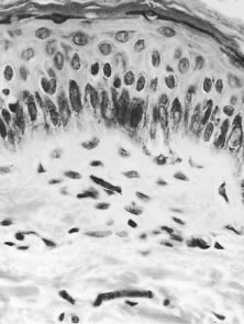
| Fig. 7. Microfilaria lying in the dermis. Although it has been cut into a number of sections, it is clearly recognizable, because the nuclei of the microfilaria are much smaller than those of the host cells. The absence of an inflammatory infiltrate around the microfilaria is characteristic (hematoxylin-eosin, ×700). |