|
|
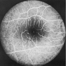
| Fig. 11. Discrete areas of retinal pigment epithelial atrophy are one of the most common fundal changes seen in onchocerciasis. Usually they occur temporal to the macula, but they may occur elsewhere. When seen through a hazy cornea, they may need to be differentiated from drusen. |