Chapter 64
Uveitis Surgery
JOSEPH B. MICHELSON, ROBERT A. NOZIK and STEVEN SANISLO
Main Menu   Table Of Contents

Search

GENERAL MEASURES
TREATMENT OF BAND KERATOPATHY
PENETRATING KERATOPLASTY IN UVEITIS
UVEITIC GLAUCOMA
CATARACT EXTRACTION IN PATIENTS WITH UVEITIS
VITRECTOMY
RETINAL DETACHMENT
RETINAL DETACHMENT AND CMV RETINITIS
MISCELLANEOUS PROCEDURES
REFERENCES

Until recently, the literature for surgical management of uveitis dealt with the many failures due to the complications of surgical inflammation preoperatively, intraoperatively, and postoperatively. One reason for the lack of success had been the viewpoint that uveitis is a single disease cluster compounded by the concept that the diagnosis of uveitis can only be determined with the greatest difficulty. There were no special surgical procedures or even major modifications of standard techniques for handling cataract, glaucoma, retinal detachment, or vitreous opacifications in the patient with uveitis.

Remarkable progress has been made recently. Using a systematic diagnostic approach, the general ophthalmologist should be able to diagnose properly up to 80% of patients with uveitis. Precise uveitic diagnosis is a necessary first step before surgical intervention is considered. Depending on the diagnostic category, the uveitis often responds in a different but predictable manner to surgical intervention. We know this very certainly for cataract extraction. Thus, patients in one category (e.g., those with Fuchs' heterochromic iridocyclitis) should be treated differently from those in another category (e.g., those with sarcoidosis) to increase the chances for success. A discussion of the choice of procedures and the modifications of those procedures for the individual cataract entities follows. Interestingly, even the use of intraocular lenses, which has undergone intensive investigation, has shown that certain uveitic entities can be candidates for intraocular lens placement while others certainly are not. Great strides have been made in dealing with the very fragile and friable retinal detachment caused by cytomegalovirus (CMV) retinitis as a frequent complication of the acquired immunodeficiency syndrome (AIDS). Many studies have shown that in the small but significant number of patients in whom retinal detachment occurs with this disease, pars plana vitrectomy and injection of 1000-centistoke silicon oil can reattach the majority of these retinas. Pars plana vitrectomy can be used to clear the optic media and manage fibrovascular proliferation and proliferative vitreoretinopathy on an inflammatory basis, remove foreign bodies, analyze microorganisms and pathologic cells in the vitreous, and treat retinal detachment. It is of increasing benefit in the treatment of uveitis.

Back to Top
GENERAL MEASURES
Before initiating surgery on patients with uveitis, some general factors must be considered because they may influence the decision of whether to operate, what procedure to use, and what medical measures to take preoperatively, during surgery, and postoperatively.

Surgery should be undertaken when the eye has been free of uveitic activity for a prolonged period. The best time is after the active inflammatory phase of the uveitis has burned out. If this is not practical, the next best time is when the uveitis has been inactive for several months without treatment with corticosteroids. If even this is not possible, then corticosteroids or even immunosuppressives should be used to quiet the eye as much as possible for at least a month before surgery.

If the primary inflammatory site is the anterior uvea, surgery through a posterior approach may be preferable. Conversely, if the primary inflammatory site is posterior, an anterior approach is optimum.

Uveitic entities that have little or no tendency to develop anterior or posterior synechiae (e.g., pars planitis, Fuchs' heterochromic iridocyclitis) respond to surgical intervention much better than “sticky” uveitic entities (e.g., sarcoid, chronic iridocyclitis of juvenile rheumatoid arthritis).

Acute, recurrent uveitic eyes handle surgery better than chronically uveitic eyes. Uveitis in association with a markedly low intraocular pressure (prephthisical), especially in the absence of a cyclitic membrane, may indicate that the eye is a poor surgical risk.

The use of preoperative, operative, and postoperative local, systemic, and/or periocular corticosteroids allows the uveitic eye to tolerate intraocular surgery better.

Back to Top
TREATMENT OF BAND KERATOPATHY
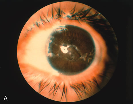
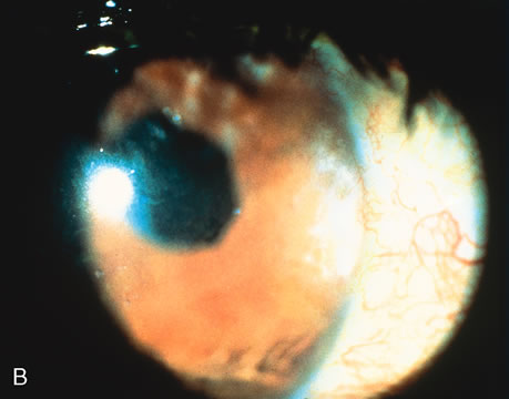
All eyes with long-standing uveitis may have band keratopathy.1 This is especially true in eyes with juvenile rheumatoid arthritis, uveitis from Toxocara canis, and phthisis (Fig. 1). To manage band keratopathy, chelating agents are used to remove the calcium deposits in the basal layers of the epithelium and in Bowman's membrane. Before the chelating agent is applied, the overlying epithelium should be removed with diluted alcohol, knife-blade scraping, or other techniques. A dilute solution of sodium ethylenediaminetetraacetatic acid (EDTA; Endrate) is then applied to the cornea.

Fig. 1. A. A 27-year-old white woman with phthisical eye, band keratopathy, secluded fibrosed pupil, and dense cataract due to juvenile rheumatoid arthritis (shown by ultrasonography). B. After removal of band keratopathy and lensectomy/vitrectomy, the vision improved from light perception to 20/80 (6/24), with a rise in intraocular pressure from 2 to 8 mm Hg. The eye is microophthalmic.

The procedure is performed with the aid of an operating microscope. After a topical anesthetic is applied, a lid speculum is placed and the epithelium is removed with a dilute solution of alcohol and a blade. A vial of sodium EDTA is diluted 1:50 with sterile balanced salt solution and applied with a sponge to the area of the band keratopathy. The chelating agent is dabbed on the cornea, not irrigated into the eye. This prevents the toxic effects of the chelating solution from injuring other ocular structures. It may take several minutes to determine whether the deposit has been chelated sufficiently to be removed with gentle scraping. If it has not been loosened sufficiently, a more concentrated solution of the chelating agent—up to a dilution of 1:10—may be used. After the procedure has been completed, an antibiotic solution, a cycloplegic, and a semipressure patch are applied to allow the epithelium to heal. Occasionally, a bandage contact lens may be necessary if there is delayed healing of the epithelium.

Back to Top
PENETRATING KERATOPLASTY IN UVEITIS
A wide variety of corneal problems are associated with ocular inflammation, the most significant of which is corneal scarring caused by herpes simplex keratouveitis. Keratoplasty can be performed in these cases if the eye is uninflamed.2,3 Studies have shown a significant recurrence of herpes in corneas after penetrating keratoplasty, but the longer the eye has been uninflamed before surgery, the better the prognosis. Therefore, in cases of active corneal and intraocular inflammation, penetrating keratoplasty should not be performed until the eye has remained quiet without medication for several months.

The usual indications for visual rehabilitation should be present. Once the eye has been quiet a reasonable length of time, the standard surgical procedure can be followed. Peripheral iridectomies should also be performed in these previously inflamed eyes. Lamellar keratoplasty in patients with herpetic scars and iridocyclitis is rarely indicated. In other forms of corneal opacification associated with ocular inflammation, the eye also should be as uninflamed as possible before keratoplasty.

Conjunctival flaps may play a role in the management of chronic, indolent, noninfectious herpetic disease. Although the bandage contact lens has revolutionized therapy for indolent ulcers recalcitrant to healing, a conjunctival flap may be indicated in such patients or in those in whom a contact lens has been ineffective.

Back to Top
UVEITIC GLAUCOMA
Secondary glaucoma, one of the serious complications of intraocular inflammation,4,5 occurs with various syndromes and may be difficult to manage. Most patients respond poorly to surgery. It is of primary importance to determine the severity of the inflammation and, if possible, the syndrome associated with it. Management includes treatment of the underlying inflammation and of the glaucoma itself. Various mechanisms produce secondary glaucoma, and it is important to identify them to institute the appropriate therapy.

CLINICAL FEATURES

In addition to the signs and symptoms of the underlying inflammation is an elevation of the intraocular pressure. If there is corneal disease or diffuse edema, the McKay-Marg tonometer or an air tonometer may be needed to measure the intraocular pressure. Gonioscopy can determine whether there are synechiae or precipitates in the angle.

PATHOLOGY

The trabecular meshwork may be blocked with inflammatory cell debris or frank peripheral anterior synechiae. Iris bombé or rubeosis is also apparent in some cases.

DIFFERENTIAL DIAGNOSIS

Table 1 is a list of some of the major uveitic syndromes with which secondary glaucoma can be associated. Glaucoma may occur in any case of severe iridocyclitis, whatever the specific syndrome. Glaucoma secondary to peripheral anterior synechiae may develop late when the inflammation is no longer active.


Table 1. Uveitic Syndromes With Which Glaucoma May Be Associated

Posner-Schlossman syndrome (glaucomatocyclitic crisis)
Herpes simplex uveitis
Herpes zoster uveitis
Severe acute iridocyclitis
Rubella iridocyclitis
Nonspecific iridocyclitis
Fuchs' heterochromic iridocyclitis
Syphilitic uveitis
Iridocyclitis secondary to toxoplasmosis


The Posner-Schlossman syndrome (glaucoma-tocyclitic crisis) is characteristically intermittent with little evidence of inflammation.6,7 Precipitates in the angle, however, may elevate the intraocular pressure considerably. Rubella, herpes zoster, and herpes simplex are often accompanied by an elevation of pressure with relatively little anterior chamber inflammation. These diseases are accompanied by characteristic changes in the cornea and skin. Glaucoma occurs in up to 50% of patients with Fuchs' heterochromic iridocyclitis.4

ETIOLOGY

The two most common causes of secondary glaucoma in patients with uveitis are severe inflammation with blockage of the trabecular meshwork and peripheral anterior synechiae with secondary closure (Table 2).


Table 2. Secondary Glaucoma: Causes, Incidence, and Treatment


Cause of Elevated Intraocular TreatmentIncidenceTopical and Systemic Treatment
Blockage of trabecula with debrisCommonTherapy for inflammation
Peripheral anterior synechiaeCommonCarbonic anhydrate inhibitors, β-adrenergic blockers, α-agonists
Iris bombéRareIridectomy
Trabeculitis (Posner-Schlossman syndromeRareCarbonic anhydrase inhibitors, β-blockers, α-agonists (rarely used), standard therapy for inflammation
Rubeosis iridisRareCarbonic anhydrase inhibitors, β-blockers, α-agonists, cyclocryotherapy
HypersecretionExtremely rareCarbonic anhydrase inhibitors, β-adrenergic blockers, α2-agonists
Sclerosis of the trabecular meshworkNot knownCarbonic anhydrase inhibitors β-adrenergic blockers, α2-agonists
Corticosteroid induced3% to 10%Discontinue corticostedroids


TREATMENT

Special consideration should be given to the management of acute intraocular inflammation with elevated intraocular pressure. If it is certain that corticosteroids are not the cause of the elevated pressure, the following regimen is suggested.

  1. A strong topical corticosteroid is instilled every hour during the day and every 2 hours during the night.
  2. Acetazolamide, 250 mg every 8 hours or 500 mg sequel every 12 hours, is prescribed.
  3. A β-blocker such as timolol maleate 0.5% solution and/or epinephrine 1% solution and/or dipivefrin (Propine; Allergan, Inc., Irvine, CA) 0.1% every 12 hours is given. Brimonidine tartrate 0.15% or latanaprost 0.005% has proven very effective.
  4. Cycloplegics are administered three or four times per day.
  5. Systemic prednisone, 60 mg, is given every morning; if inflammation is severe, a sub-Tenon's capsule injection of moderately long-acting corticosteroid is used.

Miotics are usually contraindicated in acute inflammatory glaucoma. Because epinephrine occasionally causes headaches and discomfort, a β-blocker is the drug of choice for nonspecific inflammatory glaucoma. There is some evidence that prostaglandins may aggravate ocular herpes, worsen inflammation, and cause cystoid macular edema. Their use in patients with uveitis is controversial. On the other hand, most ocular inflammations with acute secondary glaucoma due to blockage of the angle by inflammatory cells improve after just a few days of corticosteroid therapy. Prostaglandin and prostamine therapy has been noted to increase iris pigment, epithelim pigmentation, and to a lessor degree, increase inflammation in the eye. There is evidence that Xalatan (Pharmacia & Upjohn Company, Kalamazoo, MI), in particular, may worsen herptic anterior inflammation, and hence this use should be avoided in patients with uveitis.

Corticosteroid-induced glaucoma is a major differential diagnostic problem when it complicates intraocular inflammation. Usually the glaucoma does not develop until at least 4 to 6 weeks after the corticosteroid therapy is begun. If the inflammation responds well to the corticosteroid drops, sub-Tenon's capsule and subconjunctival depot preparations should be avoided. Frequently the clinician must balance the benefit conferred by the corticosteroid against the eye's limited ability to tolerate elevations of pressure. If the glaucoma cannot be controlled, it may be necessary to remove a previously injected depot preparation surgically. Glaucoma is frequently seen after anterior sub-Tenon or subconjunctival placement of depot steroid but is rare after well-placed posterior sub-Tenon injection. Often a patient with corticosteroid-induced glaucoma requires maximum medical therapy for both the glaucoma and the inflammation, and the clinician must prescribe other anti-inflammatory drugs.

SURGICAL PROCEDURES IN SECONDARY GLAUCOMA AND INFLAMMATORY GLAUCOMA

The preferred procedure for iris bombé may become surgical iridectomy if the media will not allow for laser iridotomy, done with yttrium aluminum garnet (YAG) or argon lasers, because transfixation of the iris or laser iridotomy is often followed by rapid occlusion of the small openings. Iridectomy should be done with the patient under general anesthesia or, if the eye is not too inflamed, with local anesthesia. At the same time, a sub-Tenon's capsule injection of corticosteroids may be given.

The results of filtering surgery often are not good. For this surgery, the eye should be as uninflamed as possible. Although laser trabeculoplasty may be tried (if the angle is open), results have not been encouraging8,9 and surgical intervention is usually required later. Standard trabeculectomy or other filtering procedures such as modified goniotomy, thermal sclerostomy, and posterior lip sclerotomy have afforded limited success.10–12 If the eye has been quiet for several months, however, a standard filtering procedure (thermal sclerostomy is preferred) has a greater chance of success than when the eye has been quiet for a shorter time. Krupin-Denver, Barveldt, or Molteno valves may be important as well. However, these valves may clog with inflammatory debris.13–16 A laser iridotomy may be elected to break an attack until a standard surgery can be performed.

The use of perioperative 5-fluorouracil subconjunctival injections significantly improves the success rate of filter procedures and especially trabeculectomy for uveitic glaucoma. Other antimetabolites have been considered as an adjunct to filter surgery because of the consideration of the perioperative and postoperative inflammation in the patients with uveitis. Impressive results have also been reported with the use of a single intraoperative dosage of mitomycin C to the episcleral surface at the conclusion of a filtering procedure in patients with both primary and complicated glaucoma.

The surgical strategy that seems to be most promising when facing a progressive secondary glaucoma in uveitis is the use of shunt valves. The most commonly used device is the Molteno implant. The principles that have made this implant successful include the use of a biologically inert silicon tube in the anterior chamber (Fig. 2) that drains aqueous far from the limbus to an equatorial plastic plate that is sutured in place. What makes this uniquely suited for the benefit of inflammatory glaucoma is the simple fact that the artificial material of the tube is incapable of scarring down the way a naturally fashioned surgical filter would. The surface area of this plate allows for the elaboration of a fibrotic bleb across from which there is absorption of the accumulated aqueous. These features have proven successful in the other valves such as the Schocket, Ahmed, Krupin, and Joseph models. These devices are reserved for the intractable glaucomatous patient with uveitis who has failed to improve with other medical procedures and if intercurrent or recurrent inflammation is believed to doom the prospects for a standard filter drainage procedure. In inflammatory glaucoma, these setons may become the surgical treatment of choice.

Fig. 2. Slit lamp view of two successive Molteno implants in a 34-year-old patient with Vogt-Koyanagi-Harada disease, who required the second surgery when the first tube clotted with intense fibrous reaction in and around it consequent to profound iridocyclitis.

If both filtering surgery and maximal medical therapy have failed, YAG cyclotherapy or cyclocryotherapy is recommended. Unfortunately, this procedure itself causes significant inflammation and a definite risk of phthisis bulbi. Thus it is a procedure of last resort.

Cyclocryotherapy can be performed in a number of ways. The glaucoma cryoprobe can be used with local anesthesia. The inferior 180 degrees of the eye is treated with two rows of freeze for 1 minute at –80°C in three spots per quadrant. The first row begins 2 to 3 mm behind the limbus, with the “ice ball” extending partially onto the cornea. The second row is placed immediately behind the first. By freezing two separate rows, the need to freeze and then refreeze is eliminated. The eye is patched after the instillation of a combined corticosteroid and antibiotic together with a cycloplegic. Because patients experience significant pain in the postoperative period, an effective analgesic is important.

Back to Top
CATARACT EXTRACTION IN PATIENTS WITH UVEITIS
Cataract formation is a frequent complication of both acute and chronic uveitis. Cataract extractions pose significant risks for patients with uveitis.17-21 The outcome of such surgery often depends on the particular uveitis syndrome. Approximately 50% of patients with juvenile rheumatoid arthritis,24–26 pars planitis,21 and Fuchs' heterochromic iridocyclitis20,27–30 develop visually significant cataracts. Cataract development in patients with uveitis may be a consequence of the inflammatory process or may be iatrogenic as a sequela of corticosteroid therapy. Chronic recurrent uveitis induces cataracts much more frequently than do acute episodes of short duration. Before the advent of corticosteroids in the 1960s, ocular inflammation was difficult if not impossible to control. Cataract surgery was often associated with profound complications both intraoperatively and postoperatively, including retinal detachment, choroidal detachment, vitreous loss, and intraocular hemorrhage. Postoperative complications included severe exacerbations of the intercurrent inflammation with pupillary membrane formation, ciliary body detachment with eventual phthisis, and secondary glaucoma.

The first step in the evaluation of the patients with uveitis for cataract surgery is the accurate classification of the specific disease entity causing the uveitis, the intraocular structures involved, the activity of the inflammation, and a plan for surgery that considers all of the three foregoing factors. These factors and the mode of treatment determine the potential postoperative complications and prognosis of the patient and dictate whether the surgery should be by way of the anterior segment, pars plana, or include the use of an intraocular lens and its type.

Cataract extraction poses significant risks for patients with uveitis. The less inflamed the eye at the time of surgery, the better the prognosis and the less chance of severe postoperative complications.17-22,26,28 Treating inflamed eyes with high doses of corticosteroids systemically, topically, or by sub-Tenon's capsule injection for a few days or a week before surgery is recommended. If posterior inflammation threatens the outcome, such as in recurrent retinal vasculitis, it is wise to defer surgery until the inflammation subsides either naturally or with medication. The advent of small incision phacoemulsification has lessened the intensity of postoperative inflammation and the use of iris hooks to enlarge the pupil has facilitated cataract surgery in patients with chronic uveitis.

The indications for cataract extraction in patients with ocular inflammation are visual disability, maturity of the cataract, and lens-induced inflammation. The necessity to monitor posterior disease (e.g., toxoplasmosis, Vogt-Koyanagi-Harada syndrome, sarcoid, vasculitis, cystoid macular edema) may also be considered an indication for cataract removal. If the eye has been quiet without medications and there has been no active inflammation for at least several months, a routine lens extraction can usually be performed without significant risk. But when inflammation (even low-grade) is present, special precautions and surgical techniques are necessary.37,42

If the cataract is mature and there is significant capsular involvement or liquefaction of cortical material, extraction is advisable to prevent lens-induced disorders from being superimposed on an underlying inflammatory process.

The only urgent indication for cataract surgery is the uveitis that appears to have been caused by the lens material itself. Phacoanaphylaxis, phacolytic glaucoma with cataract, and phacotoxic reactions caused by residual lens material after extracapsular lens extractions indicate that the lens or lens material should be removed to eliminate the cause of the active inflammation. If all of the lens material is not removed (both nuclear fragments and cortex), the inflammation may persist indefinitely.

The use of intraocular lens implants is controversial in patients with uveitis.19 All types of intraocular lens compositions and surfaces have been tried, including heparin-coated lenses to reduce the postoperative accumulation of inflammatory debris and fibrosis coating the new lens.33,37–40,42,46–48 Even transsclerally fixated Intraocular lens implants have been attempted in children.32 In general, the condition of the eye and the intraocular lens depends not on which type of intraocular lens implant to instill, but rather the intensity of the ocular inflammation in the postoperative period.38 It is felt that “foldable intraocular lens implantation is safe in uveitic eyes,”39 but that the intraocular lens implant biocompatibility is inversely related to the degree of inflammation present.18,19,22,36 The foldable acrylic intraocular lens implants may be preferred because these patients may have a higher potential for traction/rhegmatogenous retinal detachment on an inflammatory basis, and therefore may have to receive intraocular silicone oil.36,58 The role of which type of lens composition or surface that leads to or stimulates inflammation the least is still not concluded. Instances of intraocular lens opacification worse than the original cataract are observed and reported.37 However, even series of patients with profound inflammatory basis are noted to do well after cataract surgery if their inflammatory response can be well controlled.37,38 Intraocular lenses have been shown to be tolerated in young patients with juvenile rheumatoid arthritis.40,41 It still may prevail that even after successful cataract surgery, the vision may not improve satisfactorily because of inflammatory destruction of the retinal structures38 not appreciated before, during, or after the surgery.

SPECIFIC UVEITIC SYNDROMES

Juvenile Rheumatoid Arthritis and Iridocyclitis

Early results of extracapsular cataract extraction in patients with juvenile rheumatoid arthritis were not encouraging because of frequent vitreous loss and incarcerations in the cataract wound, development of cyclic membranes, and subsequent ciliary body detachment with eventual phthisis. One report of 15 cases, in which standard extracapsular techniques were used (e.g., aspiration), cited 11 patients with no light perception by the end of 5 years.43 A more encouraging report came from the Proctor Foundation in San Francisco: vision of 20/40 (6/12)* or better was achieved in 60% of patients.26 In the larger series in the literature, Kanski and Shan-Shin29 reported 162 eyes undergoing cataract extraction. The major complication reported in the needle aspiration group was phthisis, and results were improved when lensectomy-vitrectomy was performed. Much more encouraging results have been reported by other investigators after lensectomy-vitrectomy. With lensectomy-vitrectomy surgery, capsule remnants, cyclic membranes, and calcified particles can be removed more completely, thus significantly lessening the possible outcome of ciliary body detachment and phthisis.


*Metric equivalent given in parentheses after Snellen notation.

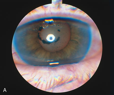
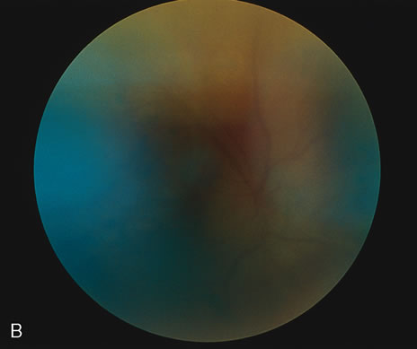
If patients with juvenile rheumatoid arthritis have progressive cataracts and extraction of the lens is indicated, combined lensectomy and subtotal vitrectomy performed through the limbus or pars plana is recommended (Fig. 3). An anterior approach for cataract extraction is recommended only when few vitreous opacities are present and the intraocular pressure is normal. If there is any suspicion of cyclic membrane with traction on the ciliary body, the treatment of choice is the posterior approach, with vitreous removal to lessen subsequent traction on the ciliary body. This may be suggested by the presence of vitreitis, vitreous opacities, severe anterior segment damage, hypotony, and an obvious cyclic membrane formation. It should be emphasized that meticulous postoperative therapy to prevent amblyopia is equally important as the surgery for full visual rehabilitation in these young patients.

Fig. 3. A 16-year-old aphakic white girl with juvenile rheumatoid arthritis. A. Up-drawn, fibrotic pupil, with dense cyclitic membrane in a hypotonous eye. B. After pupilloplasty and membranectomy/ vitrectomy, a large, round pupil is restored, along with normal ocular tension. Vision improved from counting fingers to 20/40+ (6/12+) with contact lens.

Fuchs' Heterochromic Iridocyclitis

Although early reports indicate that bleeding throughout normal angle vessels was a serious complication after cataract extraction in patients with Fuchs' heterochromic iridocyclitis,27,28,31 this was not proven to be a consistent complication in any of 29 cases reviewed by Smith and O'Connor.46 Results from the Mayo Clinic have, however, suggested a slight increase in complications after routine cataract extraction in patients with Fuchs' iridocyclitis.27 In general, however, any standard procedure for cataract surgery usually can be used without significant risk of intraoperative or postoperative complications. Transient postoperative hyphemas may occur but seldom create a long-term problem. Because Fuchs' iridocyclitis is usually unilateral, the usual indications for this type of cataract extraction apply. The patient's vision may not return to 20/20 (6/6), however, because the disease itself can cause visually impairing vitreous opacities.

Fuchs' heterochromic iridocyclitis is a low-grade, chronic, often asymptomatic anterior uveitis occurring in patients older than 40 years of age. Nearly 100% of patients develop cataracts, usually of the posterior subcapsular type, which is one of the diagnostic features of this disease. Patients with visually significant cataracts as a result of Fuchs' syndrome require relatively few special precautions and have a good prognosis after modern extracapsular cataract extraction. The favorable prognosis for successful surgery in these patients may be because of the rarity of posterior synechiae. This is the one uveitic syndrome in which active inflammation may be present and posterior chamber lens implantation (“in the bag”) appears to be well tolerated with few complications. Gee and Tabbara20 reported postoperative visual acuities of 20/40 or better in all 10 of their patients with Fuchs' heterochromic iridocyclitis undergoing extracapsular cataract extraction and posterior chamber lens implantation. The most frequently encountered postoperative complications included hyphema, vitreous hemorrhage, glaucoma, and progression to vitreous opacification, the latter two features being major causes of postoperative visual acuity less than 20/40 as well as being a diagnostic feature of the disease.20,27

Pars Planitis (Chronic Cyclitis, Peripheral Uveitis, Intermediate Uveitis)

Pars planitis is a chronic bilateral uveitis characterized by persistent vitreous cells, opacities, and low-grade anterior chamber inflammation that may last for several years. Lens opacities develop in a significant number of cases (42%), but little information is available concerning the prognosis for cataract surgery. Smith and Godfrey50 described intracapsular and extracapsular lens extractions in 21 eyes with chronic cyclitis. Of these, 13 had vision of better than 20/40 several years after surgery. Complications were not significantly higher than for those in whom there was no inflammation, and no cases of phthisis or prolonged reactivation of inflammation were noted. Diamond and Kaplan51,52 reported several cases of combined lensectomy-vitrectomy in patients with pars planitis with no significant complications.

Pars planitis does not seem to increase the risk of complications in routine cataract surgery. Postoperative vision is, however, limited by the complications of the disease itself: cystoid macular edema and vitreous opacities. Although routine cataract extraction can be performed safely, combined lensectomy-vitrectomy may be preferred for such patients since vitreous opacities are a significant problem postoperatively. Diamond and Kaplan51,52 have suggested removing the vitreous to lessen cystoid macular edema. Others have suggested that the effect of vitrectomy on cystoid macular edema is uncertain.53 More data on the effectiveness of the vitrectomy on lessening the cystoid macular edema are needed; however, anecdotal evidence seems to favor the addition of vitrectomy to the surgical regimen. Experience would dictate that eyes with insignificant inflammation in the anterior segment tolerate anterior segment cataract extraction well. Patients with significant vitreous opacities, chronic macular edema, and, of course, vitreous traction on the retina are, however, more appropriate candidates for the pars plana approach with vitrectomy-lensectomy. All patients should receive intensive corticosteroids, predominately sub-Tenon injection, before they are considered corticosteroid failures with persistent macular edema. Foster48 has repeatedly suggested a physician intolerance of any recurrent persistent inflammation preoperatively, intraoperatively, and postoperatively in these patients and suggests the implementation of a stepladder algorithm for the control of inflammation running from the routine antiinflammatory agents and corticosteroids to strict regimens of antimetabolites. Michelson and associates54 reported 60% of eyes of patients with pars planitis achieved a stable visual acuity of 20/40 or better; however, persistent but nonobservable low-grade inflammation yielded a deposition of fibrous debris over and around intraocular lens implants in these patients, forming a so-called cocoon effect of sequestering a polymethyl methacrylate posterior intraocular lens inside a network of scar tissue. Such a progressive form of scarification necessitated surgical cleaning of lenses in two patients and removal of the lens in yet another. Also, macular edema, macular hole, retinal detachment, vitreous hemorrhage, and low-grade chronic inflammation were primary complications for a 40% failure rate in the studies of Smith and colleagues,50 Mieler and coworkers,55 and Michelson and associates.54 Foster48 has advocated the importance of total ablation of all inflammation in patients who show that their uveitis is going to be chronic or recurrent. In spite of Foster's proposal for zero tolerance of inflammation postoperatively in these patients, Michelson and associates54 noted “seeming inflammatory sequelae” where no signs of inflammation were noted at the slit lamp. The results of these studies emphasize the need for appropriate case selection for surgery as well as the need for perioperative antiinflammatory therapy and the possibility of long-term antiinflammatory therapy postoperatively. Intraocular lens implantation appears to be a safe and effective alternative to other optical correction modalities in pars planitis provided that absolute absence of inflammation has been achieved and maintained after surgery.

Lens-Induced Uveitis (Phacoanaphylactic Endophthalmitis, Phacolytic Glaucoma, Phacotoxic Uveitis)

Once the diagnosis of lens-induced uveitis is established, the lens should be extracted and the residual lens material removed as soon as possible.54–57 In the case of phacoanaphylactic endophthalmitis, careful removal of all lens material is also important. Depending on the patient, an intracapsular extraction may be possible, but more often it is not. A careful and complete extracapsular extraction by means of microsurgical techniques is the alternative.

Although the lens is usually intact in patients with phacolytic glaucoma, it is often quite fragile, and attempts at intracapsular surgery may rupture the capsule. If this occurs, it is necessary to convert the procedure to an extracapsular one and to remove all lens material. Before surgery in such patients, high doses of corticosteroids, given systemically, subconjunctivally, and topically, and osmotic agents may be needed to control the intraocular pressure.

The most important aspects in the management of such patients are the correct diagnosis, control of the intraocular pressure and inflammation, and the removal of all lens material, preferably by an intracapsular procedure. These cases are considered semi-emergencies, and the patient should be operated on as soon as possible. If all lens material (nuclear and cortex) is removed, these cases may be considered appropriate candidates for intraocular lens implantation.

OTHER DISORDERS

If inflammation is damaging to intraocular structures, such as the retina, cataract surgery will not noticeably affect the ultimate visual acuity. As in Behçet's syndrome or other forms of vasculitis, the optic nerve or retinal vasculature can be so damaged that cataract surgery may not improve vision. Conversely, it is stated that cataract extraction in cases of sympathetic ophthalmia is a benign procedure.

As noted, the less inflamed the eye, the better the prognosis for cataract surgery. For example, in a case of recurrent iridocyclitis that has been totally quiescent for several years, a routine cataract extraction can be performed without significant complications. Such patients may be considered candidates for posterior chamber “in-the-bag” intraocular lenses. But in patients with low-grade chronic inflammation with progressive cataract, a more judicious and cautious approach should be taken, especially if the cause cannot be determined or the syndrome identified. Such patients are not to be considered appropriate candidates for intraocular lenses. Intraoperative and postoperative corticosteroids are indicated in such cases.

GENERAL APPROACH AND SURGICAL MANAGEMENT

If there is minimal or no active inflammation in an eye that has had previous bouts of inflammation, and if the patient is not receiving medication at the time of surgery, any standard lens extraction procedure can probably be performed without significant complications. In a young patient, careful extracapsular extraction is probably the safest procedure, if the pupil can be well dilated. The benefit of less surgical manipulation and smaller incisions is obvious with mall incision phacoemulsification. If the iris/pupil needs to be swept free of synechiae, iris hooks may prove useful to enlarge and maintain adequate dilatation.

If there is chronic active anterior segment inflammation with no posterior segment inflammation, including no vitreous cells or retinitis, an intense preoperative therapeutic regimen of topical sub-Tenon's capsule and sometimes systemic corticosteroids is recommended for 7 to 10 days before surgery. The visual indications should be considered carefully in such cases because the risk of postoperative inflammation and other complications is not well documented. If extracapsular extractions are completed with great care, the removal of all lens material should be ensured. A well-dilated pupil is necessary.

If the inflammation is anterior and posterior, with cells and opacities in the vitreous, a combined lensectomy and vitrectomy is recommended, because residual inflammatory cells and debris in the vitreous cavity may increase the frequency with which the uveitis recurs. Diamond and Kaplan,51,52 Nobe and coworkers,60 Fitzgerald,61 and Belmont and Michelson62 have reported various types of uveitis that respond well to lensectomy and vitrectomy. A pars plana approach is used with endoillumination, standard vitrectomy-lensectomy techniques, and vitrectomy instrumentation (Fig. 4). These techniques are also recommended for cases of juvenile rheumatoid arthritis, ankylosing spondylitis, and pars planitis with cataract.

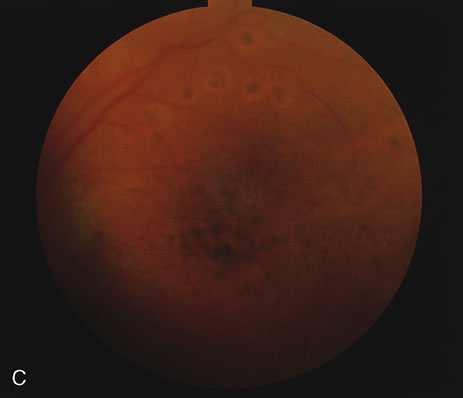
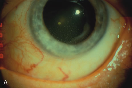
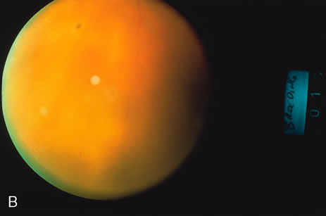
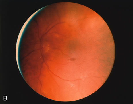
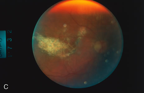
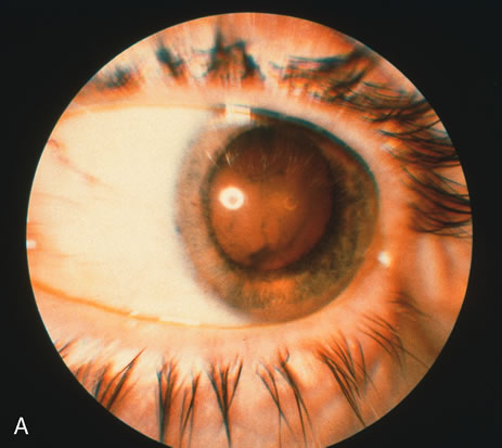
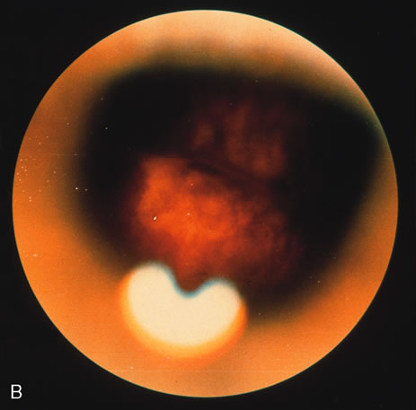
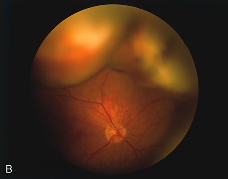
Fig. 4. A. External appearance of a patient's eye with Vogt-Koyanagi-Harada disease and chronic thick vitreous. B. Pre-operative view of the fundus demonstrates chronic, thick exudate filling vitreous gel, obscuring the view of the retina. C. View after vitrectomy shows the fundus clearly. Note argon laser scars superotemporal to the disc, where a previous ophthalmologist had rendered laser treatment for a serous retinal detachment.

SPECIAL PROBLEMS AND CONSIDERATIONS

Synechiae

Synechiae may occur not only at the pupillary margin but also in the midperiphery of the iris and between the ciliary process and the lens capsule. Whether intracapsular or extracapsular extraction is performed, gentle sweeping lysis of adhesions between the iris and lens is important. This can be performed through an iridotomy with instruments (e.g., cyclodialysis spatula) and gentle irrigation. During the intracapsular extraction, slow and careful delivery of the lens should be accompanied by lysing of the remaining synechiae. Lensectomy-vitrectomy may be preferred in such cases.

Cyclitic Membrane

Patients with chronic uveitis, especially those with juvenile arthritis and persistent sarcoid uveitis, may have a cyclitic membrane extending from the ciliary body over the back surface of the lens (Figs. 1 and 3). This membrane may be detected preoperatively by ultrasonography. If a standard procedure with a large cataract section is to be carried out, the membrane should be excised with scissors in the pupillary zone only. In closed procedures in which endosurgical techniques are used, the membrane should be amputated along with the cataract and anterior vitreous. The surgeon must be prepared to deal with these membranes and remove as much of this tissue as possible during the cataract surgery. A cyclitic membrane that pulls on the ciliary body can cause its detachment, which in turn can lead to hypotony and phthisis. A visualized membrane, sometimes extending over the front surface of the lens as well, may bleed during its isolation and excision, complicating the cataract procedure. In aphakic eyes, some cyclitic membranes can be sectioned successfully with a YAG laser, thus eliminating traction on the ciliary body. Usually the power needed is greatly in excess of a routine capsulotomy. Potentially, the YAG laser may be used to section fibrous bands over the retina that lead to tractional retinal detachment.

Inflammatory Membrane Over the Surface of the Lens and Connected to the Iris

It is common for a membrane of vascularized connective tissue to extend from the iris over the entire pupillary area. This membrane can often be separated mechanically from the iris and the anterior lens capsule. Transient bleeding that occurs during the removal of the membrane usually stops without causing significant problems. If the pupil can then be dilated, the surgeon can proceed with a routine cataract extraction.

Management of the Iris

A chronically inflamed iris tends to bleed easily during surgery, but the bleeding is often self-limited. In severe cases, bimanual, bipolar cautery must be used. Manipulation of the iris (and surgery) should be minimal.

In totally quiet eyes that have no tendency to develop synechiae, as in Fuchs' heterochromic cyclitis or pars planitis, peripheral iridectomy may be satisfactory. If the case is more complicated and if a lens extraction is to be performed without vitrectomy, a sector iridectomy is safer. If a combined lensectomy-vitrectomy is performed, there can be no postoperative synechiae between the wound and the vitreous face or lens capsule, and pupillary block cannot develop. Iris surgery, therefore, is discouraged; not only can it contribute to postoperative inflammation, but it is also unnecessary.

Vitreous Cells and Opacities

Ultrasonography in the preoperative examination may reveal vitreous opacities. If these are extensive, a combined lensectomy and vitrectomy can be performed. If a standard lensectomy alone is planned, the vitreous face should be left intact, since its stability is thought to lessen the chance of cystoid macular edema.

Back to Top
VITRECTOMY
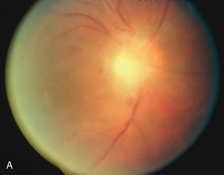
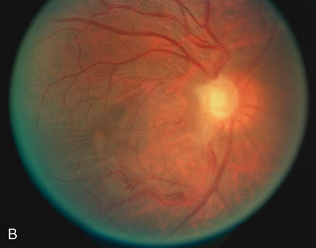
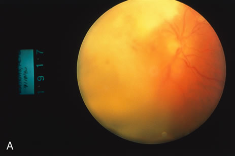
At present, the indication for vitrectomy in cases of intraocular inflammation is twofold. It is used as a controlled biopsy of the vitreous to establish the histologic diagnosis of endophthalmitis, large cell lymphoma (reticulum cell sarcoma) (Fig. 5), and other unusual infiltrations of the vitreous (Table 3, Figs. 6 through 8). It also serves as a therapeutic intervention in many cases (Figs. 9 and 10). Combined lensectomy-vitrectomy has not often resulted in a worsening of the inflammation of phthisis. Some feel that the addition of pars plana vitrectomy may lessen the burden of inflammation and the extent of edema in inflammatory cystoid macular edema.23,51–53,55,60,62

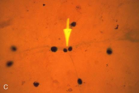
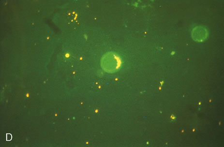
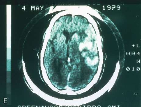
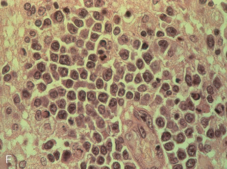
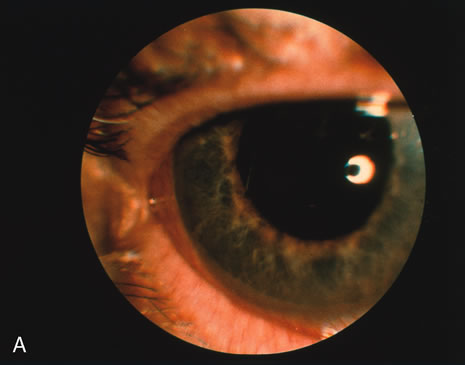
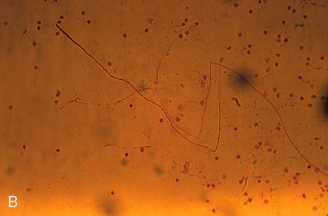
Fig. 5. A. External view of the eye of a 76-year-old man in whom reticulum cell sarcoma (large cell lymphoma) is suspected. Note large mutton-fat keratic precipitates. B. Fundus view of the same patient demonstrates 3+ vitreous cells and white subretinal infiltration. C. Vitreous aspirate demonstrates large cell lymphoma infiltrate. Note two smaller cells in mitosis (arrow). D. Vitreous cells stained for their surface immunoglobulin light chains. Fluorescein discloses IgM light chains only, as a corroboration of the monoclonal nature of this malignant infiltration. E. CT scan of same patient (different time) shows intracerebral microgliomatosis of large cell lymphoma infiltration. F. Histologic appearance of this brain tumor of “reticulum cell sarcoma” or large cell lymphoma.


Table 3. Diagnostic Paracentesis


FindingCondition or Disease Indicated
Aqueous 
  BacteriaEndophthalmitis
  FungiCandida, Aspergillus, etc.
  Tumor cellsRetinoblastoma, malignant melanoma, reticulum cell sarcoma, leukemia, metastatic cancer
  EosinophilsToxocara canis
  MacrophagesPhakolytic glaucoma
  Antibodies (enzyme-linked immunosorbent assay)Toxoplasma gondii, Toxocara canis, reticulum cell sarcoma, Behçet's disease, syphilis
  Immune complexesBehçet's disease
  Other proteins 
  Angiotensin converting enzymesSarcoid
  Lactate dehydrogenase isoenzymesRetinoblastoma
Vitreous 
  BacteriaEndophthalmitis
  FungiCandida, Aspergillus species, etc.
  Tumor cellsRetinoblastoma, malignant melanoma, r reticulum cell sarcoma, leukemia
  EosinophilsToxocara canis
  AntibodiesToxoplasma gondii, reticulum cell sarcoma, Behçet's disease, syphilis (and immune complexes)
  MacrophagesSympathetic ophthalmic, severe retinitis


Fig. 6. A. Patient with opaque media due to postoperative (cataract with posterior chamber intraocular lens) exogenous Staphylococcus aureus endophthalmitis. B. Immediate postvitrectomy view of same patient, whose retina demonstrates a small punctate hemorrhage superior to the macula, as well as profound retinal edema. The vision returned to 20/20 (6/6).

Fig. 7. A. Classic presentation of endogenous Candida endophthalmitis in a drug abuser who demonstrates prominent vitreous fluff balls amid diffuse vitreous cellularity and haze. B. Postvitrectomy view of fundus shows clear vitreous cavity with cellophane wrinkling changes in the internal limiting membrane of the retina.

Fig. 8. A. Endogenous endophthalmitis in a drug abuser. Only 3 days after injecting cocaine his vitreous filled with dense white exudate. B. Vitrectomy specimen discloses the septate hypha of Aspergillus species. C. Postoperative view of fundus demonstrates fibrous scarring of the retina with visual return to 20/70 (6/21) after intensive intravenous therapy with amphotericin B and flucytosine.

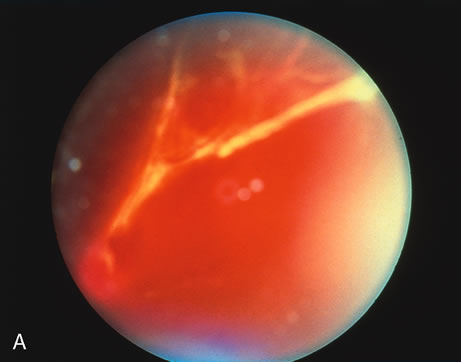
Fig. 9. A. External view of an eye demonstrating dense vitreal inflammation secondary to reactivated toxoplasmosis in which a traction retinal detachment occurred. Fundus photography was precluded by the thick exudate. B. View after pars plana vitrectomy with scleral buckling shows the scleral buckle below and one prior lesion of active toxoplasmosis at the 8 o'clock position.

Fig. 10. A. Fundus view of thick fibrous bands within profound intravitreal inflammation due to reactivated toxoplasmosis with combined traction/rhegmatogenous retinal detachment. B. After vitrectomy and scleral buckling, note the etiologic chorioretinal lesion at the 11:30 position on buckle, now inactive. (The patient's serum indirect fluorescent antibody toxoplasmosis titer was 1:1024 at surgery; the vitrectomy fluid yielded a titer of 1:4096.)

DIAGNOSTIC VITRECTOMY

Certain forms of ocular inflammation lend themselves to diagnostic vitrectomy. Patients with large cell lymphoma (reticulum cell sarcoma) are usually elderly and have bilateral chronic inflammation with a predominance of cells in the vitreous cavity62,63 and thus are suspected of having reticulum cell sarcoma. Because the treatment of this disease requires irradiation and/or chemotherapy and not corticosteroids, a diagnostic vitrectomy, performed with a standard pars plana technique, is recommended (Fig. 5). The vitreous specimens should be collected and taken immediately to a cytology laboratory. The handling of the intraocular specimens by the pathology or cytology division should be planned in advance. If possible, the specimen should be obtained by aspiration alone, because vitreous cutters can distort the cells. These patients often have a fluid vitreous, which makes aspiration feasible and extensive cutting unnecessary. These cells can then be examined both histologically and for their surface immunoglobulins.63,64 Other types of metastatic cancer cells such as adenocarcinoma, breast carcinoma, acute and chronic lymphocytic leukemia, and metastatic malignant melanoma46–48 can be found in the vitreous in rare cases.

Diagnostic vitrectomy also may be indicated in young patients presumed to have Toxocara canis endophthalmitis and in patients suspected of having infectious endophthalmitis, especially of fungal origin. Numerous eosinophils may be seen with Toxocara in the vitreous aspirate and an enzyme-linked immunosorbent assay should be performed on the specimen. In fungal endophthalmitis, hyphae and yeast forms may be identified.

Lensectomy should be avoided if a diagnostic vitrectomy is being performed; the lens is usually clear in such cases, and a mixture of lens material and vitreous contents may be very confusing to the cytologist.

The approach to suspected infectious endophthalmitis includes obtaining vitreous and aqueous specimens for smears and culture as well as intravitreal injection of antibiotics. The role of vitrectomy and systemic antibiotics was evaluated prospectively in the Endophthalmitis Vitrectomy Study (EVS).94 Immediate vitrectomy was found in the EVS to be superior to vitreous tap with antibiotic injection only in cases with vision worse than hand motion. The adjunctive use of parenteral antibiotics was not found to improve outcome regardless of assignment to vitreous tap or vitrectomy. It must be kept in mind that the EVS only evaluated cases of acute postoperative endophthalmitis following cataract surgery or secondary intraocular lens placement. Other kinds of endophthalmitis such as bleb-related or suspected fungal endophthalmitis were not evaluated. At present, most cases of endophthalmitis can be successfully managed without vitreous surgery, however vitrectomy is generally recommended for the most severe cases and for more unusual cases such as after trauma where a retained foreign body may be present or when a fungal etiology is suspected.

Technically, a total vitrectomy may not be necessary and a core vitrectomy may suffice. The inflammatory membranes that result from endophthalmitis often adhere tightly to the retina (Fig. 9), and a total vitrectomy can severely damage the retina, although it may be necessary to peel these membranes during vitrectomy.

If bacterial endophthalmitis is associated with an implanted lens, it is generally not necessary to remove the lens. During a closed vitrectomy the inflammatory membranes in the anterior chamber can be removed through an anterior incision, and the vitrectomy can be performed at the same time through the pars plana. The implant should probably be removed in cases suspected of fungal endophthalmitis.

THERAPEUTIC VITRECTOMY

There is increasing evidence that vitrectomy is of therapeutic value in various uveitic syndromes, both to eliminate the burden of inflammatory debris, threatening fibrous bands, and chronic opacification in the vitreous, as well as to ameliorate the macular edema, which is thought to be related to the vitreous and retinal inflammation (Figs. 4, 9, and 10). Diamond and Kaplan49,50 found that the cystoid maculopathy that was present in most of their patients gradually resolved in the course of a year or more with remarkable return of vision. Belmont and Michelson62 reported similar results in patients with chronic vitreal infiltration from ankylosing spondylitis. If there is an advanced cataract for any occlusion of the pupillary space in a patient with coexisting intraocular, predominantly vitreal, inflammation, a combined lensectomy and vitrectomy is practicable. Specifically, patients with chronic uveitis and dense cataract or nonspecific indolent posterior inflammation and cataract are candidates for this procedure. Diamond and Kaplan51,52 have reported that these patients tolerate lensectomy-vitrectomy well, and there seems to be no increased incidence of postoperative inflammation, phthisis, or other complications that have been regularly associated with standard cataract extraction in patients with uveitis.

Indications

What we consider an indication for lensectomy-vitrectomy in patients with intraocular inflammation is the presence of at least two of the following: need for better vision; progressive disease (hypotony, premonitory signs of phthisis, especially in the presence of cyclitic membrane); complications requiring surgery (e.g., retinal detachment, tractional or rhegmatogenous); and iris bombé (closed angle with synechiae) with hypotony (indicating the presence of cyclitic membrane)

Technicalities

It is very important to assess the visual function of these patients before surgery. An electroretinogram and the visual-evoked potential are important to determine the condition of the retina and optic nerve; ultrasonography, particularly of the anterior segment, is helpful in determining whether there is thickening of the choroid or a cyclitic membrane that could create significant problems at the time of surgery. A thickened choroid suggests engorgement of choroidal vessels, and the presence of a cyclitic membrane may make penetration of the pars plana area difficult and complications such as hemorrhage or dialysis more likely.

If the eye is hypotonous before surgery, a cyclitic membrane is probably present even if it cannot be detected by ultrasonography (Fig. 1). If there is a thickened choroid or a cyclitic membrane, a longer infusion cannula should be used in the pars plana area, and it is wise to remove as much of the lens as possible before turning on the posterior infusion cannula. In this way one can be reasonably certain that the cannula is through the pars plana and the cyclitic membrane. Lensectomy can be performed through the pars plana stab incisions with the infusion needle in one side of the lens and the vitrectomy instrument in the other side.

Synechiae should be removed or broken early. If synechiae are dense, as in patients with juvenile rheumatoid arthritis, sarcoid, or ankylosing spondylitis, a stab incision should be made through the limbus. The synechiae should then be broken with a cyclodialysis spatula or other instrument to allow dilatation of the pupil, which facilitates the surgical procedure. Flexible iris hooks can be helpful for such eyes with poorly dilating pupils.

A subtotal vitrectomy is important in most cases, but attempts to remove the organized vitreous at the vitreous base may result in an unnecessary tug on the peripheral retina. A peripheral skirt of such dense vitreous does not appear to be a significant postoperative problem and can be left in place. Careful indirect ophthalmoscopy and scleral depression at the close of the procedure are indicated to detect dialysis and retinal tears.

The use of systemic, subconjunctival, and topical corticosteroids preoperatively and sub-Tenon's capsule, systemic, and topical corticosteroids postoperatively is recommended. Any retinal complications should be repaired at the time of vitrectomy.

Many reports describe the use of vitrectomy to remove vitreous opacities secondary to ocular inflammatory disease.51,52,60–62,66–70 Fitzgerald61 successfully used pars plana vitrectomy and lensectomy in several patients with presumed ocular toxoplasmosis. Diamond and Kaplan49,50 performed combined lensectomy and vitrectomy on 15 eyes of 13 patients who had acute recurrent or chronic uveitis. Although visual improvement was noted in every eye in the latter series, maximal improvement was often limited by cystoid macular edema. Only 1 of the 15 eyes in Diamond and Kaplan's series had uveitis associated with ankylosing spondylitis. Preoperative visual acuity in this eye was light perception. After combined lensectomy and vitrectomy, the visual acuity improved to 20/400 (6/120) but remained limited by persistent optic nerve edema and macular pucker.

Vitreous opacification may occur in recurrent iridocyclitis as a consequence of posterior spillover of inflammatory cells, fibrin, and other materials from the ciliary body into the vitreous.71–73 After a single episode of iridocyclitis, inflammatory cells in the vitreous may persist for months before clearing.74 When slow clearance of inflammatory cells from the vitreous is combined with chronic iridocyclitis, progressive vitreous opacification may occur.51

The complex structural network of normal vitreous collagen provides an ideal scaffolding for accumulation and persistence of various cellular and organic mediators of acute and chronic inflammation. Many cell types may subsequently contribute to the formation of vitreous opacities, including fibrous astrocytes, fibrocytes, myofibroblasts, macrophages, various types of inflammatory cells, and hyalocytes.75 Although hyalocyte participation in vitreous collagen synthesis is known to occur,77–79 formation of collagenous vitreous opacities may actually result from participation of any or all of these cell types.75

Our own patients have demonstrated that in chronic recurrent iridocyclitis, accumulation of inflammatory cells and materials within the vitreous may lead to vitreous opacification that interferes with vision.

Chronic recurrent iridocyclitis of various etiologies may be associated with a spillover of inflammatory cells into the vitreous cavity. If allowed to continue, this can lead to gross vitreous opacification, which prevents clear visualization of posterior segment structures. Even in aphakic eyes, the scaffolding provided by the intact vitreous proves sufficient for chronic vitreous opacification to occur.

By removing the scaffolding previously provided by formed vitreous, inflammatory cells and materials entering the posterior segment through the ciliary body are free to circulate to routes of egress in the anterior segment. Despite continued chronic iridocyclitis, the vitreous cavity can often remain optically clear after vitrectomy for a long period of time.

Ocular inflammation in these eyes is usually not exacerbated by surgical intervention. Periocular administration of an intermediate-acting corticosteroid immediately before surgery often prevents postsurgical inflammation. Although chronic low-grade iridocyclitis and cystoid macular edema prevent maximum visual recovery in most of these instances, vitrectomy may result in stable visual improvement.

The posterior spillover of inflammatory materials in chronic iridocyclitis associated with ankylosing spondylitis and Reiter's disease may result in persistent vitreous opacification. Previous reports seem to overlook this finding, describing the uveitis associated with ankylosing spondylitis as acute, intermittent, and anterior.4,5,46–51 When iridocyclitis associated with ankylosing spondylitis becomes chronic, the cumulative effects of secondary involvement of the vitreous may result in visually disabling vitreous debris and opacification, making affected eyes good candidates for vitrectomy. Although vitrectomy must be chosen only after prolonged follow-up and careful consideration, it appears to offer a definitive increase in vision in certain cases.

Because of the intense episodes of recurrent inflammation, it is essential to render these eyes as quiet as possible before an operation by means of topical, periocular, or systemic corticosteroids. Diamond and Kaplan51,52 attribute much of their success to the minimal degree of inflammation present in the eyes at time of surgery. Preoperative ultrasonography is also helpful in determining the degree of vitreous opacification, thickening of the choroid, and the presence of a cyclitic membrane, which can create significant problems at surgery.

The major objective of surgery in these patients with complicated uveitic cataract and vitreous opacification is to improve vision. Our own experience (Belmont and Michelson,62 Nobe and coworkers60) compares favorably with the visual results reported by others (Diamond and Kaplan,51,52 Kanski,91 and Fitzgerald61).

Vitrectomy may favorably modify the dynamics of the uveitic process. Although lensectomy-vitrectomy does not reduce the inflammatory reaction in all cases, Diamond and Kaplan30 state that their patients noted a subjective decrease in the severity of recurrent episodes in the operated eye. Kanski89 presented a series of 77 cases of juvenile rheumatoid arthritis with uveitis and cataract treated with lensectomy and partial anterior vitrectomy. In Kanski's patients, who probably represent a very poor risk category, there was improvement in vision but no marked improvement in the course of the uveitis.

Cystoid macular edema was the major cause of decreased visual acuity after operation, although this is a common and serious complication of chronic uveitis even without surgery.51,60,77,81 In our own series, most eyes with postoperative cystoid macular edema had this complication at the time of surgery, as observed with the operating microscope. One case of cystoid macular edema resolved spontaneously several months after surgery.

Diamond and Kaplan51 suggested that vitrectomy may reduce cystoid macular edema and reported gradual resolution over the course of a year, with considerable improvement in vision in some patients. Federman91 reported complete resolution of cystoid macular edema in 20 of 22 patients who underwent vitrectomy for vitreous adhesions to the wound with persistent Irvine-Gass syndrome associated with mild vitreous inflammation. Vitrectomy itself is a rare cause of cystoid macular edema.93 Other complications observed are similar to known complications of cataract surgery, lensectomy, and vitrectomy.94,95

Back to Top
RETINAL DETACHMENT
Uveitis and retinal detachment can be related in three ways: (1) exudative retinal detachment can be a component of the underlying uveitis (Fig. 4); (2) uveitis can be a late complication of chronic rhegmatogenous retinal detachment; and (3) rhegmatogenous or retinal detachment caused by traction can be a complication of uveitis (Figs. 9 and 10).

CLINICAL FEATURES

Patients with uveitis and retinal detachment have all the clinical signs and symptoms of both disorders. If the retina is totally detached, central as well as peripheral vision is lessened; if it is only partially detached, the usual complaints are a shower of spots and floaters or, in cases of secondary traction or rhegmatogenous detachment, a cloud-like or curtain-like veil obstructing the vision.

DIFFERENTIAL DIAGNOSIS

In addition to using the customary techniques to diagnose the uveitis, the clinician should depress the sclera and examine the retinal detachment with the indirect ophthalmoscope. It is important to determine whether the retinal detachment is caused by subretinal serous exudation, traction (caused by contracting postinflammatory vitreous bands), or a break (hole) in the retina. In certain diseases, such as Vogt-Koyanagi-Harada syndrome or posterior scleritis, there are secondary exudative retinal detachments (Fig. 4). Affected patients are not surgical candidates, unless the detachment is of such a long-standing and refractory nature that retinal necrosis from ischemia is feared, in which case drainage must be considered. Table 4 is a list of conditions in which there may be a combination of intraocular inflammation (uveitis) and retinal detachment.


Table 4. Retinal Detachments and the Types of Uveitis With Which They May Be Associated


Type of DetachmentType of Uveitis
Serous (exudative)Vogt-Koyanagi-Harada syndrome
Sympathetic ophthalmia
Posterior scleritis (rheumatoid arthritis)
TractionToxoplasmic retinochoroiditis
Pars planitis
Behçet's disease
Any severe retinitis
RhegmatogenousToxoplasmic retinochoroiditis
Pars planitis
Behçet's disease
Any severe retinitis (e.g., acute retinal necrosis syndrome)


Serous or exudative retinal detachments characteristically have shifting fluid, no holes, and no significant traction. Vogt-Koyanagi-Harada syndrome is the classic inflammatory disease characterized by serous retinal detachment (Fig. 4). Rarely, sympathetic ophthalmia is accompanied by similar exudation. A localized serous elevation (as opposed to the multiloculated elevation of the Vogt-Koyanagi-Harada syndrome) may accompany the severe posterior scleritis and uveitis associated with rheumatoid arthritis. Any severe retinitis (e.g., tuberculous, syphilitic) can cause secondary retinal holes; severe toxoplasmic retinochoroiditis is sometimes associated with exudation, traction, or a rhegmatogenous detachment (Figs. 9 and 10), and pars planitis may be associated with traction or rhegmatogenous detachment. In such cases, vitrectomy is necessary to evaluate the vitreous gel (its fibrous and “sticky” components lead to traction and tear formation) as well as the necessary external scleral buckling. Some purely traction detachments are resolved with vitrectomy alone by releasing the adhesive inflammatory bands that elevate the sensory retina. Excessive tissue manipulation should be avoided because such maneuvers may contribute to the late development of massive periretinal proliferation on an inflammatory basis.

Back to Top
RETINAL DETACHMENT AND CMV RETINITIS
Retinal detachment is a frequent complication of AIDS. CMV is the most common organism to cause this complication in AIDS, because of the progressive thinning of necrotic retina in the periphery. This is usually treated with pars plana vitrectomy and the injection of 1000-centistoke silicon oil. The retina is reattached in more than 90% of patients. Frequently, when one anticipates that this type of complication may ensue, prophylactic argon laser to the less necrotic and less thinned out retina may be in order to postpone or prevent late retinal detachment from this retinal necrosis syndrome, where the retina often resembles cheesecloth. In a review of several studies, 6% to 10% of patients with CMV retinitis develop late retinal detachment. The retinal detachment occurs after a mean duration of retinitis of 24 to 36 weeks. Although visual acuities may improve postoperatively from 1 to 3 months, vision nevertheless declines thereafter because of the immutable nature of the CMV retinitis in a patient with such reduced immunocompetence. In spite of the poor prognosis for these patients and the ultimate poor durability of retinal detachment surgery with CMV retinitis, the achievement of a flattened retina back in place in the presence of such overwhelming infection is a significant surgical accomplishment.

Another progressive necrotic retinal infection that occurs in the setting of AIDS is that of the progressive outer retinal necrosis (PORN) syndrome. This is an edematous whitening of the peripheral retina seen in herpes zoster infection and has an abrupt, rapid, and devastating onset. Within only 1 to 2 weeks these patients may progress to total retinal detachment or necrosis, and treatment with acyclovir and foscarnet appears to be of temporary and limited benefit, as is silicon oil tamponade with retinal detachment surgery. Devastating recurrence appears to be the most prominent aspect of this infection. Retinal reattachment can usually be achieved with scleral buckling, vitrectomy, and silicone oil tamponade, although such patients frequently develop recurrent detachments as a result of severe proliferative vitreoretinopathy. Even with successful long-term reattachment, visual prognosis is poor.

Back to Top
MISCELLANEOUS PROCEDURES
Recent clinical studies of long-acting intraocular steroid implant have shown much promise for patients with severe uveitis.97 The device is implanted into the vitreous cavity by a pars plana incision and releases Fluocinolone acetonide for at least 2.5 years. Further clinical studies are needed but this might prove to be a highly effective treatment for patients with severe, chronic uveitis.97

The use of cryopexy and photocoagulation has found limited application in the control of intraocular inflammation. Although Aaberg and associates93 have advocated its use in the treatment of the pars plana in chronic cyclitis, this method has gained limited acceptance except when the snowbanks neovascularize. Similarly, the use of cryopexy98,99 and photocoagulation100,101 for active toxoplasmic retinochoroiditis lesions has not gained widespread acceptance. New immunologic treatments, such as plasmapheresis, have been reported to eliminate the burden of immune complex deposition from the blood in cases of known immune-mediated vasculitis.102 The increasing pursuit of new applications of intraocular microsurgery coupled with the ever-expanding vista of immunologic mechanisms should make the future ever more promising for both the patient with uveitis and the ophthalmologist.

Back to Top
REFERENCES

1. O'Connor OR: Calcific band keratopathy. Trans Am Ophthalmol Soc 70:58, 1972

2. Langston R, Pavan-Langston D, Dohlman CH: Penetrating keratoplasty for herpetic keratitis. Trans Am Acad Ophthalmol Otolaryngol 79:577, 1975

3. Cobo LM, Coster DJ, Rice NS, et al: Prognosis and management of corneal transplantation for herpetic keratitis. Arch Ophthalmol 98:1755, 1980

4. Intraocular Inflammation, Uveitis, and Ocular Tumors. Ophthalmology Basic and Clinical Science Course (Section 3), San Francisco, American Academy of Ophthalmology, 1981–1982

5. Smith RE, Nozik RM: Uveitis: A Clinical Approach.Baltimore, Lippincott Williams & Wilkins, 1983

6. De Roetth A Jr: Glaucomatocyclitic crisis. Am J Ophthalmol 69:370, 1970

7. Kass MA, Becker B, Kolker AE: Glaucomatocyclitic crisis and primary open-angle glaucoma. Am J Ophthalmol 75:668, 1973

8. Lieberman MF, Hoskins HD, Hetherington J: Laser trabeculoplasty and the glaucomas. Ophthalmology 90:790, 1983

9. Robin AL, Pollack IP: Argon laser trabeculoplasty in secondary forms of open-angle glaucoma. Arch Ophthalmol 101:382, 1983

10. Singh D, Singh M: Pretrabecular filtration for secondary glaucoma. Trans Ophthalmol Soc UK 98:96, 1978

11. Herschler J, Davis EB: Modified goniotomy for inflammatory glaucoma: Histologic evidence for the mechanism of pressure reduction. Arch Ophthalmol 98:684, 1980

12. Sugar HS: Limbal trephination: A twenty-year follow-up. Int Ophthalmol Clin 21:29, 1981

13. Molteno ACB: A new implant for glaucoma: Clinical trial. Br J Ophthalmol 53:606, 1969

14. Krupin T, Rodos SM, et al: Valve implants in filtering surgery. Am J Ophthalmol 81:232, 1976

15. Schlaegel TF Jr : Essentials of Uveitis, chaps 4–12. Boston, Little, Brown and Company, 1969

16. Ceballos EM, Beck MA, Lynn MJ: Trabeculectoomy with antiproliferative agents in uveitic glaucoma. J Glaucoma 11:189, 2002

17. Foster , CS, Barrett F: Cataract development and cataract surgery in patients with juvenile rheumatoid arthritis-associated iridocyclitis. Ophthalmology 100:809, 1993

18. Foster CS, Fong LP, Singh G: Cataract Surgery and Intraocular Lens Implantation in Patients with Uveitis. Ophthalmology 96:281, 1989

19. Foster RE, Lauder CY, Meisler DM, et al: Extracapsular cataract extraction with posterior chamber intraocular lens implantation in uveitis patients. Ophthalmology 99:1234, 1992

20. Gee SS, Tabbara KF: Extracapsular cataract extraction in Fuchs' heterochromic iridocyclitis. Am J Ophthalmol 108:310, 1989

21. Kaufman AH, Foster CS: Cataract extraction in pars planitis patients. Ophthalmology 100:1210, 1993

22. Seamone CD, Deschenes J, Jacksoon WB: Cataract extraction in uveitis: comparison of aphakia and posterior chamber lens implantation. Can J Ophthalmol 27:120, 1992

23. Kaplan HJ, Diamond JG, Brown SA: Vitrectomy in experimental uveitis: I. Operative technique in rabbits. Arch Ophthalmol 97:331, 1979

24. Key SN III, Kimura SJ: Iridocyclitis associated with juvenile rheumatoid arthritis. Am J Ophthalmol 80:425, 1975

25. Wolf MD, Lichter PR, Ragsdale CG: Prognostic factors in the uveitis of juvenile rheumatoid arthritis. Ophthalmology 94:1242, 1987

26. Hooper PL, Rao NA, Smith RE: Cataract extraction in uveitis patients. Surv Ophthalmol 35:120, 1990

27. Franceschetti A: Heterochromic cyclitis (Fuchs' syndrome). Am J Ophthalmol 39:50, 1955

28. Jain IS, Gupta A, Gangwar DN, et al: Fuchs' heterochromic cyclitis: Some observations on clinical picture and on cataract surgery. Ann Ophthalmol 15:640, 1983

29. Kanski JJ, Shan-Shin GA: Systemic uveitis syndromes in childhood: An analysis of 340 cases. Ophthalmology 91:1247, 1984

30. Wong IG: Fuchs' Heterochromic uveitis. eMedicine 2002 topic 432.htm

31. Jones NP: Fuchs' Heterochromic uveitis: An update. Surv Ophthalmol 37:253, 1993

32. Osman AT, Dogru M, Erturk H, et al: Transclerally fixated Intraoccular lenses in children. Ophthal Surg Lasers 33:394, 2002

33. Hazari A, Sangwan VS: Cataract surgery in uveitis. Indian J Ophthalmol 50:103, 2002

34. Abela-Formanek C, Amon M, Schauerberger J, et al: Uveal and capsular biocompatibility of two foldable acrylic intraocular lenses in patients with uveitis or pseudo-exfoliation syndrome. J Cataract Refract Surg 28:1160, 2002

35. Abela-Formanek C, Amon M, Schauerberger J, et al: Inflalmmation after implantation of hydrophilic acrylic, hydrophobic acrylic or silicone intraocular lenses in eyes with cataract and uveitis. J Cataract Refract Surg 28:1153, 2002

36. Abela-Formanek C, Amon M, Schauerberger J, et al: Results of hydrophilic acrylic, hydrophobic acrylic or silicone intraocular lenses in uveitis eyes with cataract. J Cataract Refract Surg 28:1141, 2002

37. Kim CY, Kang SJ, Lee SJ, et al: Opacification of a hydrophilic acrylic intraocular lens with exacerbation of Behcets' uveitis. J Cataract Refract Surg 28:1276, 2002

38. Kadayifcilar S, Gedik S, Elden B, et al: Cataract surgery in patients with Behcets' disease. J Cataract Refract Surg 28:316, 2002

39. Suresh PS, Jones NP: Phacoemulsification with intraocular lenses in patients with uveitis. Eye 15(Pt 5):621, 2001

40. Probst, LE, Holland EJ: Intraocular lens implantation in patients with juvenile rheumatoid arthritis. Am J Ophthalmol 122:225, 1996

41. Holland, GN Intraocular lens implantation in patients with Juvenile Rheumatoid Arthritis associated uveitis: and unresolved management issue. Am. J. Ophthalmol. 1996 Aug; 122(2) 161–70

42. Gierek-Lipinska A, Mrukwa-Kominek E, Gierek-Ciociura S, Complicated cataract, Part 1; complicated cataract in uveitis patients. Klin Oczna 2001; 103(1)59–63.

43. Ridley H: Cataract surgery in chronic uveitis. Trans Ophthalmol Soc UK 85:519, 1965

44. Ward DM, Hart CT: Complicated cataract extraction in Fuchs' heterochromic uveitis. Br J Ophthalmol 51:530, 1967

45. Norn MS: Cataract extraction in Fuchs' heterochromia. Acta Ophthalmol 46:685, 1968

46. Smith RE, O'Connor GR: Cataract extraction in Fuchs' syndrome. Arch Ophthalmol 91:39, 1974

47. Liesengang TJ: Clinical features and prognosis in Fuchs' uveitis syndrome. Arch Ophthalmol 100:1622, 1982

48. Foster CS: Cataract surgery and intraocular lens implantation in patients with pars planitis. Proceedings of the Third International Symposium on Uveitis. In Dernouchamps JP, Verougstraete C, Caspers-Velu L, Tassignon MJ (eds): Recent Advances in Uveitis, pp 593–595. Amsterdam, Kugler Publications, 1993

49. Fogla R, Biswas J, Ganesh SK, et al: Evaluation of cataract surgery in intermediate uveitis. Ophthal Surg Lasers 3:191, 1999

50. Smith RE, Godfrey WA, Kimura SJ: Complications of chronic cyclitis. Am J Ophthalmol 82:277, 1976

51. Diamond JG, Kaplan HJ: Lensectomy and vitrectomy for complicated cataract secondary to uveitis. Arch Ophthalmol 96:1798, 1978

52. Diamond JG, Kaplan HJ: Uveitis: Effect of vitrectomy combined with lensectomy. Ophthalmology 86:1320, 1979

53. Dugel PU, Rao NA, Ozler S, et al: Pars plana vitrectomy for intraocular inflammation-related cystoid macular edema unresponsive to corticosteroids: A preliminary study. Ophthalmology 99:1535, 1992

54. Michelson B, Friedlander MN, Nozik RA: Lens implant surgery in pars planitis. Ophthalmology 97:1023, 1990

55. Mieler WF, Will BR, Lewis H, et al: Vitrectomy in the management of peripheral uveitis. Ophthalmology 95:859, 1988

56. Wirostko E, Spalter HF: Lens-induced uveitis. Arch Ophthalmol 78:1, 1967

57. Riise P: Endophthalmitis phacoanaphylactica. Am J Ophthalmol 60:911, 1965

58. Easom HA, Zimmerman LE: Sympathetic ophthalmia and bilateral phacoanaphylaxis: A clinicopathologic correlation of the sympathogenic and sympathizing eyes. Arch Ophthalmol 72:9, 1964

59. de Veer JA: Bilateral endophthalmitis phacoanaphylactica. Arch Ophthalmol 49:607, 1953

60. Nobe JR, Kokoris N, Diddie KR, et al: Lensectomy-vitrectomy in chronic uveitis. Retina 3:71, 1983

61. Fitzgerald CR: Pars plana vitrectomy for vitreous opacity secondary to presumed toxoplasmosis. Arch Ophthalmol 98:321, 1980

62. Belmont JB, Michelson JB: Vitrectomy in uveitis associated with ankylosing spondylitis. Am J Ophthalmol 94:300, 1984

63. Michelson JB, Michelson PE, Bordin GM, et al: Ocular reticulum cell sarcoma: Presentation as retinal detachment with demonstration of monoclonal immunoglobulin light chains on the vitreous cells. Arch Ophthalmol 99:1409, 1981

64. Char DH: Immunology of Uveitis and Ocular Tumors. New York, Grune & Stratton, 1978

65. Michelson JB: Color Atlas of Uveitis, 2nd ed. St. Louis, Mosby, 1992

66. Engel HM, Green WR, Michels RG, et al: Diagnostic vitrectomy. Retina 1:121, 1981

67. Robertson DM, Wilkinson CP, Murray JL, et al: Metastatic tumor to the retina and vitreous cavity from primary melanoma of the skin. Ophthalmology 88:1296, 1981

68. Cottingham AJ Jr, Forster RK: Vitrectomy in endophthalmitis: Results of study using vitrectomy, intraocular antibiotics, or a combination of both. Arch Ophthalmol 94:2078, 1978.

69. Eichenbaum DM, Jaffe NS, Clayman HM, et al: Pars plana vitrectomy as a primary treatment for acute bacterial endophthalmitis. Am J Ophthalmol 86:167, 1978

70. Forster RK: Etiology and diagnosis of bacterial postoperative endophthalmitis. Trans Am Acad Ophthalmol Otolaryngol 85:320, 1978

71. Forster RK, Zachary IG, Cottingham AJ Jr, et al: Further observations on the diagnosis, cause and treatment of endophthalmitis. Am J Ophthalmol 81:52, 1976

72. Michelson JB, Freedman SD, Boyden DG: Aspergillus endophthalmitis in a drug abuser. Ann Ophthalmol 14:1051, 1982

73. Ohno S, Kimura SJ, O'Connor GR, et al: HLA antigens and uveitis. Br J Ophthalmol 61:62, 1977

74. Pettit TH, Olson RJ, Foos RY, et al: Fungal endophthalmitis following intraocular lens implantation: A surgical epidemic. Arch Ophthalmol 98:1025, 1988

75. Peyman GA: Antibiotic administration in the treatment of bacterial endophthalmitis: II. Intravitreal injections. Surv Ophthalmol 21:332, 1977

76. Belmont JB, Irvine AR, Benson WE, et al: Vitrectomy in ocular toxocariasis. Arch Ophthalmol 100:1912, 1982

77. Halger WS, Pollard ZF, Jarrett WH, et al: Results of surgery for ocular Toxocara canis. Ophthalmology 88:1081, 1981

78. Puig-Llano M, Irvine AR, Stone RD: Pupillary membrane excision and anterior vitrectomy in eyes after uveitis. Am J Ophthalmol 98:533, 1979

79. Michels RG, Ryan SJ: Results and complications of 100 consecutive cases of pars plana vitrectomy. Am J Ophthalmol 80:24, 1975

80. Peyman GA, Huamonte FU, Goldberg MF: Vitrectomy treatment of vitreous opacities. Trans Am Acad Ophthalmol Otolaryngol 81:394, 1976

81. Ballantyne AJ, Michaelson IC: Textbook of the Fundus of the Eye, 2nd ed. Baltimore, Lippincott Williams & Wilkins, 1970

82. Hogan MJ, Zimmerman LE (eds): Ophthalmic Pathology: An Atlas and Textbook, 2nd ed, Philadelphia, WB Saunders, 1962

83. Yanoff M, Fine BS: Ocular Pathology. Hagerstown, MD, Harper & Row, 1975

84. Saari KM, Solja J, Hakli J, et al: Genetic background of acute anterior uveitis. Am J Ophthalmol 91:711, 1981

85. Hultsch E: Vitreous structure and ocular inflammation. In Silverstein AM, Michels RG, Green WR, et al: (eds): Immunology and Immunopathology of the Eye, pp 97–102. New York, Masson USA, 1979

86. Kampik A, Kenyon KR, Michels RG, et al: Epiretinal and vitreous membranes: Comparative study of 56 cases. Arch Ophthalmol 99:1445, 1981

87. Gartner J: Elekonenmikroskopische Untersuchungen uber Glaskoperrindenzellen und Zonulafasern. Z Zellforsch 66:737, 1965

88. Newsome DA, Linsenmayer TF, Trelstad RL: Vitreous body collagen: Evidence for a dual origin from the neural retina and hyalocytes. J Cell Biol 71:59, 1976

89. Gloor BP: Mitotic activity in the cortical vitreous cells (hyalocytes) after photocoagulation. Invest Ophthalmol 8:633, 1969

90. O'Connor GR: Basic mechanisms responsible for the initiation and perpetuation of anterior segment inflammation. Trans Am Acad Ophthalmol Otolaryngol 79:56, 1975

91. Kanski J: Uveitic cataracts in children. Presented at the 11th Cambridge Ophthalmologic Symposium, St. John's College, Cambridge, England, September 10–11, 1981

92. Federman JL, Annesley WH Jr, Sarin LK, et al: Vitrectomy and cystoid macular edema. Ophthalmology 87:622, 1980

93. Aaberg TM, Van Horn DL: Late complications of pars plana vitreous surgery. Ophthalmology 85:126, 1980

94. Peyman GA, Huamonte FU, Goldberg MF, et al: Four hundred consecutive pars plana vitrectomies with vitrophage. Arch Ophthalmol 96:45, 1978

95. Jaffe WE: Cataract Surgery and Its Complications, p 341. St. Louis, Mosby, 1976

96. Results of the Endophthalmitis Vitrectomy Study. A randomized trial of immediate vitrectomy and of intravitreous antibiotics for the treatment of postoperative bacterial endophthalmitis. Endophthalmitis Vitrectomy Study Group. Arch Ophthalmol 113:1479. 1995

97. Jaffe GJ, Ben-Nun J, Guo H, et al: Fluocinolone acetonide sustained drug delivery device to treat severe uveitis. Ophthalmology 107:2024, 2000

98. Aaberg TM, Cesarz TJ, Flikinger RR: Treatment of peripheral uveoretinitis by cryotherapy. Am J Ophthalmol 75:685, 1973

99. Dobbie JG: Cryotherapy in the management of Toxoplasma retinochoroiditis. Trans Am Acad Ophthalmol Otolaryngol 72:364, 1968

100. Ghartey KN, Brockhurst RJ: Photocoagulation of active toxoplasmic retinochoroiditis. Am J Ophthalmol 89:858, 1980

101. Spalter HF, Campbell CJ, Noyori KS, et al: Prophylactic photocoagulation of recurrent toxoplasmic retinochoroiditis: A preliminary report. Arch Ophthalmol 75:21, 1966

102. Michelson JB, Chisari FV: Behçet's disease: A review. Surv Ophthalmol 26:190, 1982

Back to Top