|
|
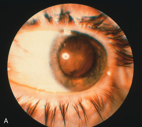
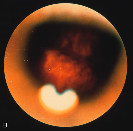
| Fig. 9. A. External view of an eye demonstrating dense vitreal inflammation secondary to reactivated toxoplasmosis in which a traction retinal detachment occurred. Fundus photography was precluded by the thick exudate. B. View after pars plana vitrectomy with scleral buckling shows the scleral buckle below and one prior lesion of active toxoplasmosis at the 8 o'clock position. |